
Mandibular bone transport reconstruction plate

a mandibular bone and reconstruction plate technology, applied in the field of mandibular bone transport reconstruction plate, can solve the problems of difficult task, no complete satisfaction, and segmental bone loss, and achieve the effect of appropriate stability

Active Publication Date: 2007-02-27
CRANIOTECH ACR DEVICES
View PDF6 Cites 30 Cited by
  • Summary
  • Abstract
  • Description
  • Claims
  • Application Information

AI Technical Summary

Benefits of technology

"The device described in this patent is a tool used to create new bone to fill gaps in the mandible after surgery. It uses a special plate that fixes the bone stumps on either side of the gap. The plate also has a track that allows a transport unit to move along the plate and create space for the new bone. The transport unit is attached to a disc that can be easily removed after the bone is formed. The technical effect of this device is that it simplifies the process of creating new bone in the mandible and reduces the risk of complications."

Problems solved by technology

Definitive treatment of many of the more common benign tumors may also require segmental resection due to high incidence of local recurrence after simple curettage or intra-lesional excision.
Blast injuries and high impact trauma to the mandible may lead to segmental bone loss either directly or due to surgical debridement.
This has always been a challenging task.
In spite of the wide variety of reconstruction methods, none of them is completely satisfactory.
Others, however, prefer not to do the definitive bony reconstruction at all.
Although good aesthetic results were reported with reconstruction plate alone with no bone replacement, this deprives the patient of having any restoration of occlusion, not even a removable lower denture.
This step is often technically difficult due to extensive fibrosis, loss of anatomical planes and landmarks, and the possibility of vascular injury that can result in serious bleeding or injury to the oral mucosa.
The procedure is lengthy and highly demanding especially when microvascular techniques are used.
The overall outcome of this technique, with either primary or secondary bone reconstruction was less than satisfactory.
Disadvantages of free non-vascularized bone graft include graft resorption, high incidence of failure due to resistant infection, especially with primary reconstruction, insufficient amount of bone for large gaps and donor site morbidity.
Except for iliac crest graft, the harvested bone mass was not sufficient either for osseointegrated implant insertion or carrying a removable lower denture.
Although the mesh does have to be removed after reconstruction, it still requires the use of large amount of autogenous bone graft, and has a high failure rate mainly due to resistant infection necessitating removal of the prosthesis.
Again, rehabilitation of jaw function was not achieved by this method.
However, achieving acceptable results with primary reconstruction methods was never an easy task.
The use of non-vascularized bone graft during primary reconstruction is not advised due to high incidence of failure.
In spite of their promising success rate, free vascularized flaps have high rates of complications, especially medical complications, which may be as severe as postoperative death.
These highly demanding techniques, which require a specialized surgical team, definitely add to the longevity and complications of the primary surgery.
Other disadvantages of vascularized tissue transfer include donor site morbidity.
Additionally, the characteristics of each flap design limited its use to only specific defects.
However, in addition to the technical problems associated with device assembly and application, the distraction devices had a completely independent function to the reconstruction plate.
In addition, the length range of the regenerate is limited by the length of the device, which makes it unable to reconstruct large mandibular defect especially those crossing the midline.
Yet, its disadvantages included poor control of the transport disc, pin-tract infection, pin extrusion, and retrusion of the newly formed bone in case of anterior reconstruction.

Method used


Image

  • Mandibular bone transport reconstruction plate
  • Mandibular bone transport reconstruction plate
  • Mandibular bone transport reconstruction plate

Examples


Embodiment Construction

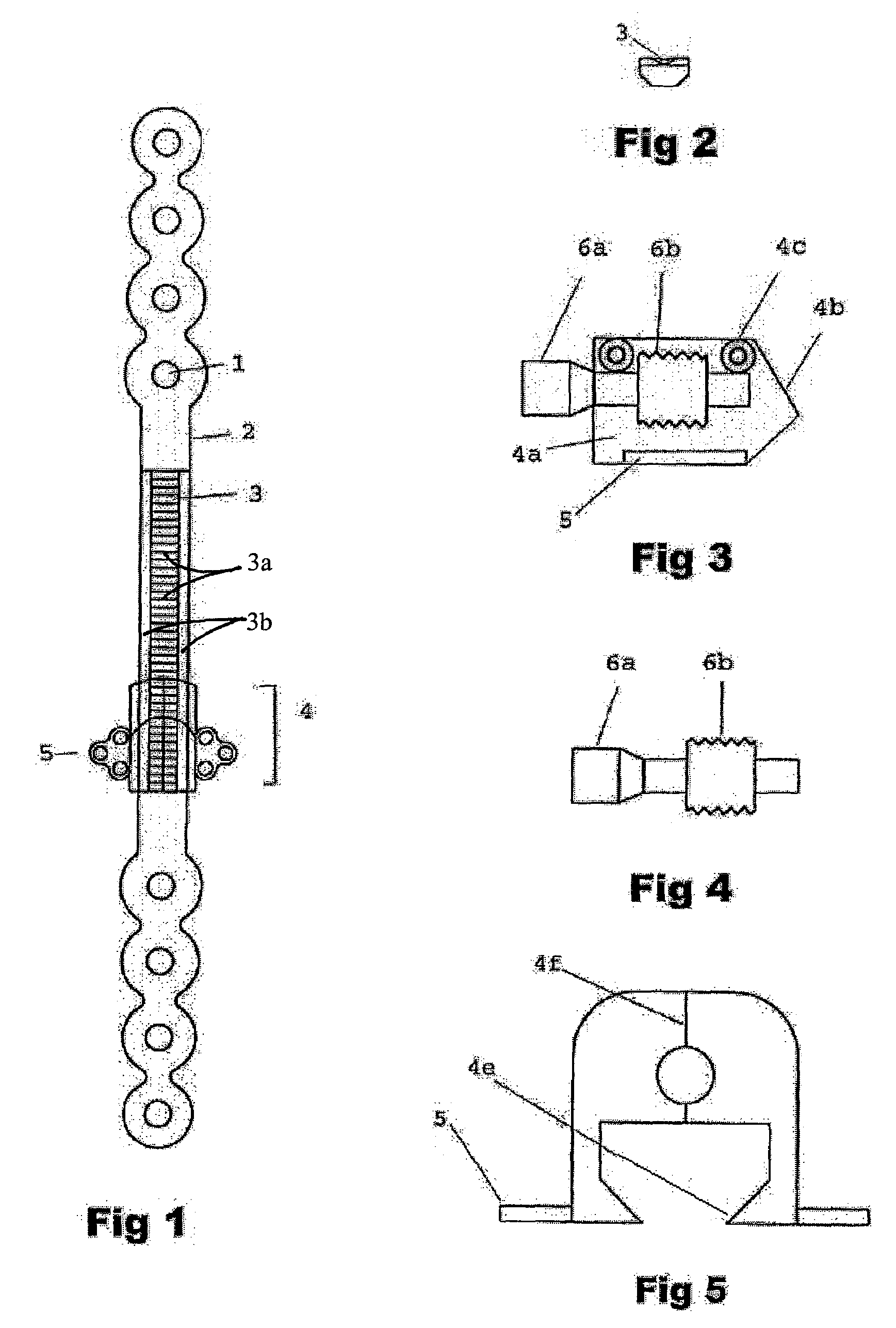
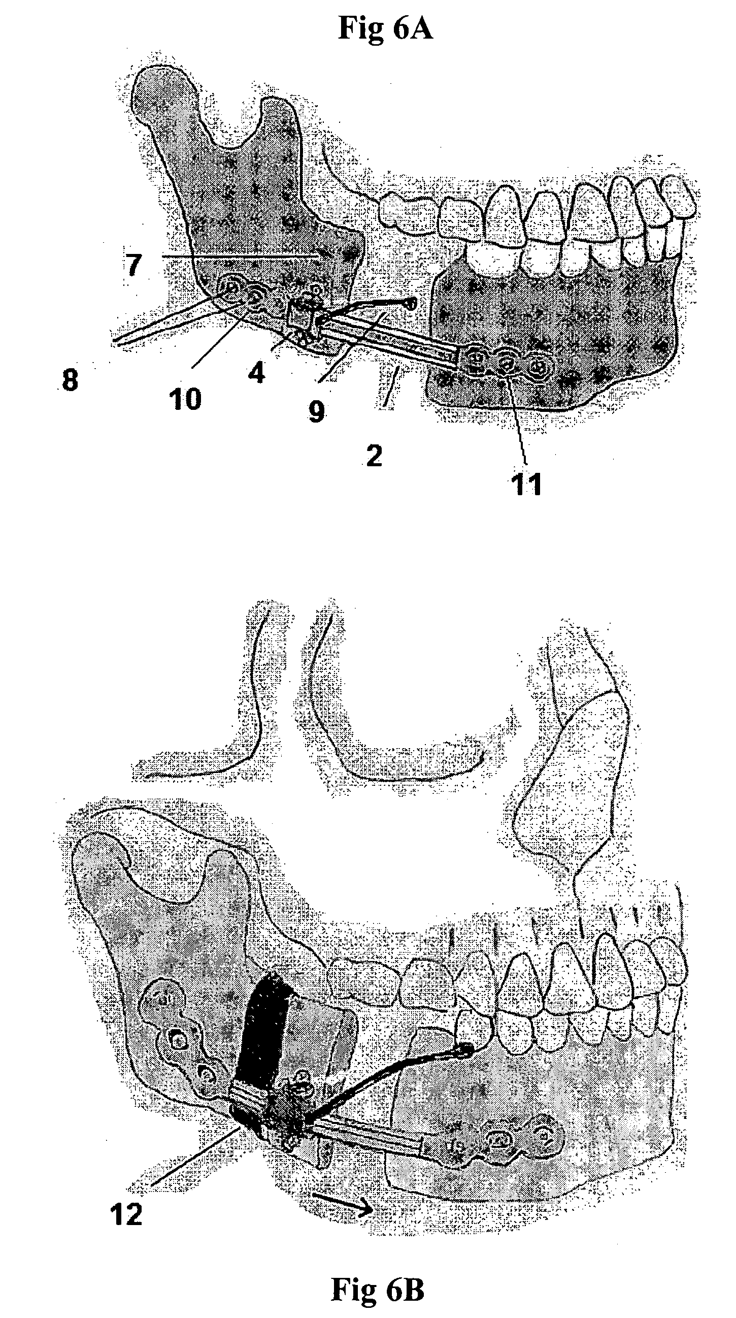
[0027]This device can be used to create new bone to fill a gap in the mandible after surgical excision. It uses the rigid bone reconstruction plate as distraction device. The reconstruction plate fixes the bone stumps on both sides of the bone gap by large (2.3–2.7 mm, bicortical titanium bone screws). In the middle segment of the plate overlying the bone gap, the transport bone disc is carried on a transport unit that moves along a rail on the outer surface of the reconstruction plate when a screw within the unit is activated.

[0028]The device is composed of a traditional titanium mandibular reconstruction plate (FIG. 1) with a middle segment (2) that functions as a straight track over which a transport unit can be moved from one end of the track to the other. The middle segment of the plate will be overlying the bone gap that resulted from surgical bone removal for any reason. The two ends of the plate (10 and 11) will be stabilized to the bone segments on both sides of the gap wit...


PUM


Abstract

This device can be used to create new bone to fill a gap in the mandible after surgical excision. It uses a bone reconstruction plate as a distraction device. The reconstruction plate fixes the bone stumps on both sides of the bone gap. In the middle segment of the plate overlying the bone gap, the transport bone disc is carried on a transport unit that moves along a rail on the outer surface of the reconstruction plate.

Description

PRIORITY[0001]This application claims the benefit of U.S. provisional application No. 60 / 552,272 file Mar. 11, 2004.FIELD OF THE INVENTION[0002]This device can be used to create new bone to fill a gap in the mandible after surgical excision. It uses a bone reconstruction plate as a distraction device. The reconstruction plate fixes the bone stumps on both sides of the bone gap. In the middle segment of the plate overlying the bone gap, the transport bone disc is carried on a transport unit that moves along a rail on the outer surface of the reconstruction plate.BACKGROUND OF THE INVENTION[0003]Segmental resection is a basic component of the surgical treatment of malignant mandibular tumors. Incidence of oral cancer in the US is 7.7 / 100,000 (30,000 case / year). Surgical resection may be followed by radiotherapy or chemotherapy. Definitive treatment of many of the more common benign tumors may also require segmental resection due to high incidence of local recurrence after simple curet...

Claims


Application Information

Patent Timeline
Patent Type & Authority Patents(United States)
IPC IPC(8): A61F2/28A61B17/58A61B17/80
CPCA61B17/8004A61B17/8071Y10S606/915
Inventor ELSALANTY, MOHAMMED E.MULONE, TIMOTHY D.
Owner CRANIOTECH ACR DEVICES
Learn More