Screening for papilloma viruses
a papilloma virus and papilloma virus technology, applied in the field of papilloma virus screening, can solve the problems of laborious and time-consuming, conflicting results of polyclonal antibody studies on the e4 proteins of mucosal viruses, and requires considerable experience to interpret results correctly
- Summary
- Abstract
- Description
- Claims
- Application Information
AI Technical Summary
Problems solved by technology
Method used
Image

Examples
example 1
Preparation of Anti-E4 Monoclonal and Polyclonal Immunoglobulins
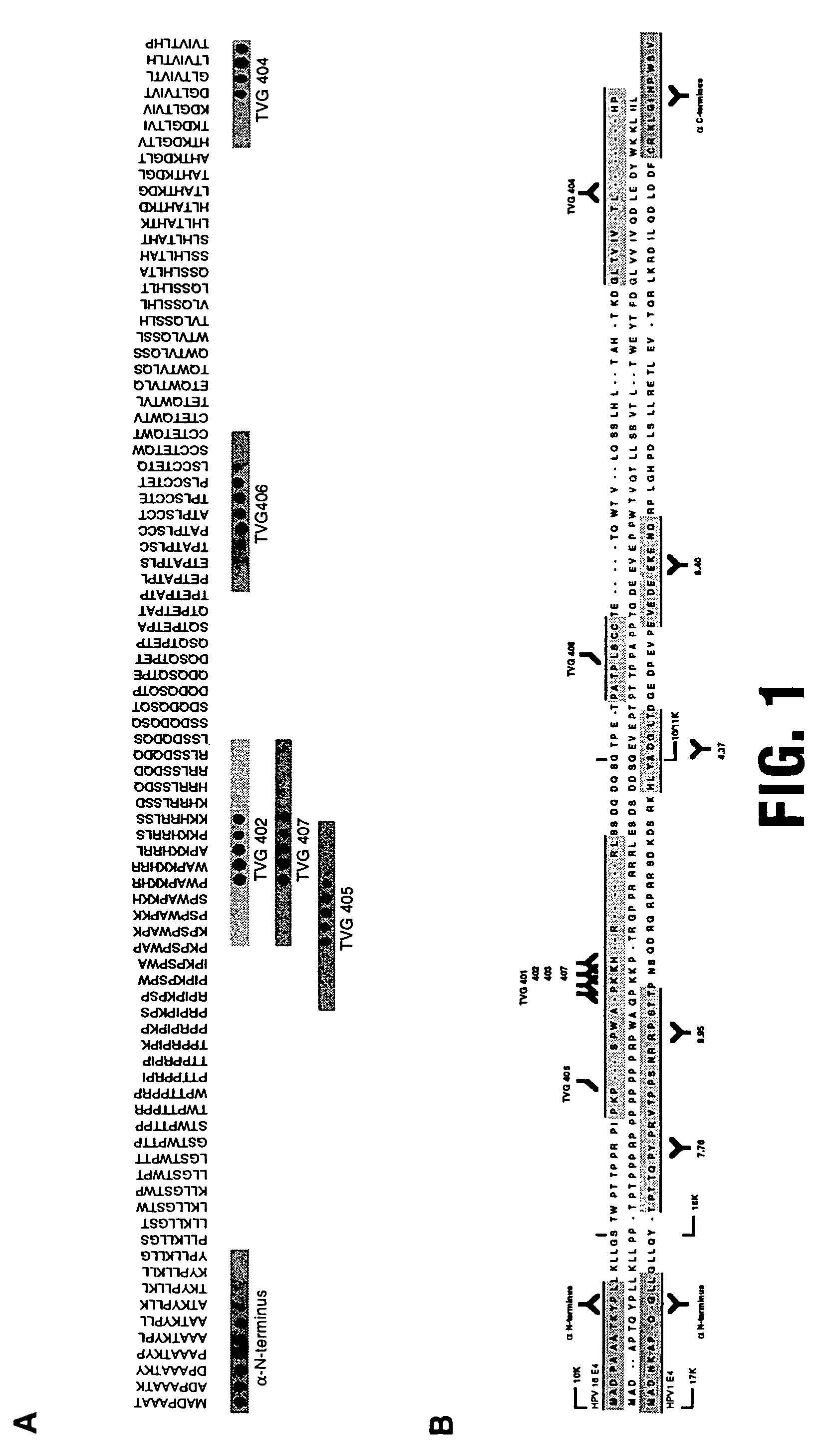
[0066]Although Mabs against HPV16 E1^E4 have been described previously (TVG401, 402, 403; Doorbar et al, 1992) these reagents recognise a single overlapping epitope at the major antigenic site of E4, and have been reported not to detect the protein in archival tissue biopsies (Doorbar et al, 1992).
[0067]Although these results suggest that E4 may not be a candidate for immunological detection of HPV, further antibodies are generated targeted at the N and C termini of HPV16 E4.
[0068]The generation of further Mabs by standard hybridoma technology results in the isolation of TVG404, an IgM which recognises an epitope at the very C-terminus of the protein.
[0069]To complement this reagent polyclonal antiserum to the N-terminus of the protein is raised against an N-terminal synthetic peptide (−E4 N term). Polyclonal antibodies (to HPV16 and HPV63 E4 proteins) are prepared by immunisation of rabbits with maltose binding protein...
example 2
Preparation of Synthetic Immunoglobulins
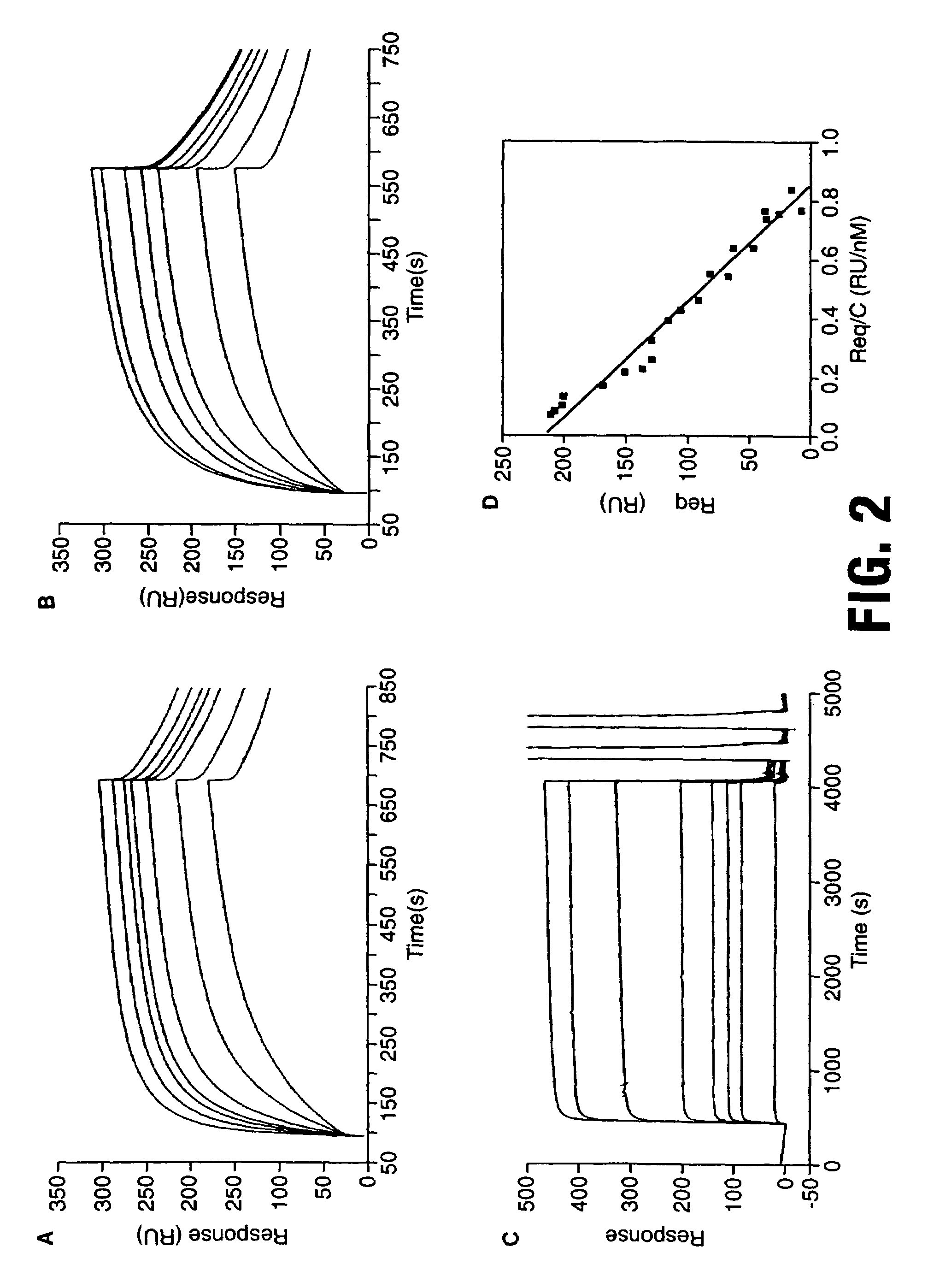
[0074]Fabs are isolated from a synthetic antibody displayed on fd bacteriophage (Griffiths et al, 1994) as described below. Immunotubes (Life Technologies, Paisley, UK) are coated overnight at 4° C. with either MBP-E4 or GST-E4 at a concentration of 0.1 g / ml. These are subsequently blocked at 37° C. for 1 hour in PBS / 2% marvel™ prior to incubation in the presence of 1011 phage on a blood tube rotator (37° C.). Unbound phage are poured off and tubes are washed 10× with PBS / 0.1% Tween 20. Binders are eluted with 100 mM triethylamine pH 11.0 (1 ml) and immediately neutralised with 1M Tris (pH8.0) before being reintroduced into E. coli TG1 cells. The enriched library is grown up and the selection procedure repeated three more times.
[0075]Phage selections are carried out alternately against GST 16 E1^E4 and MBP 16 E1^E4 in order to prevent isolation of antibodies to MBP or GST protein, using a repertoire of 6.5×10¹⁰ (Griffiths et al, 1994). MBP 16 ...
example 3
Preparation of Anti-E4 Peptides
[0086]A commercially available two-hybrid screening kit is purchased from ClonTech and employed for identifying naturally occurring E4-binding peptides, according to the instructions given by the manufacturer. A HeLa cDNA library, obtained from the same supplier, is screened. By this method, seven DNA sequences are isolated which encode E4-binding polypeptides, of which three are identified after sequencing.
[0087]The first peptide is ferritin. (SEQ ID NO: 1)
[0088]The second peptide is a keratin filament binding protein, which has the sequence set forth in (SEQ ID NO: 2).
[0089]The third polypeptide is a novel polypeptide recognised as a member of the DEAD box family of proteins, which contain the characteristic sequence motif DEAD (SEQ ID NO: 129). The sequence of the third polypeptide is shown in (SEQ ID NO: 3).
[0090]In order to identify the site of interaction between these polypeptides and E4, a series of overlapping peptides of between 10 and 20 ami...
PUM
| Property | Measurement | Unit |
|---|---|---|
| Hydrophilicity | aaaaa | aaaaa |
Abstract
Description
Claims
Application Information
 - R&D
- Intellectual Property
- Life Sciences
- Materials
- Tech Scout
- Unparalleled Data Quality
- Higher Quality Content
- 60% Fewer Hallucinations
Browse by: Latest US Patents, China's latest patents, Technical Efficacy Thesaurus, Application Domain, Technology Topic, Popular Technical Reports.
© 2025 PatSnap. All rights reserved.Legal|Privacy policy|Modern Slavery Act Transparency Statement|Sitemap|About US| Contact US: help@patsnap.com