
Detecting receptor oligomerization

a cell surface and receptor technology, applied in the field of cell surface membrane receptors, can solve the problems of lack of flexibility, lack of convenient and sensitive techniques, and difficult application of techniques for measuring such interactions, so as to reduce background, facilitate multiplexing, and improve the effect of sensitivity

Active Publication Date: 2006-09-12
VIROLOGIC INCORPORATED
View PDF42 Cites 70 Cited by
  • Summary
  • Abstract
  • Description
  • Claims
  • Application Information

AI Technical Summary

Benefits of technology

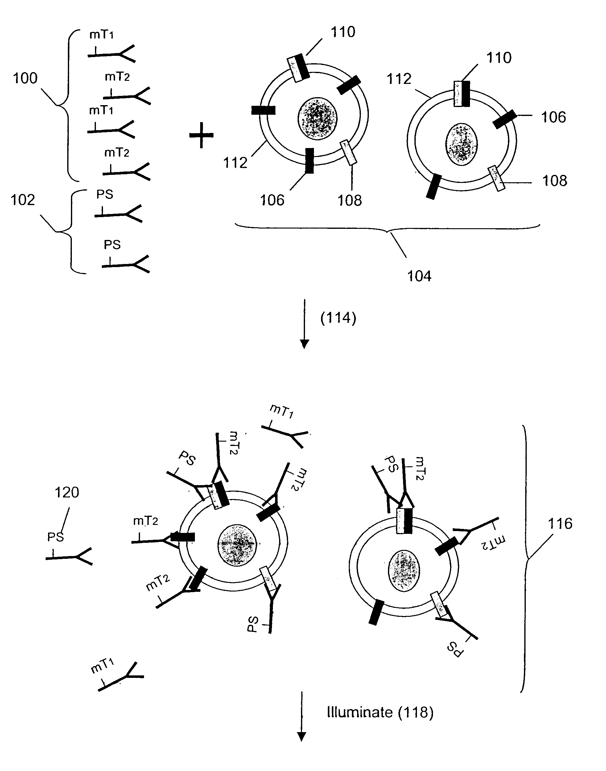
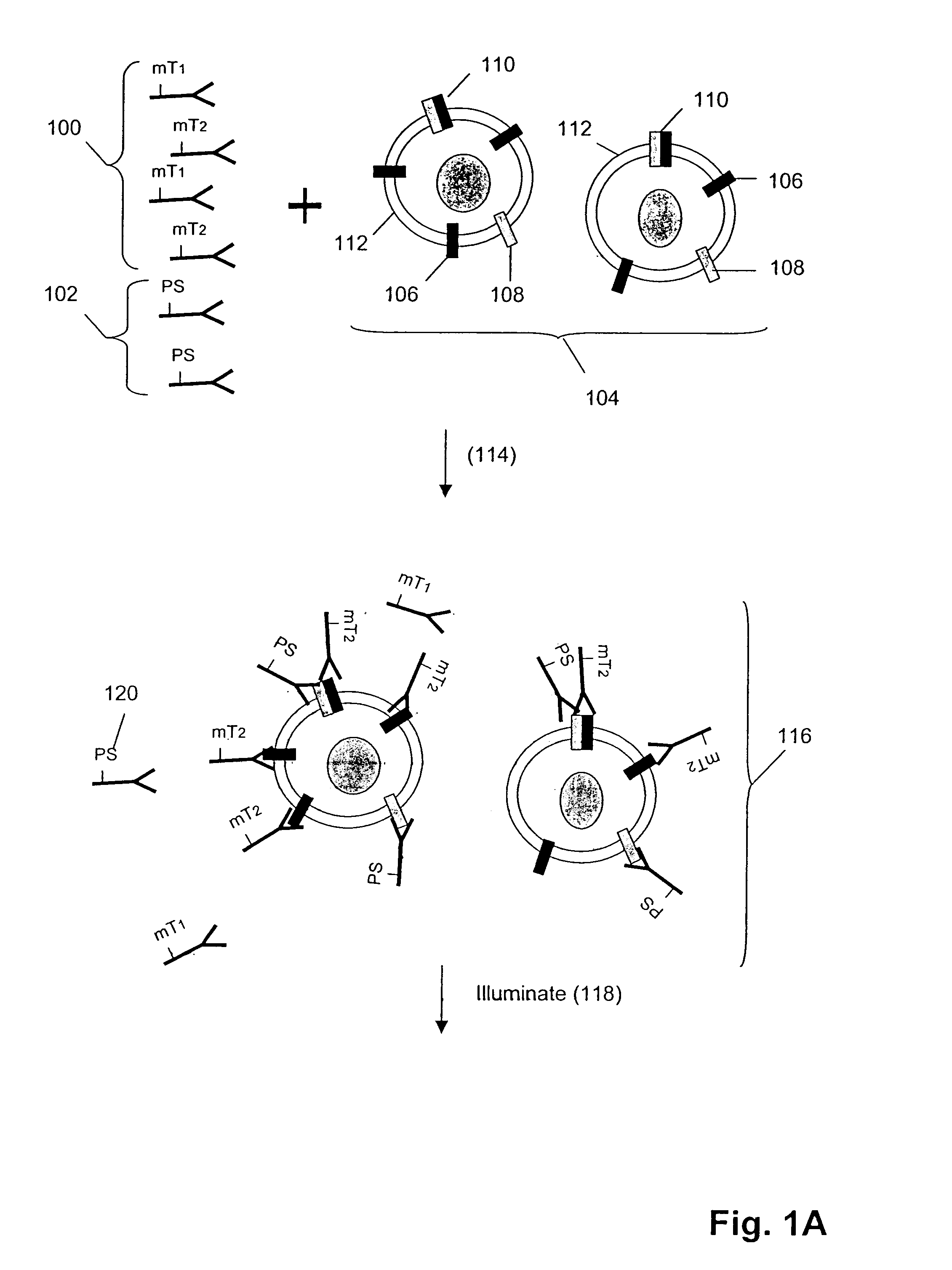
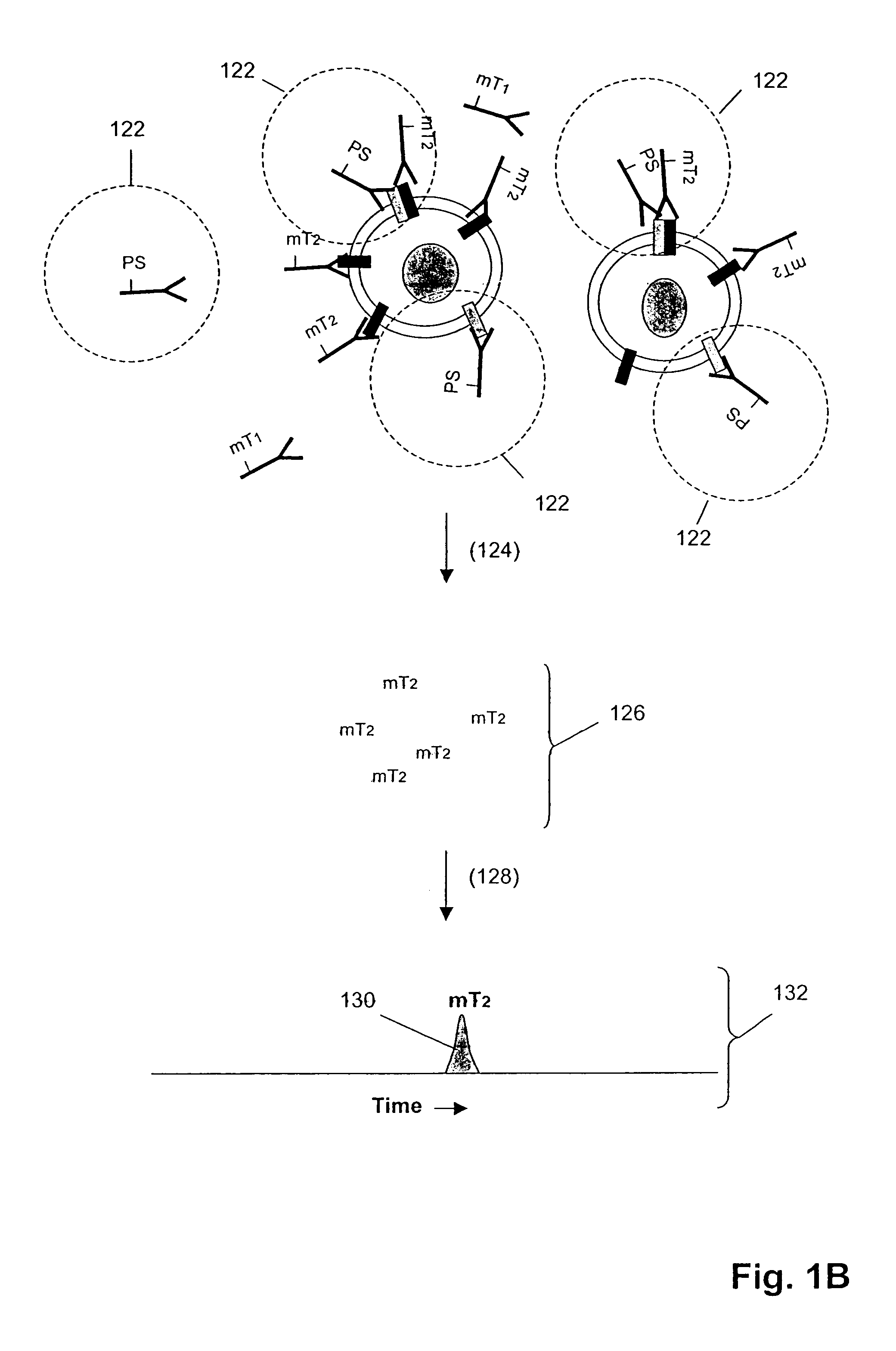
[0011]The present invention provides a method of detecting or measuring the dimerization or oligomerization of membrane-associated analytes that has several advantages over current techniques including, but not limited to, (1) the detection and / or measurement of molecular tags that are separated from an assay mixture provide greatly reduced background and a significant gain in sensitivity; and (2) the use of molecular tags that are specially designed for ease of separation and detection thereby providing convenient multiplexing capability.

Problems solved by technology

Unfortunately, despite the importance of receptor dimerization and oligomerization in signal transduction processes, the techniques for measuring such interactions are difficult to apply, lack flexibility, and lack sensitivity.
The lack of a convenient and sensitive technique for analyzing the oligomerization of cell surface molecules has greatly increased the difficulty of developing new therapeutics or diagnostic methods based on such phenomena.

Method used


Image

  • Detecting receptor oligomerization
  • Detecting receptor oligomerization
  • Detecting receptor oligomerization

Examples


example 1

Assay for Monitoring GABABR1 / GABABR2 Hetero-oligomerization

[0224]The γ-aminobutyric acidB (GABA)B receptor, a G-protein coupled receptor (GPCR), mediates stimulation of high-affimity GTPase activity in brain membranes by GABA to regulate potassium and calcium channels. The active form of this receptor, localized to the cell surface, has been shown to be a hetero-oligomer comprising the two receptors GABABR1 and GABABR2, which are both class III GPCRs and share 35% sequence identity (Jones, et al., 1998, Kaupmann, et al., 1998, White, et al., 1998, Milligan, 2001).

[0225]GABAB receptor hetero-oligomerization can be monitored using the methods of the present invention. First, epitope-tagged GABABR1 and GABABR2 receptors are generated and transfected into HEK293T cells as described by White, et al. (1998).

[0226]Protocol for transfection of HEK293T cells with GABABR1 and GABABR2 coding sequence[0227]1. cDNA encoding a Myc epitope (used with monoclonal antibody 9E10) is fused in-frame to ...

example 2

Analysis of Cell Lysates for Her-2 Heterodimerization and Receptor Phosphorylation

[0237]In this example, Her1–Her2 and Her2–Her3 heterodimers and phosphorylation states are measured in cell lysates from several cell lines after treatment with various concentrations of epidermal growth factor (EGF) and heregulin (HRG). Measurements are made using three binding compounds and a cleaving probe as described below.

Sample Preparalion:

[0238]1. Serum-starve breast cancer cell line culture overnight before use.[0239]2. Stimulate cell lines with EGF and / or HRG in culture media for 10 minutes at 37° C.

[0240]Exemplary doses of EGF / HRG are 0, 0.032,0.16, 0.8,4,20,100 nM for all cell lines (e.g. MCF-7, T47D, SKBR-3) except BT20 for which the maximal dose is increased to 500 nM because saturation is not achieved with 100 nM EGF.[0241]3. Aspirate culture media, transfer onto ice, and add lysis buffer to lyse cells in situ.[0242]4. Scrape and transfer lysate to microfuge tube. Incubate on ice for 30 ...

example 3

Analysis of Tissue Lysates for Her2 Heterodimerization and Receptor Phosphorylation

[0266]In this example, Her1–Her2 and Her2–Her3 heterodimers and phosphorylation states are measured in tissue lysates from human breast cancer specimens.

Sample Preparation:

[0267]1. Snap frozen tissues are mechanically disrupted at the frozen state by cutting.[0268]2. Transfer tissues to microfuge tube and add 3× tissue volumes of lysis buffer (from appendix I) followed by vortexing to disperse tissues in buffer.[0269]3. Incubate on ice for 30 min with intermittent vortexing to mix.[0270]4. Centrifuge at 14,000 rpm, 4° C., for 20 min.[0271]5. Collect supernatants as lysates and determine total protein concentration with BCA assay (Pierce) using a small aliquot.[0272]6. Aliquot the rest for storage at -80C until use.

Assay design:[0273]1. The total assay volume is 40 ul.[0274]2. The lysates are tested in serial titration series of 40, 20, 10, 5,2.5, 1.25, 0.63, 0.31 ug total-equivalents and the volume is...


PUM


Abstract

Methods are provided for detecting formation of oligomeric complexes of molecules on the surface of cell membranes. These methods employ pairs of tagged probes and cleaving probes, each of which binds specificly to a cell surface molecule. The tagged probe includes a molecular tag that is linked to a first binding compound through a cleavable linkage, and the cleaving probe includes a second binding agent and a cleavage-inducing moiety that can cleave the linkage when within a defined proximity thereto. Binding of the two probes to cell surface molecules that have formed an oligomeric complex results in release of the molecular tag from the binding compound, providing a measure of formation of the complex.

Description

[0001]This patent application claims priority from U.S. Provisional Application Ser. No. 60 / 398,724 filed Jul. 25, 2002, which is incorporated herein by reference in its entirety.FIELD OF THE INVENTION[0002]The present invention relates to methods for measuring oligomerization of cell surface molecules, particularly cell surface membrane receptors.BACKGROUND OF THE INVENTION[0003]The interactions of cell surface membrane components play crucial roles in transmitting extracellular signals to a cell in normal physiology, and in disease-conditions. In particular, many types of cell surface receptors undergo dimerization or oligomerization in connection with the transduction of an extracellular event or signal, e.g. ligand-receptor binding, into a cellular response, such as proliferation, increased or decreased gene expression, or the like, e.g. George et al, Nature Reviews Drug Discovery, 1: 808–820 (2002); Mellado et al, Ann. Rev. Immunol., 19: 397–421 (2001); Schlessinger, Cell, 103:...

Claims


Application Information

Patent Timeline
Patent Type & Authority Patents(United States)
IPC IPC(8): G01N33/53C12Q1/68G01NG01N27/447G01N33/483G01N33/542G01N33/543G01N33/556G01N33/566G01N33/567G01N33/68
CPCG01N33/542G01N33/566G01N2333/726G01N2333/71
Inventor CHAN-HUI, PO-YINGSHI, YININGPIDAPARTHI, SAILAJADUA, RAJIVSINGH, SHARAT
Owner VIROLOGIC INCORPORATED
Learn More