
X-ray therapy electronic portal imaging system and method for artifact reduction

a portal imaging and x-ray therapy technology, applied in the field of x-ray imaging and dosimetric measurements, can solve the problems of intensity artifacts generated in the resulting images, and achieve the effect of reducing pulse rate artifact variations, reducing or eliminating non-linearities

Inactive Publication Date: 2006-07-11
SIEMENS MEDICAL SOLUTIONS USA INC
View PDF2 Cites 19 Cited by
  • Summary
  • Abstract
  • Description
  • Claims
  • Application Information

AI Technical Summary

Benefits of technology

This approach results in stable and accurate dosimetric measurements by aligning and correcting pulse variation artifacts, improving the quality of x-ray images and reducing their impact on therapy verification.

Problems solved by technology

The scanning period is longer than the pulse rate period, so intensity artifacts are generated within the resulting images.

Method used


Image

  • X-ray therapy electronic portal imaging system and method for artifact reduction
  • X-ray therapy electronic portal imaging system and method for artifact reduction
  • X-ray therapy electronic portal imaging system and method for artifact reduction

Examples


Embodiment Construction

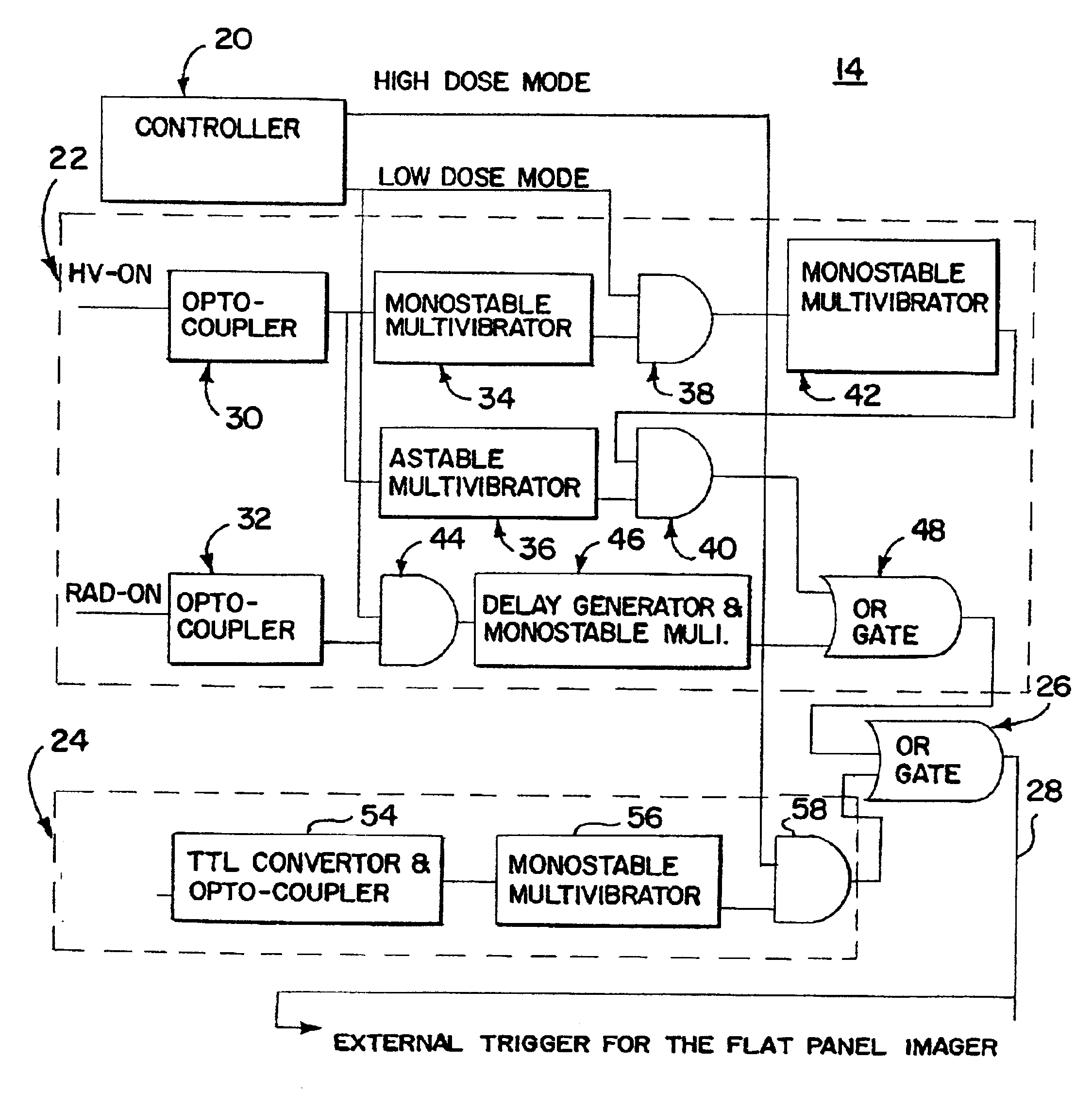
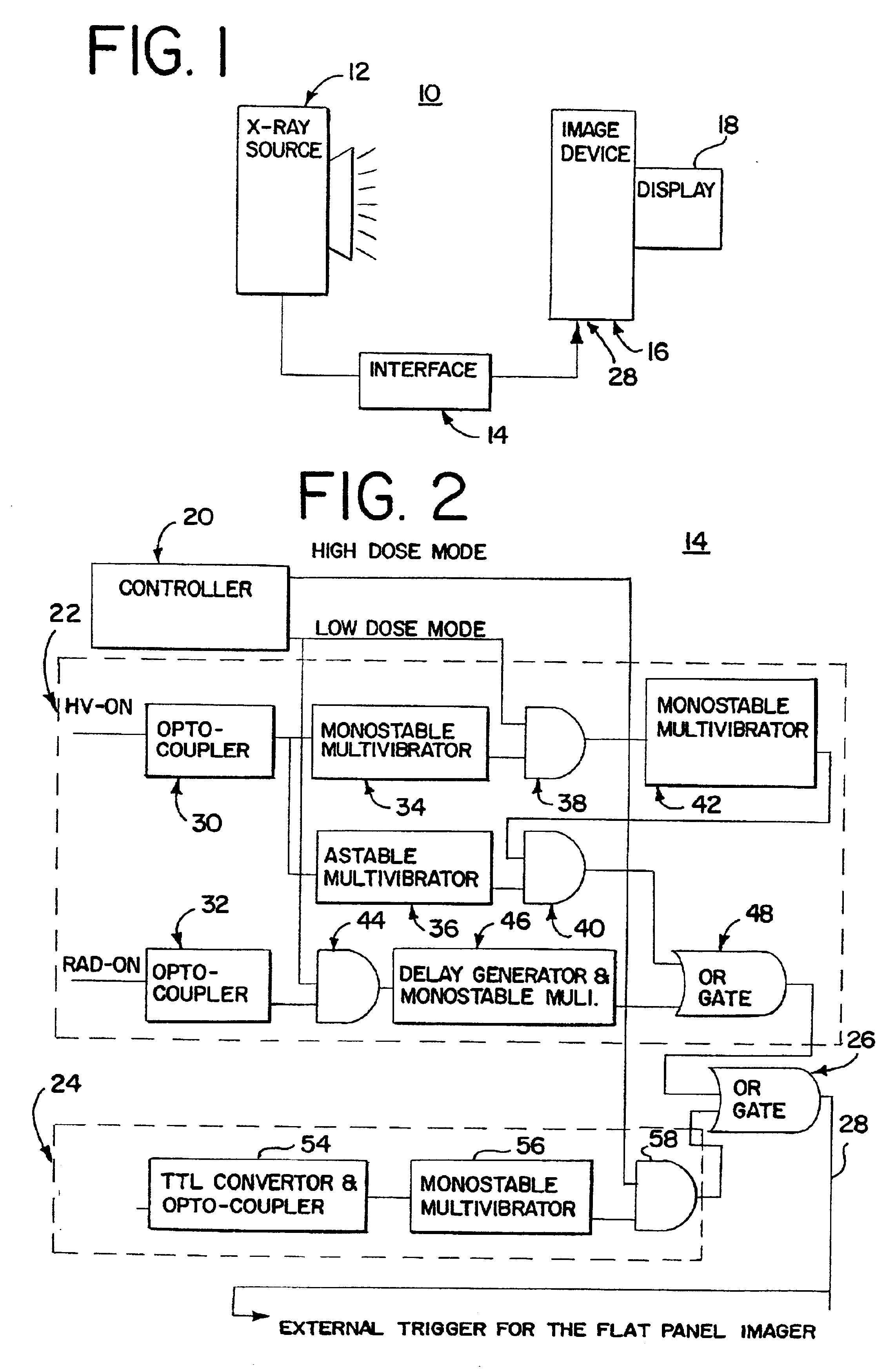
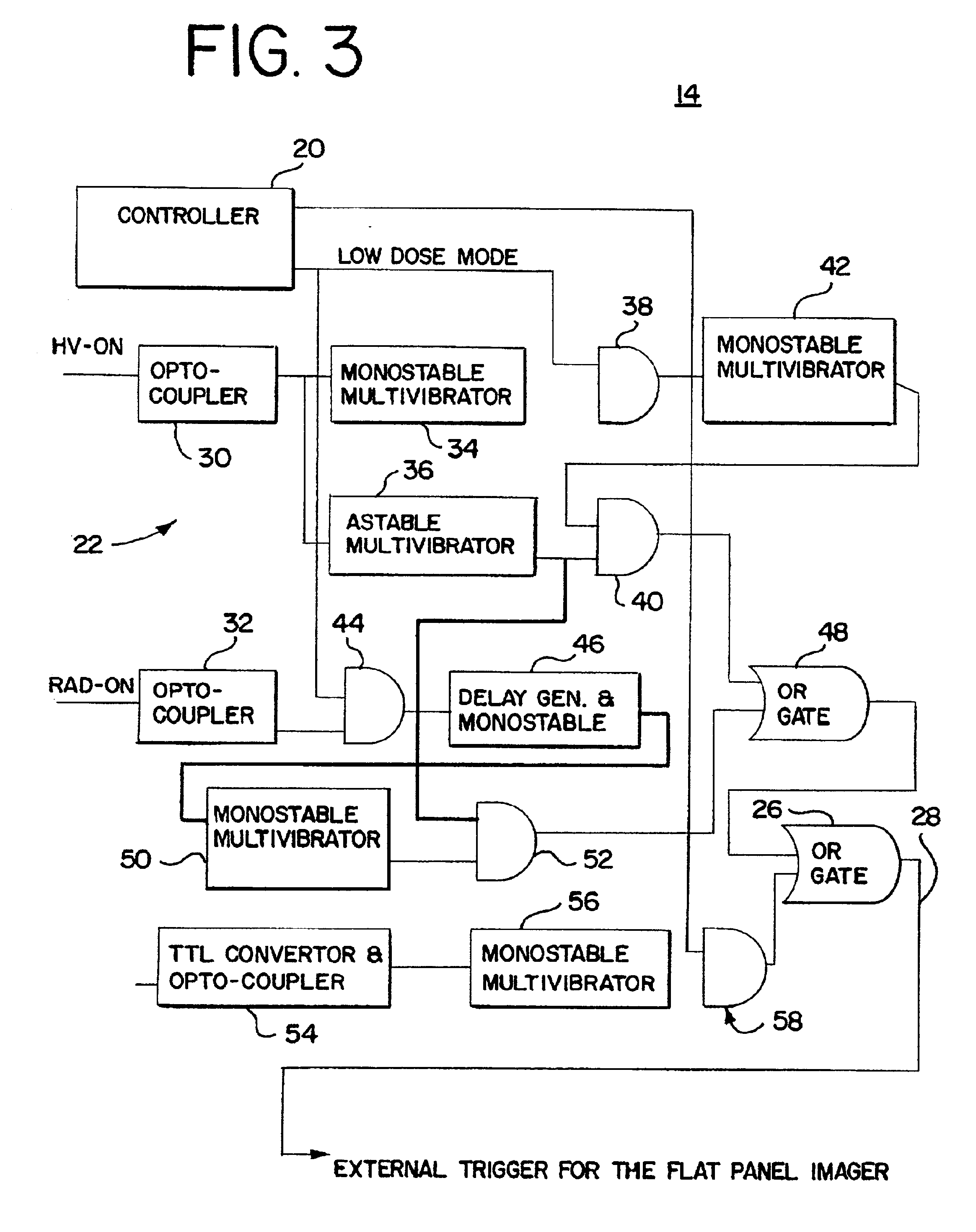
[0018]Image scans of a digital x-ray imaging device are synchronized with pulses of x-rays from an x-ray source. Synchronization aligns the resulting pulsing artifacts within multiple images. Location of the artifacts is known, and the combination of multiple images results in distinct artifact patterns. The artifacts are associated with linear positions within the images, such as horizontal lines across images. By controlling gain as a function of line, the x-ray pulse variation artifacts are removed or reduced. Electronic readouts provide information for stable, accurate dosimetric measurements.

[0019]FIG. 1 shows an x-ray therapy system 10 of one embodiment. The x-ray therapy system 10 includes an x-ray source 12, and interface 14, and an imaging device 16 with a display 18. Additional components, such as a patient bed, motors, components for Intensity Modulation Radio Therapy (IMRT) or other therapeutic x-ray components, may be included. In alternative embodiments, the interface ...


PUM


Abstract

X-ray therapy EPI artifact reduction and control of imaging is provided. Scanning of images is synchronized with the pulse rate of the x-rays. The scanning period is longer than the pulse rate period, so artifacts are generated within the resulting images. Due to the synchronization, the pulse variation artifacts are aligned across multiple images. The synchronization and resulting alignment of linear artifacts allows for gain correction as a function of lines within the image. Such gain correction reduces or removes non-linearities associated with pulse rate variation.

Description

BACKGROUND[0001]The present invention relates to x-ray imaging and dosimetric measurements. In particular, reduction of linear accelerator pulsing artifacts for electronic imaging devices of x-rays are provided.[0002]X-ray treatment uses the therapeutic application of x-ray energy to destroy tumor tissue or for other therapy. X-rays generated by a megavoltage or other high voltage sources generate x-ray pulses or periodically vary the amplitude of the x-rays output, such as every 5 milliseconds.[0003]X-ray imaging detectors output signals responsive to the incident x-rays. The variation in the x-rays results in image artifacts. Where multiple images are combined or averaged, the pulse rate variation artifacts are randomly averaged or combined. The resulting combined image also undesirably includes artifacts.[0004]X-ray images generated digitally are used for dosimetric treatment verification. The pulse rate variation artifacts introduce a non-linearity within the images. The artifac...

Claims


Application Information

Patent Timeline
Patent Type & Authority Patents(United States)
IPC IPC(8): A61N5/10A61B6/00H04N5/32H04N5/325H04N5/357H05G1/60
CPCA61B6/00A61B6/4233A61B6/488H05G1/60H04N5/325H04N5/357H04N5/32H04N25/30H04N25/68H04N25/60
Inventor GHELMANSARAI, FARHAD ABBASICOLLINS, WILLIAM F.
Owner SIEMENS MEDICAL SOLUTIONS USA INC
Learn More