
Method of forming vascularized kidney tissue

a vascularized kidney and tissue technology, applied in the field of new kidney engineering in vitro, can solve the problems of inability to promote the proliferation and branching morphogenesis of isolated ub in vitro, no known soluble factor or set of factors had been able to induce ub branching morphogenesis in vitro, and a variety of defects in the kidney and urinary collecting system

Inactive Publication Date: 2006-07-11
RGT UNIV OF CALIFORNIA
View PDF2 Cites 14 Cited by
  • Summary
  • Abstract
  • Description
  • Claims
  • Application Information

AI Technical Summary

Benefits of technology

[0017]A further, most preferred object is to provide a bank of embryonic organs and tissues capable of replacing diseased, or otherwise incapacitated vital organs and tissues, minimizing the need for matching donors and / or immunosuppressive drugs.

Problems solved by technology

Further, no known soluble factor or set of factors had been able to induce UB branching morphogenesis in vitro.
Nevertheless, recent studies have also shown that glial cell line derived neurotrophic factor (GDNF) is necessary for early UB outgrowth, but on its own, it fails to promote proliferation and branching morphogenesis of isolated UB in vitro.
It is now clear that a variety of defects in the kidney and urinary collecting system are the result of abnormal development of these structures in the fetus.
The molecular basis for these diseases is poorly understood; however, it appears, in many instances, that the problem lies in defective morphogenesis of the ureteric bud.
Since normal kidney development depends critically upon mutual inductive interactions between the UB and the metanephric mesenchyme (MM), inefficient or defective branching morphogenesis of the UB would be expected to result in various aplastic, hypoplastic, and perhaps dysplastic diseases.
In addition, several targeted gene-deletion experiments have resulted in phenotypes that may be best characterized as resulting form defective UB morphogenesis (directly or indirectly).
This may well be the result of defective branching morphogenesis during development of the urinary collecting system, because the degree of ureteric bud branching during collecting system development determines the number of nephrons in the adult kidney.
Furthermore, no known soluble factor or set of factors had been able to induce UB branching morphogenesis in vitro.
Nevertheless, recent studies have demonstrated that glial cell line derived neurotrophic factor (GDNF) is necessary for early UB outgrowth, but as the inventors have shown, on its own, it fails to promote proliferation and branching morphogenesis of isolated UB in vitro (FIG. 6).

Method used


Image

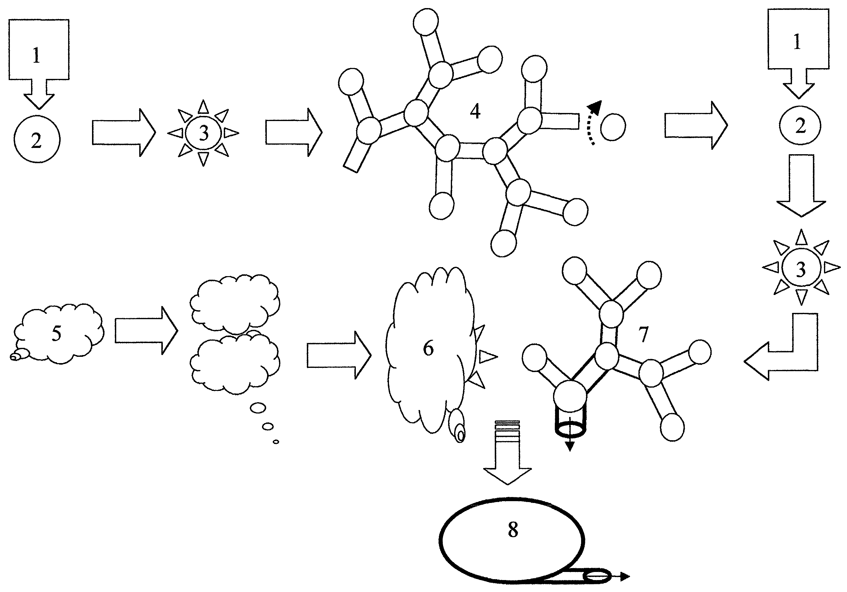
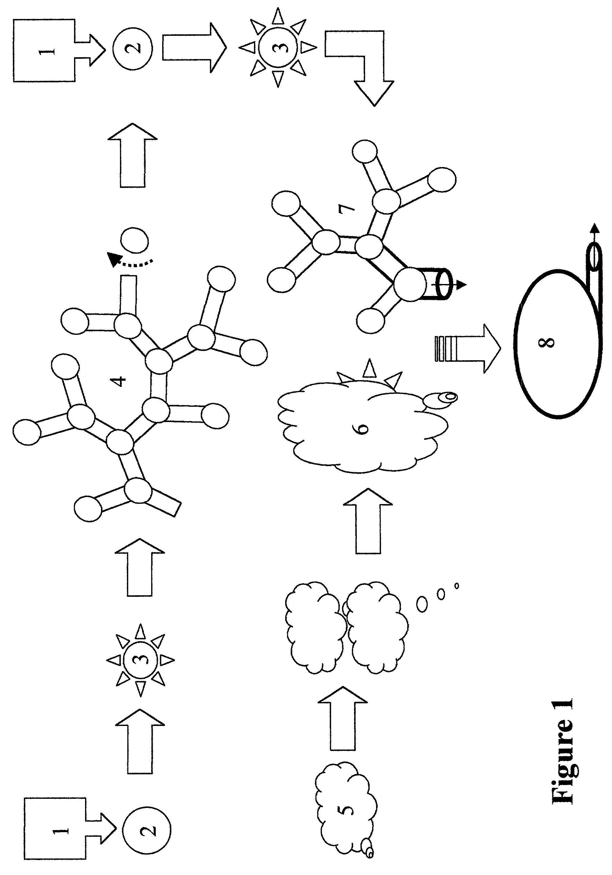
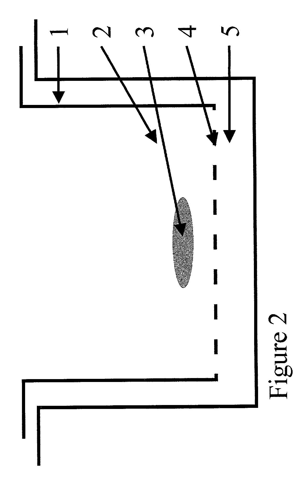
  • Method of forming vascularized kidney tissue
  • Method of forming vascularized kidney tissue
  • Method of forming vascularized kidney tissue

Examples


example 1

[0041]Isolation of ureteric bud (UB) epithelium and UB culture (Proc. Natl. Acad. Sci., 86, 7330–7335, 1999 incorporated herein by reference): Kidney rudiments were dissected from timed pregnant Sprague Dawley rats at gestation day 13. (The plug day was designated as day 0). The UB was isolated from mesenchyme by incubating kidney rudiments in 0.1% trypsin in the presence of 50 U / ml DNAase at 37° C. for 15 minutes, and by mechanical separation with two fine-tipped minutia pins. For culture, Transwell tissue culture plates and a polycarbonate membrane insert with 3 um pore size were used. The extracellular matrix (ECM) gel (a mixture of type I collagen and Matrigel) was applied on top of the Transwell insert. Isolated UB was suspended in the ECM gel and cultured at the interface of air and medium. All cultures were carried out at 37° C. with 5% CO2 and 100% humidity in DMEM / F12 supplemented with 10% Fetal Calf Serum (FCS). Growth factors were added as indicated elsewhere. Culture med...

example 2

[0042]Cells and conditioned media: The BSN cell line was derived from day 11.5 mouse embryonic kidney metanephric mesenchyme originally obtained from a mouse line transgenic fro the early region of SV-40 / large T antigen. As described elsewhere, the BSN cells express the mesenchymal protein marker vimentin, but not classic epithelial marker proteins such as cytokeratin, ZO-1 and E-cadherin. Differences in the expression patterns of 588 genes in BSN cells have been analyzed by the inventors on commercially available cDNA grids (Am. J. Physiol.—Renal Physiol., 277, F:650–F663, 1999), and confirmed the largely non-epithelial character of BSN-cells, though it remains to be determined whether they are mesenchymal or stromal, or have characteristics of both cell types. The SV-40 / large T antigen transformed UB cell line and murine inner-medulla collecting duct (mIMCD) cells have been extensively characterized before. To obtain conditioned media, a confluent cell monolayer was washed with se...

example 3

[0043]The ECM gel mix: The ECM gel mix was composed of 50% type I collagen (Collaborative Biomedical Product) and 50% growth factor-reduced Matrigel® (Collaborative Biomedical Product). The procedure for gelation has been previously described in detail and is incorporated herein.


PUM


Abstract

A method for constructing a stable bioactive mammalian embryonic kidney is described herein. A kidney so constructed requires no artificial support, nor porous man made membranes or tubing to effectuate its biological function of filtering body fluids. A single donor embryonic kidney, or fragment thereof, can produce a great number of functional kidneys suitable for treating subjects with various kidney disorders. It is anticipated that said in vitro produced kidney would be less, or not at all, antigenic when transplanted into a subject, because of its embryonic character and artificial propagation in culture. This method of producing a functional organ can be useful in cloning other organ structures containing inducible epithelial tissues.

Description

BACKGROUND OF THE INVENTION[0001]1. Field of the Invention[0002]The present invention generally concerns a new method of engineering a kidney in vitro.[0003]The present invention particularly concerns a new method and procedure for propagating cloned kidney members from embryonic ureteric bud tips grown in vitro under specific culture conditions.[0004]2. Description of Related Art[0005]Branching tubulogenesis is an essential mechanism by which epithelial tissues such as kidney, salivary gland and prostate develop (Proc. Natl. Acad. Sci. USA, 96, 7330–7335, 1999 incorporated herein by reference). Largely based on the classical studies of Brobstein and coworkers, direct interactions between mesenchymal and epithelial components of embryonic tissue have been thought to be crucial for branching morphogenesis in most epithelial tissues. During kidney development, for example, direct cell-cell interactions between the metanephric mesenchyme and the epithelial component, the ureteric bud (...

Claims


Application Information

Patent Timeline
IPC IPC(8): C12N5/06C12N5/071
CPCA61L27/3604A61L27/3641A61L27/3839C12N5/0686C12N2501/105C12N2533/90C12N2501/115C12N2501/12C12N2501/13C12N2502/256C12N2533/54C12N2501/11A61L27/3895
Inventor NIGAM, SANJAY KUMARQIAO, JIZENGSAKURAI, HIROYUKI
Owner RGT UNIV OF CALIFORNIA
Learn More