
Method and apparatus for reducing electroporation-mediated muscle reaction and pain response

a technology of electroporation and muscle reaction, applied in electrotherapy, heart stimulators, therapy, etc., can solve the problems of bleomycin, inability to effectively penetrate the membranes of certain cancer cells, and inability to effectively remove bleomycin, so as to reduce pain and reduce discomfort.

Inactive Publication Date: 2006-05-30
INOVIO PHARMA
View PDF4 Cites 231 Cited by
  • Summary
  • Abstract
  • Description
  • Claims
  • Application Information

AI Technical Summary

Benefits of technology

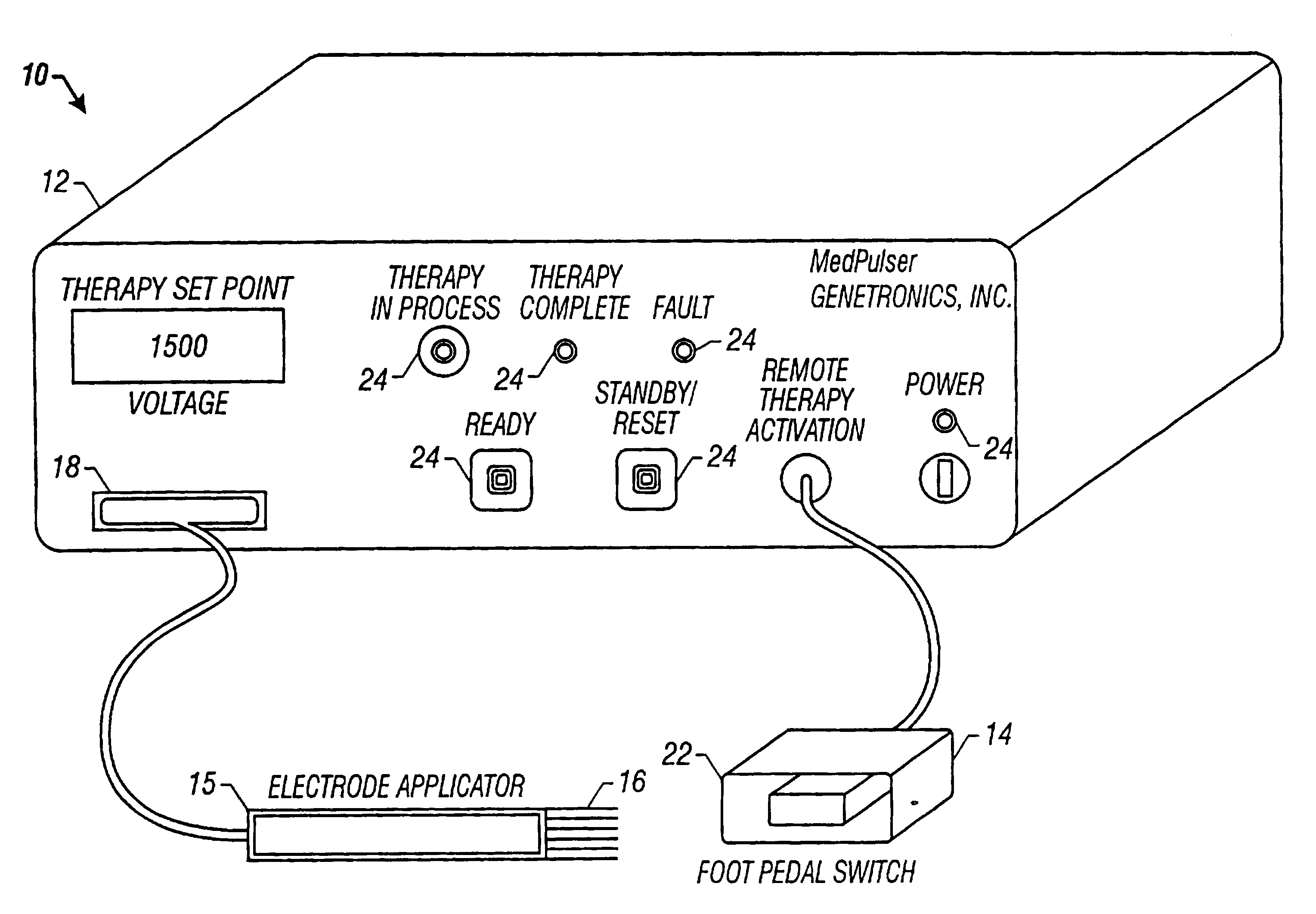
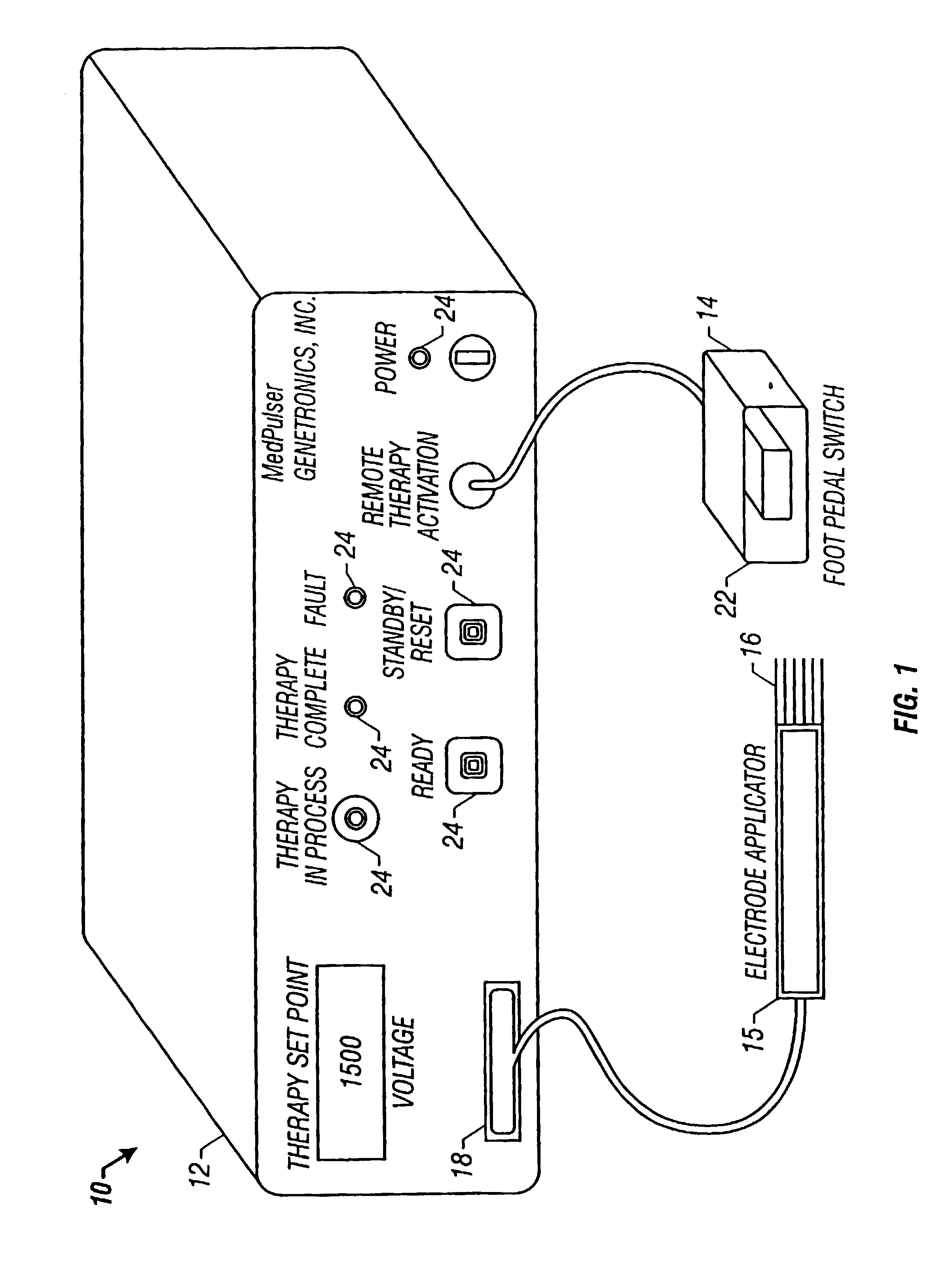
[0011]The present invention is based on the discovery of apparatuses, instruments and methods for reducing pain often associated with clinical use of electroporation and delivery of agents to cells. The invention provides such apparatus and instruments which allow administration of electric pulses to a patient while reducing the level of discomfort.

Problems solved by technology

Electroporation in vivo is often limited to tissue or cells that are close to the skin of the organism where the electrodes can be placed.
Therefore, tissue which would otherwise be treatable by systemic drug delivery or chemotherapy, such as a tumor, is generally inaccessible to electrodes used for electroporation.
Some of the anticancer drugs, for example, bleomycin, normally cannot penetrate the membranes of certain cancer cells effectively.
When large or internal tumors are to be treated, it is not easy to properly locate electrodes and measure the distance between them.
Despite the suitability of the epidermis as a target tissue for gene therapy or DNA vaccination, there are significant barriers to safe, easy, efficient, and economical gene delivery.
However, the electrical signals which are typically used for electroporation cause considerable discomfort to a patient.
There is often enough discomfort that patients are given general anesthesia before receiving the electroporation treatment.

Method used


Image

  • Method and apparatus for reducing electroporation-mediated muscle reaction and pain response
  • Method and apparatus for reducing electroporation-mediated muscle reaction and pain response
  • Method and apparatus for reducing electroporation-mediated muscle reaction and pain response

Examples


example 1

[0177]Six volunteers were each exposed to four different electroporation signals. Each electroporation signal had a bipolar square waveform and a different frequency. The volunteers were asked to rate their degree of discomfort on a scale of 0 to 10 where 0 meant no pain and 10 was unbearable. The responses related to the same electroporation signals were averaged. FIG. 15 plots the averaged discomfort level versus the frequency of the electroporation signals. Increasing the signal frequency more than halved the discomfort level.

example 2

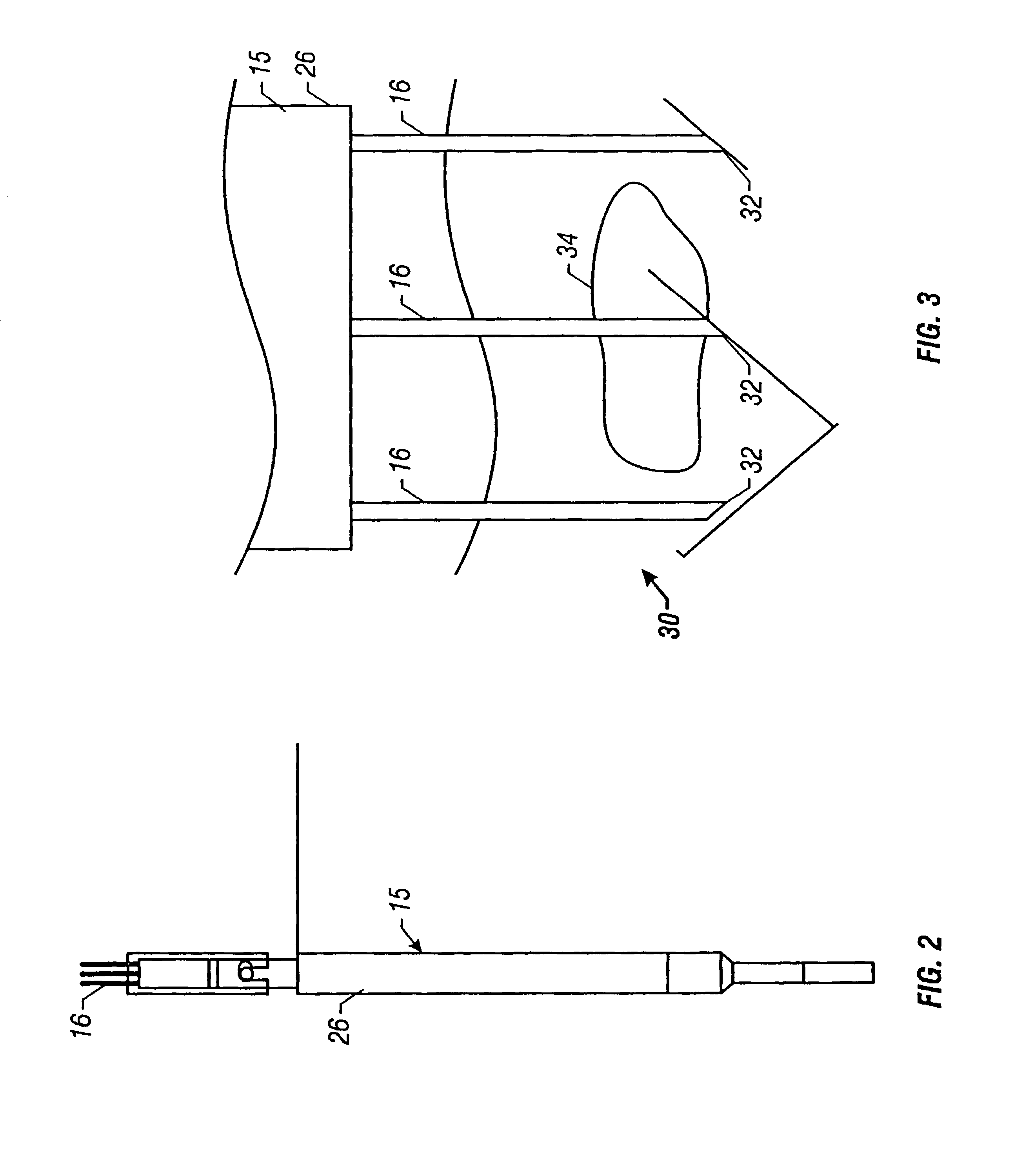
[0178]HEp-2 (Epidermoid carcinoma of human larynx, ATCC CCL-23, passage no 350) were obtained from American Type Culture Collection, Rockville, Md. Cells were grown in Eagle's MEM (Gibco, BRL) supplemented with 10% FCS, 0.1 mM Non-essential Amino Acids, 1.0 mM Sodium pyruvate, and 1% L-glutamine in a 5% CO2—air atmosphere at 37° C. Cells growing in an exponential phase were harvested by trypsinization and viability determined by trypan blue dye exclusion test. A cell suspension in culture medium was prepared at a concentration of 222,000 cells / ml and cells seeded in a 96 well plate at a final concentration of 40,000 cells per well. Cells were pulsed using 0.5 cm 6-needle hexagonal array electrodes 16 connected to a prototype bipolar pulse generator. The needle array was inserted in the well of a 96-well microplate and pulsed with the selected experimental parameters. The plates were incubated for 44 hrs in a 5% CO2-air atmosphere at 37° C. before carrying out the XTT cell survival a...

example 3

[0180]BALB / c A nu / nu mice were surgically implanted with HEp-2 tumors in a subcutaneous sac made in the right flank of nude mice. The tumors 34 were allowed to grow and were treated when their average size was about 80 mm3. The drug, bleomycin, 0.5 units dissolved in 0.15 ml of saline, was injected in each mouse intratumorally using a 30 gauge needle. The drug was injected very slowly at the tumor base and the needle direction rotated (fanning technique) for uniform drug distribution in the tumor 34. The mice in the control were only pulsed D−E+; D=Drug, E=Electric field, + / −denote presence or absence, respectively) while those in the treated group were pulsed and received drug (D+E+). A time lapse of 10+ / −1 minute was maintained between the drug injection and the application of electric pulse to allow bleomycin to spread uniformly throughout the tumor 34. The electrical pulses, generated by the prototype bipolar square wave pulse generator, were delivered to the tumor 34 through a ...


PUM


Abstract

A method for delivery of an agent to a cell using electroporation is disclosed. The method includes positioning a first electrode and a second electrode such that an electrical signal passed between the first electrode and the second electrode passes through the cell. The method also includes passing an electrical signal between the first electrode and the second electrode, the electrical signal having a frequency greater than about 10 kHz. In one embodiment of the method, the electrical signal has a bipolar waveform. In another embodiment of the method, the electrodes are positioned at a treatment site, e.g., a tumor, for in vivo delivery of an agent.

Description

[0001]This is a divisional of U.S. patent application Ser. No. 09 / 535,683, filed Mar. 23, 2000, which is a conversion of U.S. Provisional Patent Application No. 60 / 126,953 and of PCT / US00 / 07972, filed Mar. 23, 2000, all of which are incorporated disclosure of this application.RELATED APPLICATION[0002]This application relies for priority under 35 U.S.C. §119(e)(1) on provisional application Ser. No. 60 / 126,953, filed Mar. 25, 1999.FIELD OF THE INVENTION[0003]The present invention relates generally to the use of electric pulses to increase the permeability of cell, and more specifically to a method and electroporation therapy apparatus for the application of controlled electric fields for delivery of agents into cells by electroporation.BACKGROUND[0004]In the 1970=s it was discovered that electric fields could be used to create pores in cells without causing permanent damage. This discovery made possible the insertion of large molecules into cell cytoplasm. It is known that genes and ...

Claims


Application Information

Patent Timeline
IPC IPC(8): A61N1/362A61N1/32
CPCA61N1/327A61N1/325A61N1/08A61N1/306
Inventor DIMMER, STEVEHOFMANN, GUNTERHOLT, DANIELNANDA, GURVINDERNOLAN, EDWARD M.
Owner INOVIO PHARMA
Learn More