[0011] Methods and devices of the present invention generally facilitate diagnosis, and in some cases treatment, of
discogenic pain. More specifically, methods and devices of the invention help determine whether one or more intervertebral discs in a patient are actually causing the patient's back pain, and also help pinpoint which disc or discs are causing the pain. In one embodiment, a
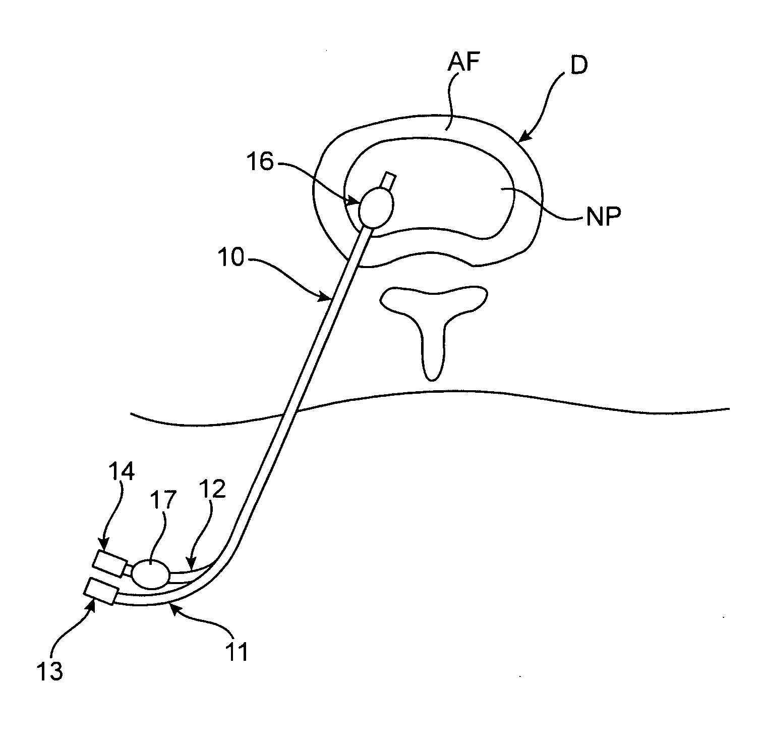
distal portion of a
catheter device is positioned in an
intervertebral disc that is thought to be the cause of the patient's pain. One or more anchoring members are then used to maintain the
distal portion in the disc. The patient is asked to assume a position or perform a task, such as bending over, which typically causes the patient to experience back pain. Using the
catheter device, one or more substances, such as an
anesthetic or
analgesic, are injected into the disc. The patient then reports whether the
anesthetic has alleviated the pain. Optionally, additional intervertebral discs may be tested, one or more
placebo injections may be used, a conventional discography may be added to the procedure and / or the like. Based on the patient's response to the introduction of substance(s) into the disc (or discs), a determination may be made as to whether one or more specific discs are causing the patient's pain. Diagnostic and treatment decisions may be based on such a determination.
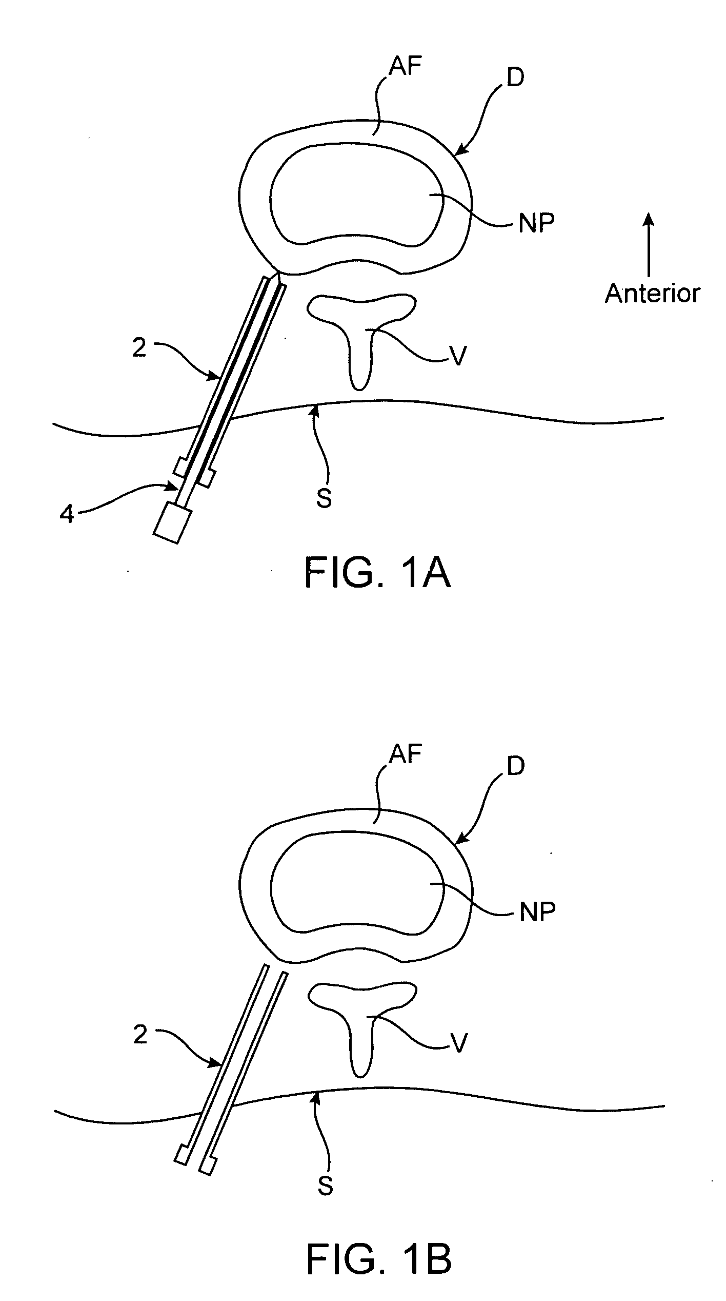
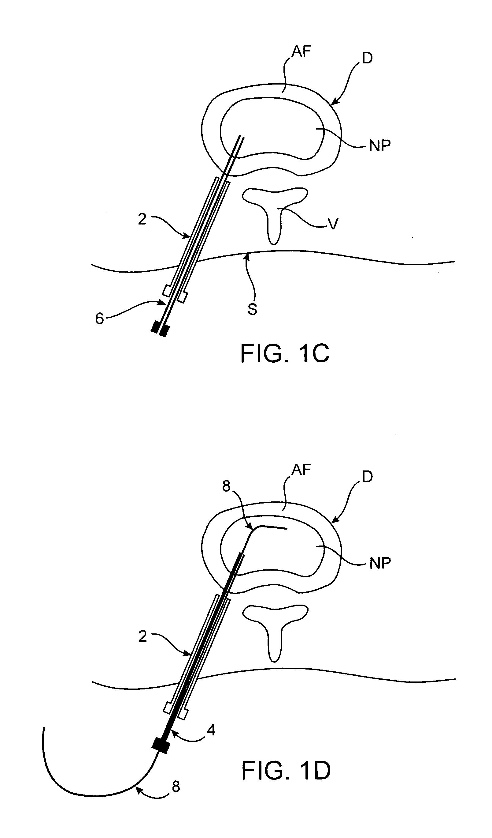
[0015] In various embodiments in which an introducer device is used, the introducer device may either be advanced to a position within the disc or to a position just outside the disc. In either case, the
catheter device may have a distal end or portion configured to facilitate advancement of the distal end through the annulus
fibrosis. In some embodiments, the catheter device is passed over a guidewire. In some cases, the catheter is passed over the guidewire within the needle, while in alternative embodiments the introducer is removed over the guidewire before the catheter device is passed over the guidewire. In some embodiments, positioning the distal portion is facilitated by visualizing at least one radiopaque marker or material at or near the distal portion to assess its location.
[0020] In some embodiments, the method further involves, before introducing the substance, causing the patient to assume a position in which substantial spinal pain is experienced. In such embodiments, the substance introduced is often an anesthetic or
analgesic, and determining whether the patient feels the spinal pain after introduction of the substance helps determine whether pain is caused by that particular disc. Such a method may optionally further include positioning a distal portion of a second catheter device in a second
intervertebral disc, anchoring the distal portion of the second catheter device to maintain the distal portion in the second disc, and introducing at least one substance into the second disc through the second catheter device. The method may also involve, before introducing the at least one substance into the second disc, causing the patient to assume a position in which substantial spinal pain is experienced, wherein the at least one substance includes at least one anesthetic or analgesic. In some embodiments, the method involves determining which of the discs into which the at least one substance was introduced is causing the patient's spinal pain. Such methods may optionally further include performing a discography procedure on the
intervertebral disc before positioning the distal portion of the catheter device in the disc. Alternatively, in other embodiments the discography procedure may be performed on the intervertebral disc after introducing the at least one anesthetic.
[0027] A number of features may facilitate passage of the catheter body through an introducer device. For example, in one embodiment the catheter body includes a friction resistant outer surface. In some embodiments, the outer
diameter of the catheter body is less than 2 mm. Also in some embodiments, a cross-sectional
diameter of the catheter body is larger near a proximal end of the body than near a distal end of the body. Optionally, the catheter body may also include an outer surface having one or more markings for indicating depth of
insertion of the catheter device into a patient's body. In alternative embodiments, the catheter body may include an outer surface having two or more different colors for indicating depth of
insertion of the catheter device into a patient's body. The catheter body may further include at least one radiopaque marker or material for facilitating
visualization of the catheter device in a patient.
[0035] In various embodiments, the kit may also include one or more additional devices, such as but not limited to an introducer device for facilitating positioning of the catheter device in the disc, a pointed stylet removably disposed within a lumen of the catheter device for piercing through the annulus
fibrosis and / or a guidewire passable through the needle.
