Reovirus clearance of ras-mediated neoplastic cells from mixed cellular compositions
- Summary
- Abstract
- Description
- Claims
- Application Information
AI Technical Summary
Benefits of technology
Problems solved by technology
Method used
Image
Examples
example 1
Reovirus Induced Oncolysis and Apoptosis in Breast Cancer Cells
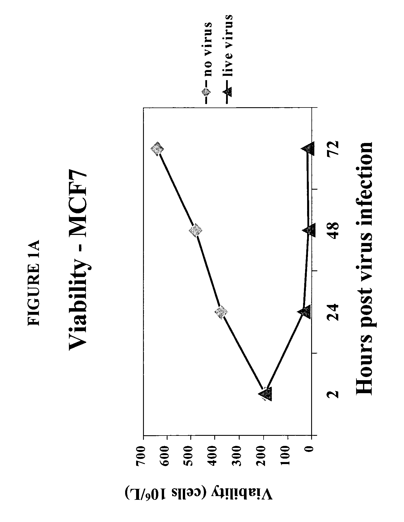
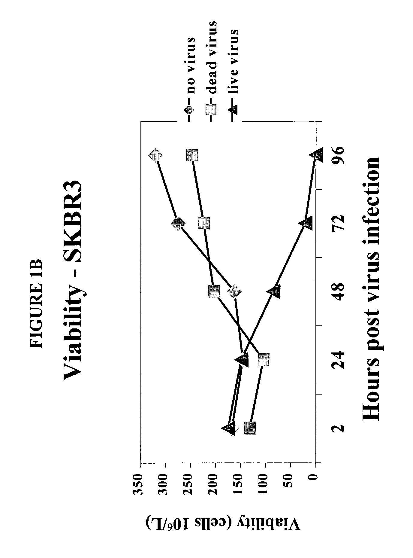
[0085]To determine the effect of reovirus on the viability of neoplastic cells, we first used three breast cancer model systems, MCF7 (ATCC number HTB-22), SKBR3 (ATCC number HTB-30) and MDA MB 468 (ATCC number HTB 132). Cells of each cell line were grown to 50–60% confluency and infected with reovirus serotype 3, strain Dearing, at a multiplicity of infection of 40. Reovirus was obtained and maintained as described in U.S. Pat. No. 6,136,307. Reovirus infected and non-infected cells were harvested at 0, 24, 48 and 72 hours after infection and the viability was determined.
[0086]The results are shown in FIGS. 1A–1D. Viable cell count in reovirus-infected MCF7 (FIG. 1A), SKBR3 (FIG. 1B) or MDA MB 468 cells (FIG. 1C) dropped significantly after the infection, while the cells infected with dead virus or no virus proliferated as expected. Reovirus treatment caused MCF7 (FIG. 1D) and SKBR3 viability to drop from 93% to 16% by ...
example 2
Reovirus Selectively Inhibited Protein Synthesis in Cancer Cells But Not CD34+ Stem Cells
[0088]For further proof of selective viral infection of cancer cells, 35S labeling / SDS / PAGE of viral proteins was undertaken. Viral protein synthesis was evident after 1–2 days in MCF7 cells infected with reovirus, while cellular protein synthesis decreased at the same time, indicating that reovirus had taken over the cellular machinery. At 4 days after infection, no protein synthesis could be detected anymore, suggesting that all the cells had been killed. In the control experiments where cells were infected with dead reovirus or no virus, there was no viral protein synthesis, whereas cellular protein synthesis was at the normal level. In contrast, 35S labeling of CD34+ stem cells in the presence or absence of reovirus showed no viral protein synthesis up to 72 hours after the addition of virus. Therefore, reovirus selectively infect MCF7 cells but not CD34+ stem cells.
example 3
Reovirus Treatment Neither Inhibited Cell Proliferation Nor Altered Differentiation Potential of CD34+ Cells
[0089]Consistent with the protein synthesis results, viable cell count indicated that reovirus treatment did not decrease the number of viable cells in CD34+ cells (FIG. 3A) as compared to the no virus control.
[0090]While the number of CD34+ cells was unaffected by reovirus infection, there remained the question whether reovirus changed the potential of CD34+ stem cells to differentiate into all the hematopoietic lineages in the appropriate proportion. If this was the case, reovirus treated stem cells would not be a good candidate for the reconstitution of the whole hematopoietic system. To investigate this possibility, CD34+ cells were incubated with reovirus for 2, 24, 48 or 72 hours, respectively. The reovirus was then removed and the cells were diluted and cultured in fresh media for 14 days to allow colonies to form. Each colony was examined to determine if it belongs to ...
PUM
| Property | Measurement | Unit |
|---|---|---|
| Composition | aaaaa | aaaaa |
| Freezing point | aaaaa | aaaaa |
| Strain point | aaaaa | aaaaa |
Abstract
Description
Claims
Application Information
 - R&D
- Intellectual Property
- Life Sciences
- Materials
- Tech Scout
- Unparalleled Data Quality
- Higher Quality Content
- 60% Fewer Hallucinations
Browse by: Latest US Patents, China's latest patents, Technical Efficacy Thesaurus, Application Domain, Technology Topic, Popular Technical Reports.
© 2025 PatSnap. All rights reserved.Legal|Privacy policy|Modern Slavery Act Transparency Statement|Sitemap|About US| Contact US: help@patsnap.com


