
System for cell-based screening

a cell-based screening and molecular biochemical technology, applied in the field of fluorescence-based cell and molecular biochemical assays for drug discovery, can solve the problems of inability to detect functional data, inefficient, and inefficient, and achieve the effects of reducing the number of false positives

Inactive Publication Date: 2006-01-17
CELLOMICS
View PDF26 Cites 56 Cited by
  • Summary
  • Abstract
  • Description
  • Claims
  • Application Information

AI Technical Summary

Benefits of technology

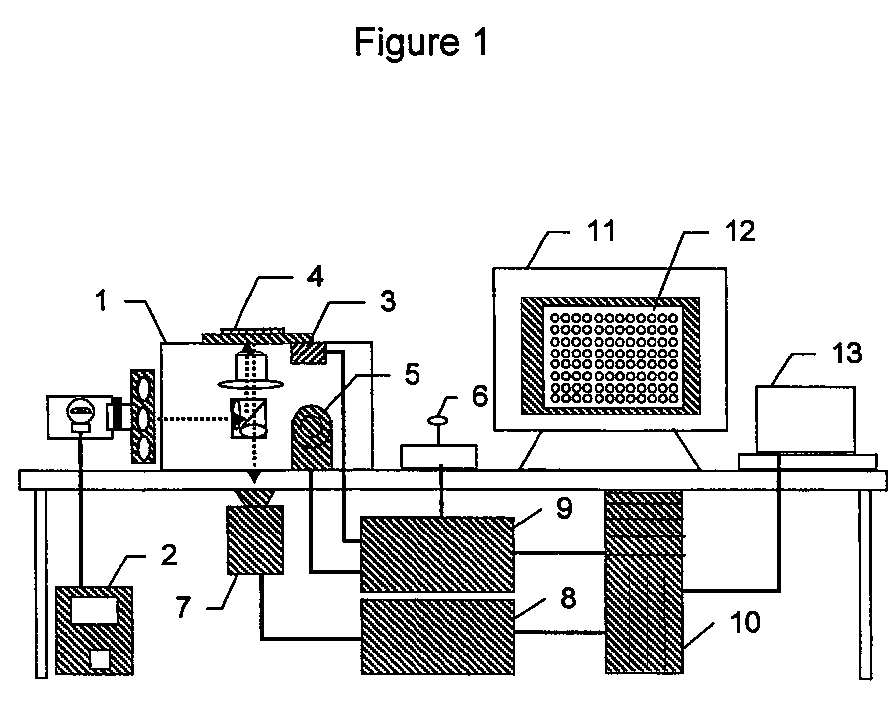
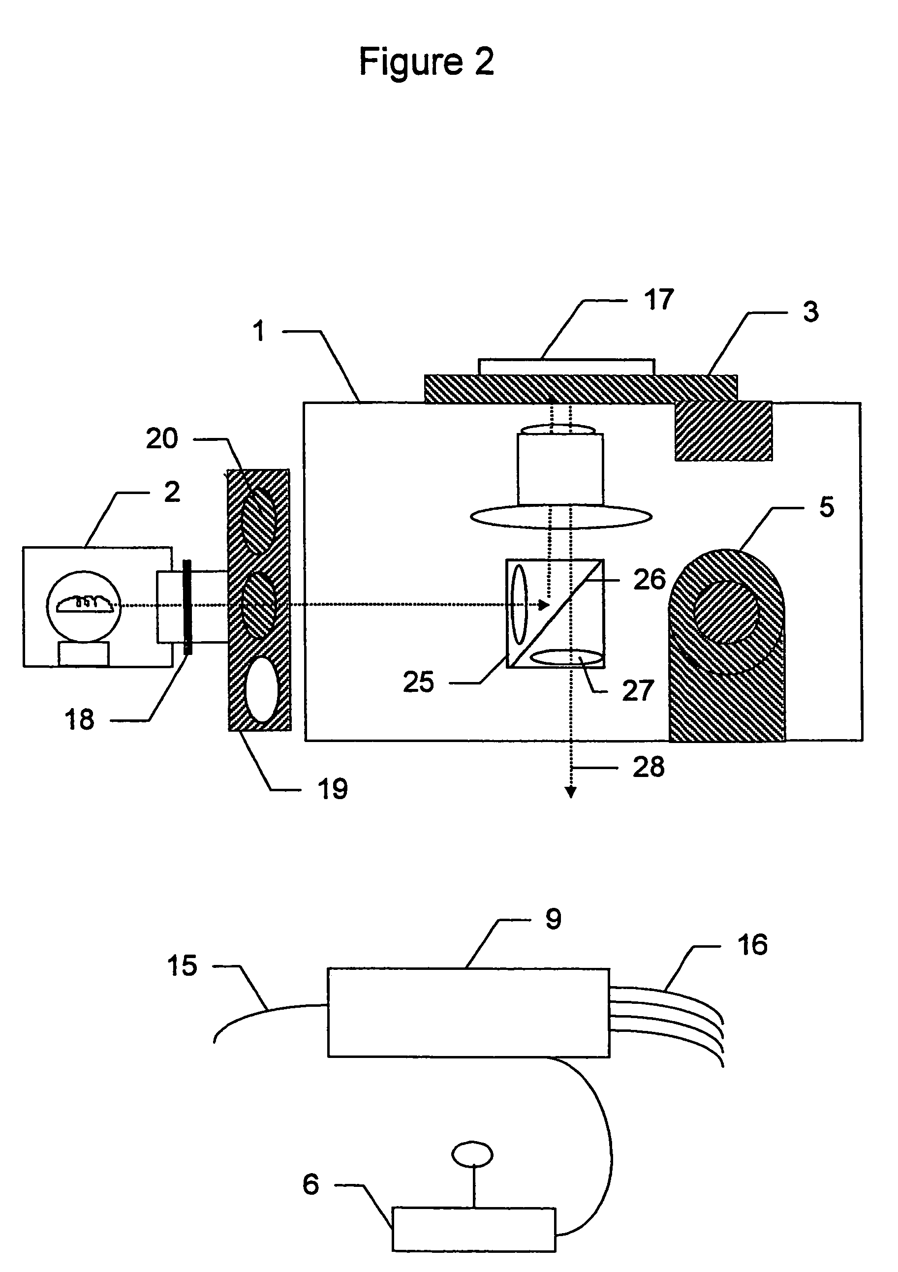
"The present invention is related to a method for analyzing cells using a microtiter plate or microchip with fluorescent reporter molecules. The method involves treating the cells with reagents, imaging them with a fluorescence optics, and analyzing the optical data to determine the distribution, environment, or activity of the fluorescent molecules in the cells. The system can also include a computer means for receiving, processing, and displaying the data, as well as a confocal scanning illumination and detection system. The invention allows for high-throughput screening of large numbers of compounds for their effects on specific biological functions. The patent also provides a machine-readable storage medium containing a program for executing the procedures of the cell screening system. Various automated cell screening methods are also provided."

Problems solved by technology

Historically, drug discovery is a slow and costly process, spanning numerous years and consuming hundreds of millions of dollars per drug created.
Validation of genomic targets as points for therapeutic intervention using the existing methods and protocols has become a bottleneck in the drug discovery process due to the slow, manual methods employed, such as in vivo functional models, functional analysis of recombinant proteins, and stable cell line expression of candidate genes.
Choosing a subset of DAS as drug screening targets is “random”, and thus extremely inefficient, without functional data to provide a mechanistic link with disease.
However, the next level of biological complexity is the cell.
J. Cell. Physiol. 141:410; Giuliano, (1996) Cell Motil. Cytoskel. 35:237)) as well as the high spatial and temporal frequency of chemical and molecular information present within cells, makes it impossible to extract high-content information from populations of cells using existing whole microtiter plate readers.
No existing high-content screening platform has been designed for multicolor, fluorescence-based screens using cells that are analyzed individually.
Similarly, no method is currently available that combines automated fluid delivery to arrays of cells for the purpose of systematically screening compounds for the ability to induce a cellular response that is identified by HCS analysis, especially from cells grown in microtiter plates.
Furthermore, no method exists in the art combining high throughput well-by-well measurements to identify “hits” in one assay followed by a second high content cell-by-cell measurement on the same plate of only those wells identified as hits.

Method used


Image

  • System for cell-based screening
  • System for cell-based screening
  • System for cell-based screening

Examples


example 1

Cytoplasm to Nucleus Translocation Screening

a. Transcription Factors

[0149]Regulation of transcription of some genes involves activation of a transcription factor in the cytoplasm, resulting in that factor being transported into the nucleus where it can initiate transcription of a particular gene or genes. This change in transcription factor distribution is the basis of a screen for the cell-based screening system to detect compounds that inhibit or induce transcription of a particular gene or group of genes. A general description of the screen is given followed by a specific example.

[0150]The distribution of the transcription factor is determined by labeling the nuclei with a DNA specific fluorophore like Hoechst 33423 and the transcription factor with a specific fluorescent antibody. After autofocusing on the Hoechst labeled nuclei, an image of the nuclei is acquired in the cell-based screening system and used to create a mask by one of several optional thresholding methods, as des...

example 2

Automated Screen for Compounds that Modify Cellular Morphology

[0170]Changes in cell size are associated with a number of cellular conditions, such as hypertrophy, cell attachment and spreading, differentiation, growth and division, necrotic and programmed cell death, cell motility, morphogenesis, tube formation, and colony formation.

[0171]For example, cellular hypertrophy has been associated with a cascade of alterations in gene expression and can be characterized in cell culture by an alteration in cell size, that is clearly visible in adherent cells growing on a coverslip.

[0172]Cell size can also be measured to determine the attachment and spreading of adherent cells. Cell spreading is the result of selective binding of cell surface receptors to substrate ligands and subsequent activation of signaling pathways to the cytoskeleton. Cell attachment and spreading to substrate molecules is an important step for the metastasis of cancer cells, leukocyte activation during the inflammato...

example 3

Dual Mode High Throughput and High-Content Screen

[0187]The following example is a screen for activation of a G-protein coupled receptor (GPCR) as detected by the translocation of the GPCR from the plasma membrane to a proximal nuclear location. This example illustrates how a high throughput screen can be coupled with a high-content screen in the dual mode System for Cell Based Screening.

[0188]G-protein coupled receptors are a large class of 7 trans-membrane domain cell surface receptors. Ligands for these receptors stimulate a cascade of secondary signals in the cell, which may include, but are not limited to, Ca++ transients, cyclic AMP production, inositol triphosphate (IP3) production and phosphorylation. Each of these signals are rapid, occuring in a matter of seconds to minutes, but are also generic. For example, many different GPCRs produce a secondary Ca++ signal when activated. Stimulation of a GPCR also results in the transport of that GPCR from the cell surface membrane to...


PUM

PropertyMeasurementUnit
volumeaaaaaaaaaa
volumeaaaaaaaaaa
diameteraaaaaaaaaa

Abstract

The present invention provides systems, methods, screens, reagents and kits for optical system analysis of cells to rapidly determine the distribution, environment, or activity of fluorescently labeled reporter molecules in cells for the purpose of screening large numbers of compounds for those that specifically affect neurite outgrowth.

Description

CROSS REFERENCE[0001]This application is a continuation of U.S. patent application Ser. No. 09 / 632,552 filed Aug. 4, 2000, which claims priority from U.S. Provisional Patent Application Ser. No. 60 / 147,443 filed Aug. 5, 1999; Ser. No. 60 / 176,589 filed Jan. 18, 2000; and Ser. No. 60 / 205,696 filed May 19, 2000. This application is related to co-pending U.S. Applications for patent Ser. No. 09 / 398,965 filed Sep. 17, 1999; Ser. No. 09 / 031,271 filed Feb. 27, 1998; Ser. No. 09 / 380,259 filed Feb. 27, 1998; 09 / 352,171 filed Jul. 12, 1999; Ser. No. 09 / 598,347 filed Jun. 21, 2000; Ser. No. 09 / 293,209 filed Apr. 16, 1999; 09 / 293,210 filed Apr. 16, 1999; Ser. No. 09 / 398,965 filed Sep. 17, 1999; Ser. No. 09 / 430,656 filed Oct. 29, 1999; Ser. No. 09 / 513,783 filed Feb. 25, 2000; and Ser. No. 09 / 569,508 filed May 12, 2000.FIELD OF THE INVENTION[0002]This invention is in the field of fluorescence-based cell and molecular biochemical assays for drug discovery.BACKGROUND OF THE INVENTION[0003]Drug disc...

Claims


Application Information

Patent Timeline
Patent Type & Authority Patents(United States)
IPC IPC(8): G01N33/53G01N33/566G01N21/76
CPCG01N21/6428G01N21/6452G01N21/6458G01N33/5058G01N33/582G01N33/5029Y10T436/11
Inventor GHOSH, RICHIK N.DEBIASIO, ROBBINJANARDHAN, PREM
Owner CELLOMICS
Learn More