However, in some patients, no matter what physical adjustments are made or the particular oral airway which is inserted,
mask ventilation cannot be successfully achieved.
Such cases are literally life-threatening as
hypoxemia and death can quickly ensue if the patient's blood is deprived of
oxygen due to a lack of ventilation.
Most significantly,
soft tissue structures in the hypopharynx (the area between where conventional oral airways end and the
glottis opens into the trachea) collapse inwardly and obstruct
airflow.
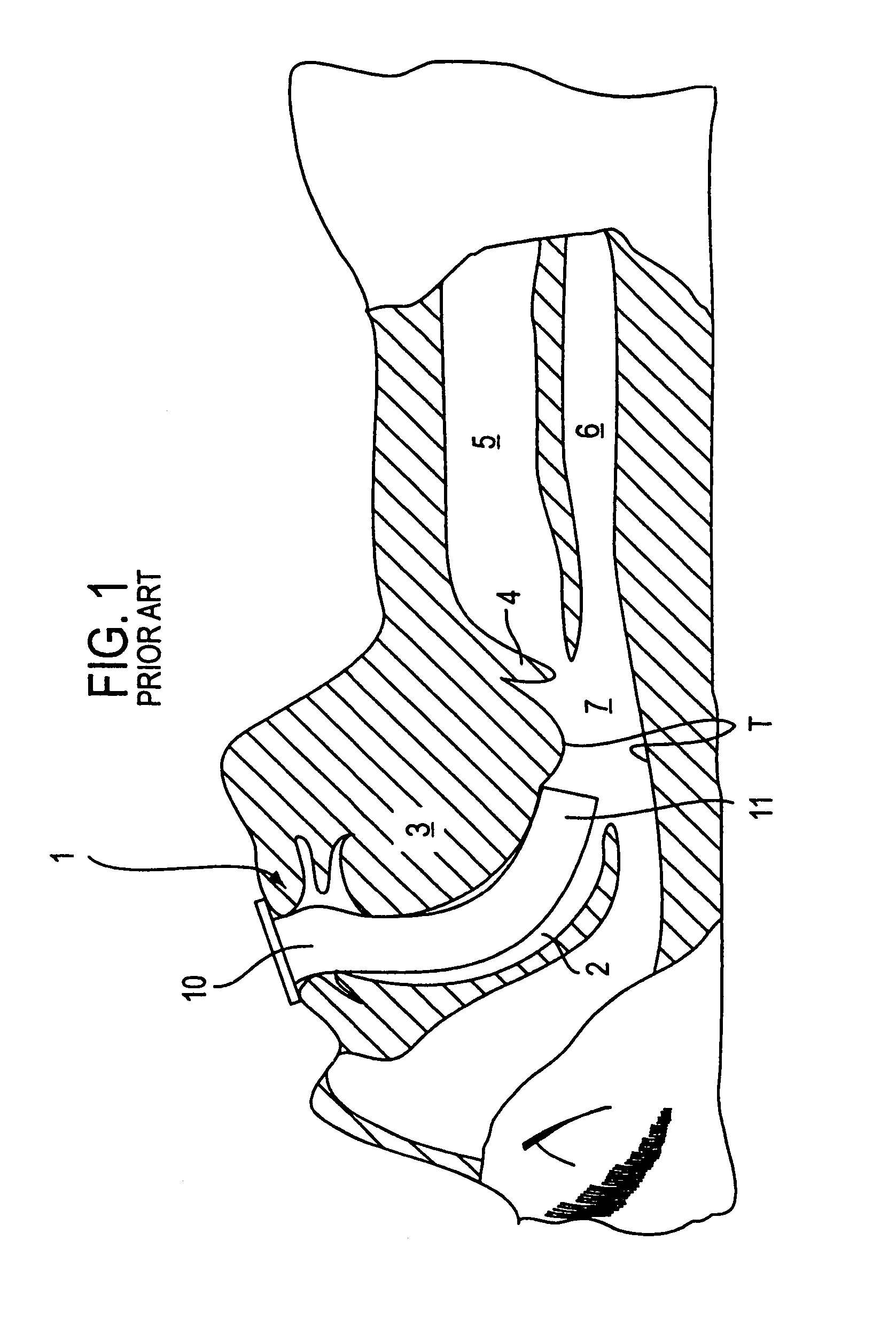
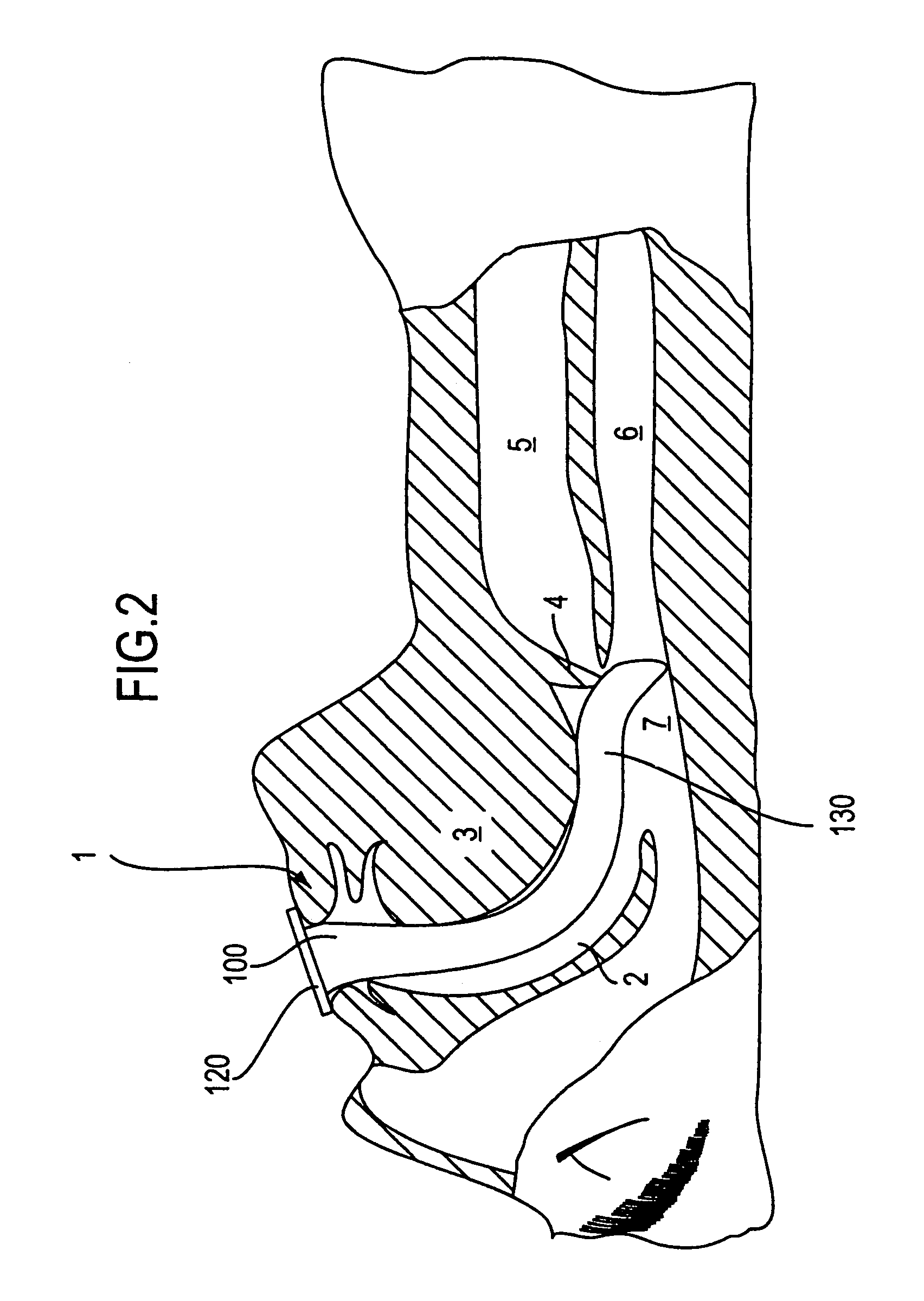
Unfortunately, all oral airways which have been introduced into practice to date end bluntly well above the
epiglottis (the cartilaginous structure just above the
glottis or laryngeal opening) and
glottis and thus place patients at risk for significant
airway obstruction.
Another mechanism of
airway obstruction which occurs while using oral airways is the patient having large lips covering the outside opening of the oral airway with subsequent inadequate
airflow through the
nasal passages (due to the
solid posterior wall of the airway limiting passage of air into the airway at the level of the nasopharynx).
As a result, the distal end (i.e., the end which first enters the mouth and passes down into the
pharynx of the patient) often bruises or otherwise damages soft mucosal surfaces of the patient during
insertion or once the oral airway has been seated in place.
For this reason, the Baildon oral airway suffers from the problems discussed above in that it fails to provide any structure to prevent the collapse of
soft tissue structures in the hypopharynx.
However, the airway of Moses likewise suffers from the problems discussed in detail above in that the blunt-shaped end terminates well above the glottis, thereby allowing possible
soft tissue obstruction to occur.
Moreover, the device of Augustine functions as a guide for placing an
endotracheal tube in a “blind” manner and is neither designed for nor could it possibly function to allow
mask ventilation to be carried out.
While providing a seal with which to administer
positive pressure ventilation, there are several potential problems when using an LMA.
First, the device is easily malpositioned so that ventilation is not possible, for example, by virtue of the
epiglottis bending back over the glottis and thereby obstructing air flow.
Second, by directly covering the glottic aperture, trauma to the glottic structures (arrhytenoid cartilages, vocal cords) can occur.
In addition, the cost of this product (over $200) becomes a factor when limitations to reuse occur due to physical damage of the device or accidental loss.
Third, as a reusable product, the
hazard of cross-
contamination from one patient to another cannot be completely eliminated.
However, this device has several significant limitations which prevent it from functioning adequately.
First, the airway suffers from the problems of those previously discussed in that it ends well above the glottis, thereby allowing soft tissue obstruction to impair the flow of
oxygen to the lungs.
Second, with the
cuff placed so far proximally in the oro-
pharynx, the device tends to push itself out of the patient's mouth, thereby requiring that the device be secured in place by means of a strap placed around the patient's head.
These devices end well above the glottic opening and thus function poorly in terms of reliably directing the end of an
endotracheal tube into the glottis with blind passage.
As a result, the hard distal end of the
endotracheal tube may be directed against the structures which surround the glottic opening (arytenoid, cuneiform and corniculate cartilages,
epiglottis, aryepiglottic folds) and cause damage to these structures or their soft tissue surfaces.
Further, that damage may result in hemorrhage which obscures vision if subsequent placement of the endotracheal tube by means of a fiberoptic device is attempted.
However, these structures have hard advancing surfaces which can likewise cause trauma.
However, in addition to sharing the above discussed problems common to all LMA's, this device relies on precise positioning so that a movable flap raises an obstructing epiglottis out of the way of an advancing endotracheal tube.
However, LMA's occupy a somewhat variable and inconsistent position within the hypopharynx in relation to the precise
anatomic location of the glottis (due to anatomic variability among patients as well as the distensible nature of the proximal epiglottis and hypopharynx where it resides).
As a result, blind
intubation with an endotracheal tube with this device can also result in
tissue trauma by virtue of its advancing end being misdirected.
Significant reductions in body temperature (which are the common and usual course following induction of general
anesthesia) in patients undergoing
surgery are associated with an increased incidence of cardiac morbidity, increased rates of
wound infection, impaired
wound healing and alterations in blood coagulation status.
In addition, although rare, a patient undergoing general anesthesia may have a sudden and dramatic rise in body temperature due to an abnormal acceleration of
metabolic rate in a condition termed malignant
hyperthermia.
No supraglottic airway or oral airway which is currently in use provides measurement of
core temperature by a temperature sensor incorporated into that device.

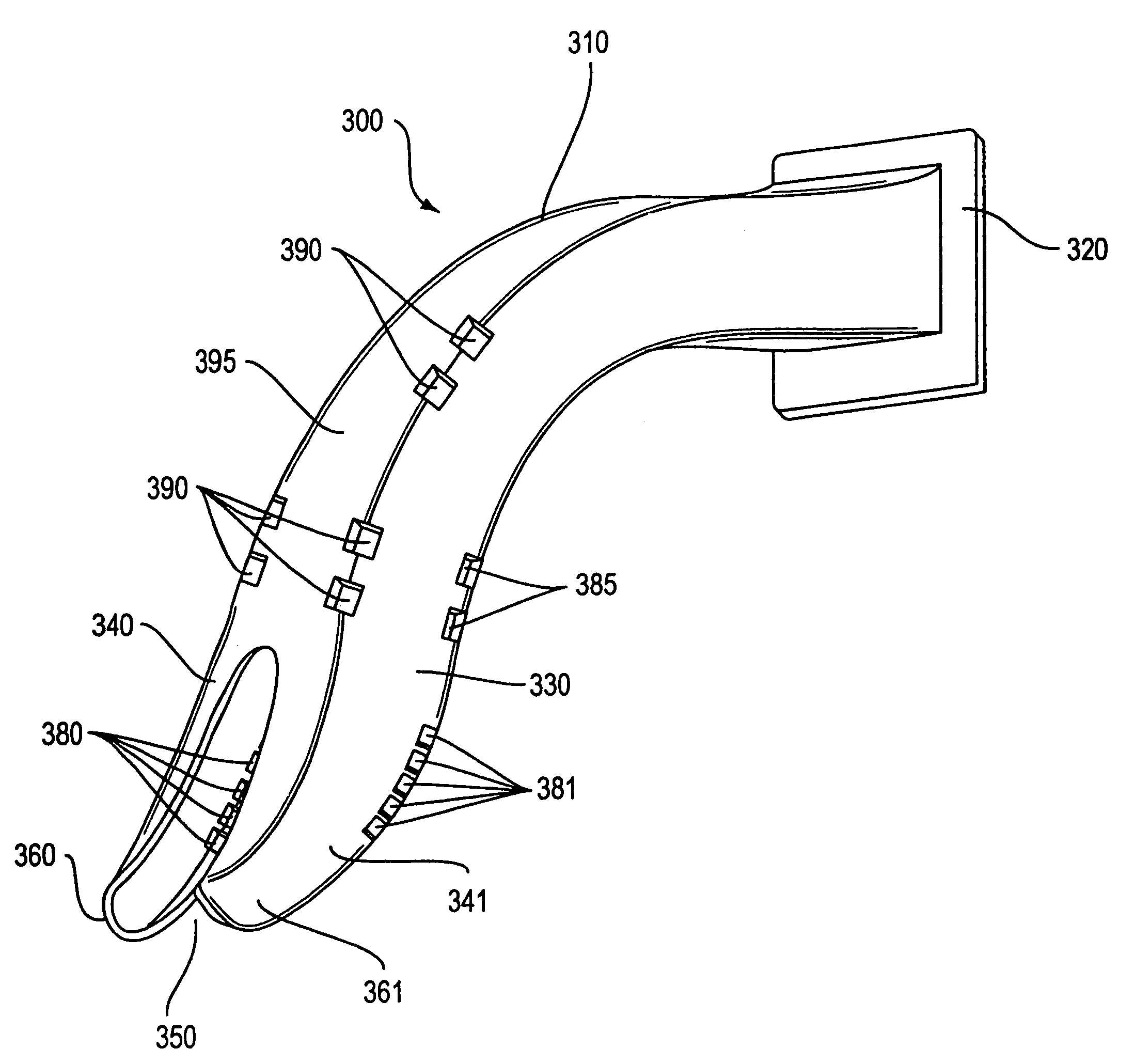