Method and device for accurately positioning a patient in radiotherapy and/or radiosurgery

Inactive Publication Date: 2005-03-08
BRAINLAB
View PDF33 Cites 225 Cited by
  • Summary
  • Abstract
  • Description
  • Claims
  • Application Information

AI Technical Summary

Benefits of technology

It is the object of the present invention to propose a method and a device for accurately positioning a patient for radiotherapy and / or radiosurgery, wherein the above disadvantages of the prior art are not present. In particular, a greater flexibility in producing images and a cost-effective system are to be made available.
The advantageous nature of the present invention is based, among other things, upon the fact that the x-ray images are produced on a single image recorder. Through this, the overall construction is naturally more cost-effective, and gains above all in flexibility, because a single image recorder can much more easily be placed right where it is most effective. In particular, it can also be provided mobile, which opens additional possibilities in recording technology for the overall method.
Furthermore, repositioning as proposed in accordance with the invention offers a very quick and simple way of achieving very accurate target irradiation. Determining the positioning error electronically and / or with computer guidance considerably increases accuracy as compared to manual methods. Spatially localising the x-ray image allows even this input value to be evaluated with sufficient accuracy, such that errors and delays in repositioning may also be avoided from this side.
Within the framework of the method in accordance with the invention, there exists the possibility of producing the x-ray images at an oblique angle on an image recorder spatially arranged horizontally, and of projecting them back onto each respectively defined normal plane, the corresponding reconstructed images being likewise produced in these normal planes. Such images in the normal plane do not suffer from distortions, and are therefore easier to interpret visually.
In a preferred embodiment of the present invention, the image recorder is positioned on a support for a movable patient table. In this way, one or more image recorders rigidly installed in the floor of the treatment room may be dispensed with, resulting in very high flexibility. The treatment room can then easily be used for other purposes as well, without disruptive image recorders in the floor. Moreover, an image recorder positioned on a support for a movable patient table is much more easily accessible, and can therefore also be more easily serviced. The image recorder may be vertically portable together with the patient table and the support, while it is securely arranged horizontally. In other words, the patient table may be moved horizontally, independent of the image detector. If the two x-ray sources are then arranged respectively over a patient table, in particular fixed to the ceiling, and to the side, shifting the patient table sideways while the image recorder remains secured in this direction may be used to ensure that even when the radiation target is on the patient's side, an image of the radiation target always appears on the image intensifier, and in a substantially central position in the image recorder.

Problems solved by technology

Although dosage planning is, as mentioned, relatively successful, the use of high doses administered in a few or a single fraction is often obstructed by the fact that the patient and / or the body section to be irradiated can be positioned only relatively imprecisely.
Disadvantageously, this way of positioning the patient is imprecise, for the following reasons alone:
A three-dimensional spatial shift from two-dimensional images and without computer assistance is only possible to a limited extent, and requires a very experienced user.
A disadvantage of this method is the high computational demands, since such DRRs initially have to be generated at random, and a great many DRRs have to be compared with the actual x-ray image.
These systems are inflexible and costly in terms of apparatus, and therefore also expensive.

Method used


Image

  • Method and device for accurately positioning a patient in radiotherapy and/or radiosurgery
  • Method and device for accurately positioning a patient in radiotherapy and/or radiosurgery
  • Method and device for accurately positioning a patient in radiotherapy and/or radiosurgery

Examples


Embodiment Construction

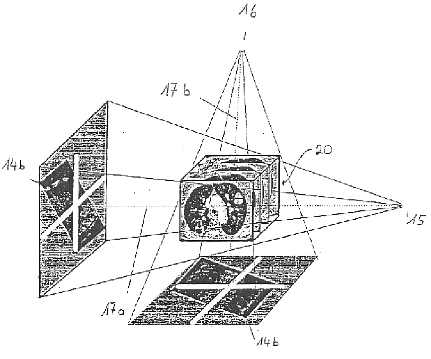
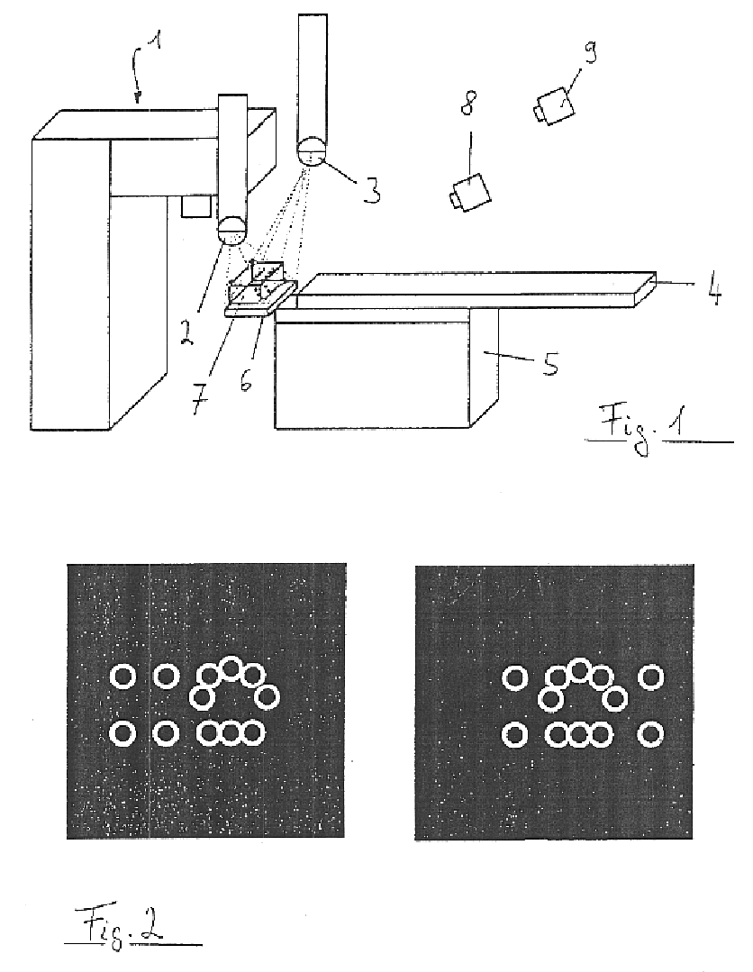
Referring to the figures mentioned above, those components of the device in accordance with the invention will now be described which are necessary for carrying out the preferred embodiment of the invention described here. The device comprises two x-ray tubes 2, 3 mounted to the ceiling of a radiotherapy room, which in other embodiments may also optionally be fixed in or on the floor.

Furthermore, an x-ray detector 6 (image recorder) made of amorphous silicon is provided, fixed to a support 5 for a patient table 4. The x-ray detector can be moved vertically using the support 5, the patient table 4 can however be moved horizontally, independent of the detector 6. In other embodiments, the detector can consist of another material, or can be an image intensifier; it can also be fixed to the floor or to the ceiling, according to the location of the x-ray tubes.

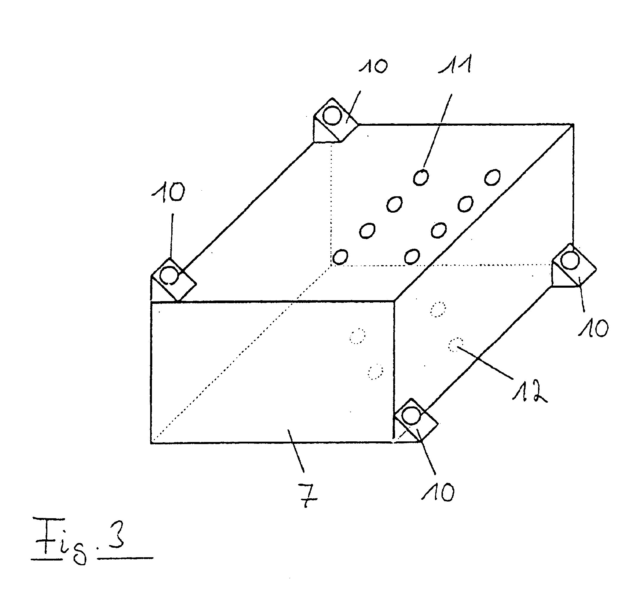
The device further includes an infrared tracking system with cameras 8, 9 for tracking passive markers 10, 13 (FIG. 3 and FIG. 7)...


PUM


Abstract

A method for accurately positioning a patient for radiotherapy and / or radiosurgery, comprising the following steps: the patient is pre-positioned as accurately as possible with respect to a linear accelerator; at least two x-ray images of the patient and / or one of the parts of his body in the vicinity of the radiation target point are produced from different respective recording angles on a single image recorder; the x-ray image is spatially localized; at least one reconstructed image, corresponding to each x-ray image and deriving from a three-dimensional patient scan data set, is produced, the reconstructed images containing the desired image contents of the x-ray images when the patient is correctly positioned; and the real x-ray images are superimposed, and the positioning error is determined electronically and / or with computer guidance by way of particular landmarks and / or the intensity gradient or the contours in the two images; and the position of the patient is corrected by way of the determined positioning error.

Description

BACKGROUNDGreat progress has been made in radiotherapy and radiosurgery recently in dosage planning. People are striving to move treatment more and more in the direction of radiosurgery, i.e. working with high radiation dosages applied in a few, and preferably in just a single, radiation treatment to a target volume, so for example to a tumour. Although dosage planning is, as mentioned, relatively successful, the use of high doses administered in a few or a single fraction is often obstructed by the fact that the patient and / or the body section to be irradiated can be positioned only relatively imprecisely. In order to avoid significant damage to healthy tissue, one therefore falls back in most cases on conventional fractionated radiotherapy, in which repeated irradiation with small doses is applied.In order to improve positioning, one is currently still making do with a very imprecise “manual” method, whereby an x-ray image of a body section of the patient is produced on the linear...

Claims


Application Information

Patent Timeline
IPC IPC(8): A61N5/10A61B6/08A61B6/12A61B6/00
CPCA61B6/547A61N5/1049A61B6/08A61B6/12A61N2005/1062A61N5/1069A61N2005/1059A61B6/583A61N2005/1076
Inventor BLUMHOFER, ANDREASFROHLICH, STEPHANLACHNER, RAINERSCHLOSSBAUER, CORNEL
Owner BRAINLAB
Why Eureka
Try Eureka