In vitro activation of mammalian oocytes

a technology of mammalian oocytes and in vitro activation, which is applied in the field of in vitro activation of mammalian oocytes, can solve the problems of high rate of parthenogenic activation, difficult activation of metaphase ii bovine oocytes, and much lower development rate, so as to improve the development of nuclear transplanted cells, short time period, and faster turn around time

Inactive Publication Date: 2004-01-20
ADVANCED CELL TECH INC
View PDF3 Cites 16 Cited by
  • Summary
  • Abstract
  • Description
  • Claims
  • Application Information

AI Technical Summary

Benefits of technology

The present invention is further directed to a process for parthenogenically activating a 10-52 hour mammalian oocyte comprising increasing intercellular levels of divalent cations in the oocyte by introducing a divalent cation into the oocyte cytoplasm, and reducing phosphorylation of cellular proteins in the oocyte wherein phosphorylation of cellular proteins is reduced by adding an effective phosphorylation inhibiting amount of a serine-threonine kinase inhibitor to the oocyte.
The present invention allows nuclear transfer processes to proceed with younger oocytes such as a 24-hour oocyte, which may produce healthier embryonic cells. There is evidence indicating that early oocyte activation allows for better development of the nuclear transplanted cell. The 24-hour oocyte is the approximate age of an in vivo oocyte during natural fertilization.
Another advantage to activating younger oocytes is the ability, in the laboratory, to obtain a faster turn around time. Within the procedure of the current art, a typical oocyte is a 41-43 hour oocyte. Therefore, the oocyte used in nuclear transfer technology is typically 17-19 hours older than an oocyte used within the process of the present invention which, for example, allows a 24-hour oocyte to be activated.
The younger oocyte potentially allows for tests to be performed in a shorter time period. Further, the laboratory is operated more efficiently with faster turn around of test results. In industry, the use of a younger oocyte will allow progeny to be produced in less time.

Problems solved by technology

A major imbalance in the components of the multi-molecular complex which is required for cell cycle arrest may be responsible for the increasing sensitivity of oocytes to activation stimuli during aging.
Parthenogenic activation of metaphase II bovine oocytes has proven to be more difficult than mouse oocytes.
These procedures that lead to high rates of parthenogenic activation and development of mouse oocytes do not activate young bovine oocytes and / or lead to a much lower development rate.

Method used


Image

  • In vitro activation of mammalian oocytes
  • In vitro activation of mammalian oocytes
  • In vitro activation of mammalian oocytes

Examples


experiment 1

determined the effect of sequential exposure of oocytes to ionomycin and DMAP. One level of ionomycin (5 .mu.M) and DMAP (1.9 mM) was used throughout all studies. The oocytes were cultured for 5 hours after ionomycin incubation with or without DMAP. Control oocytes were cultured in embryonic cell development medium alone. Activation was determined after 5 hours of culture.

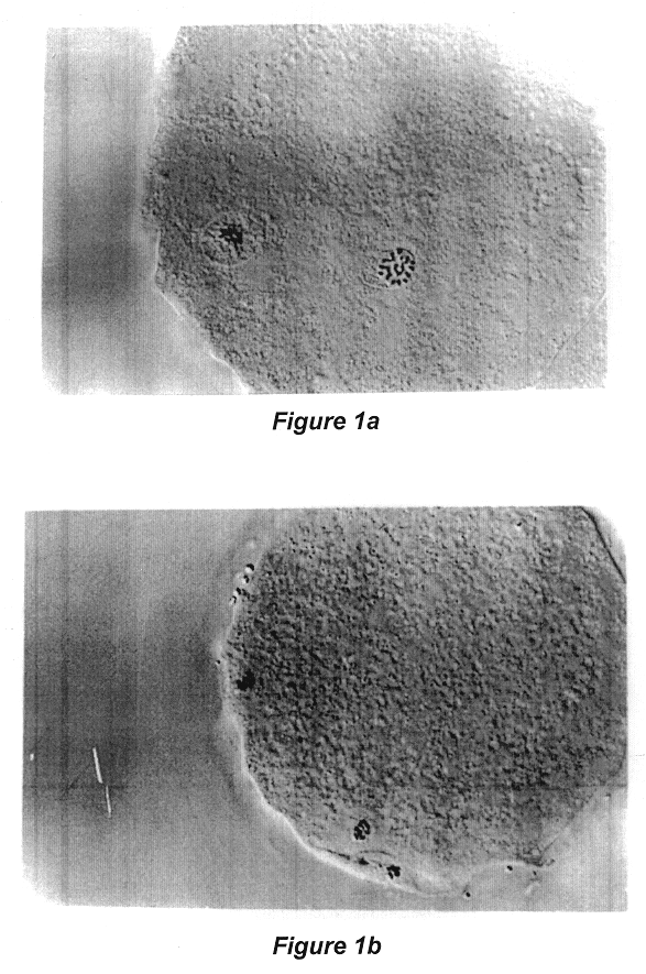
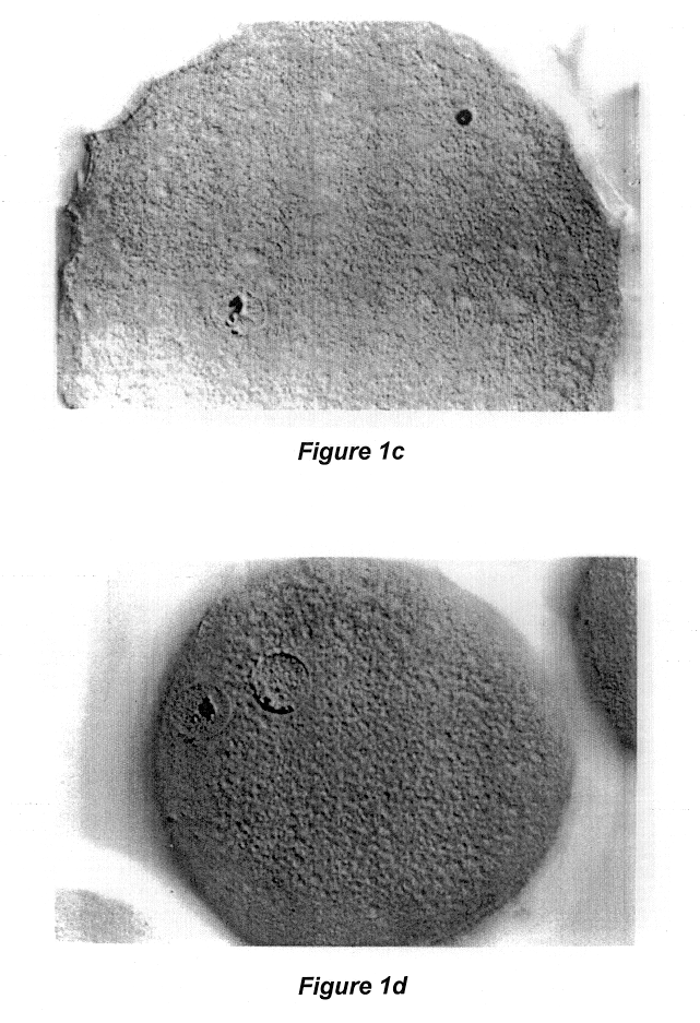
Reference is made to Table 1 following, which illustrates the effect of culture in ionomycin and DMAP for 5 hours on parthenogenic activation of the bovine oocytes matured for 24 hours, and to FIGS. 1a, 1b, and 1c to illustrate the results of Experiment 1.

TABLE 1 The effect of culture in Ionomycin and DMAP for 5 hours on parthenogenic activation of the bovine oocytes matured for 24 hours..sup.1 Treatment N % Activation % Pronuclear formation Control 83 1.4 .+-. 1.4.sup.a 0.sup.a Ionomycin (5 .mu.M) 80 57.8 .+-. 7.8.sup.b 8.9 .+-. 7.3.sup.a DMAP (2 mM) 93 7.8 .+-. 5.5.sup.a 7.8 .+-. 5.5.sup.a Ionomycin + DMAP 91 80....

experiment 2b

sign except that oocytes were incubated for 2 hours, 3 hours, 4 hours, or 5 hours in DMAP.

The results of Experiment 2a are shown in the following Table 2, which illustrates the effect of time of incubation in DMAP on activation, pronuclear formation, initial cleavage and parthenogenic development:

TABLE 2 The effect of time of incubation of DMAP on activation, pronuclear formation, initial cleavage and parthenogenic development (r = 4).sup.1 Pronuclear Development Length of 6-DMAP Activation Formation Cleavage to Blastocyst Incubation (min) N (.+-. se) (.+-. se) N (.+-. se) (.+-. se).sup.2 0 76 58.2.sup.a,b (4.6) 1.4.sup.a (1.4) 95 3.4.sup.a (3.4) 0.sup.a 15 70 52.4.sup.a,b (11.0) 1.4.sup.a (1.4) 88 7.2.sup.a (3.2) 0.sup.a 30 80 37.8.sup.b,c (11.4) 2.5.sup.a (2.5) 103 4.4.sup.a (2.1) 0.sup.a 60 69 22.6.sup.b,c (4.8) 5.4.sup.a (5.2) 111 2.3.sup.a (1.5) 0.7.sup.a (0.7).sup.3 150 79 32.9.sup.b,c (6.3) 31.6.sup.b (6.5) 105 30.9.sup.b (10.4) 9.9.sup.b (4.3).sup.4 300 71 76.6.sup.a (9.8) 7...

experiment 3

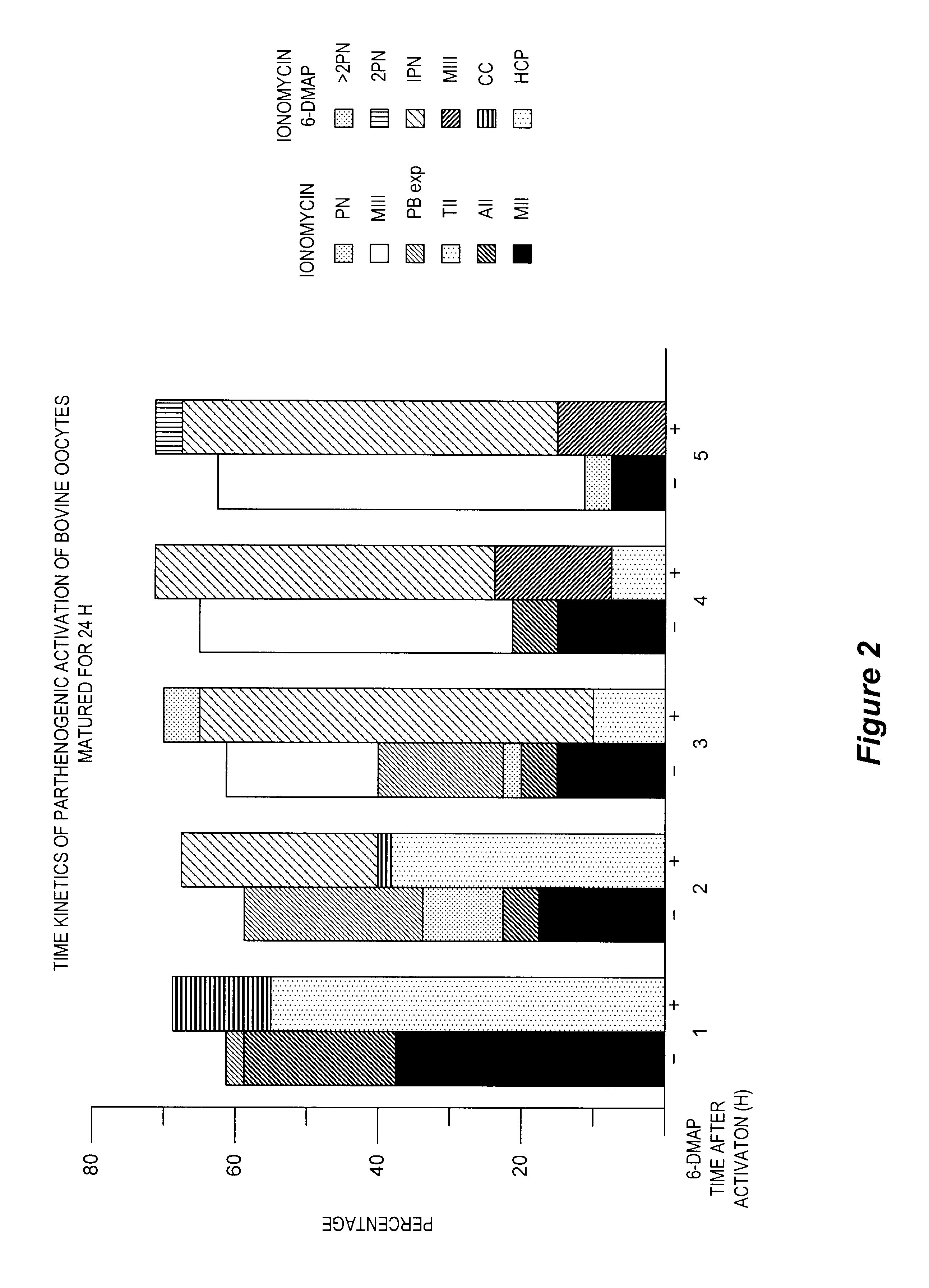
determine the kinetics of activation when oocytes were exposed to ionomycin alone or to ionomycin followed by a 3 hour incubation in DMAP. At the end of the 3 hour incubation, oocytes in DMAP were diluted in TL-HEPES (1 mg / ml Fraction V BSA) and moved to embryonic cell development medium until mounting. A sample of oocytes from both treatments were mounted at 1, 2, 3, 4 and 5 hours after exposure to ionomycin to determine the activation state.

The results of Experiment 3 are illustrated in FIG. 2, which is a graph illustrating the kinetics of parthenogenic activation of the bovine oocyte (matured for 24 hours) by ionomycin and 6-DMAP. When the oocytes were treated with ionomycin alone, they had resumed meiosis by 1 hour and there was evidence of polar body expulsion by 2 hours. By 4-5 hours, the oocytes were arrested in metaphase III. In contrast, when the oocytes were exposed to the sequential treatment of ionomycin+DMAP, there was evidence of pronuclear formation by 2 hours and max...


PUM

PropertyMeasurementUnit
timeaaaaaaaaaa
inner diameteraaaaaaaaaa

Abstract

A process of parthenogenic activation of mammalian oocytes which includes increasing intercellular levels of divalent cations in the oocyte; and reducing phosphorylation of cellular proteins in the oocyte. One method of accomplishing this is by introducing Ca.sup.2+ free cation, such as ionomycin, to the oocyte and then preventing phosphorylation of the cellular proteins within the oocyte by adding a serine-threonine kinase inhibitor, such as 6-dimethylaminopurine (DMAP).

Description

FIELD OF THE INVENTIONThe present invention is generally directed to an improved process for cloning or multiplying mammalian embryonic cells and to an improved process for transferring the nuclei of donor embryonic cells into enucleated recipient oocytes. The present invention is specifically directed to a process for parthenogenically activating mammalian oocytes and to the use of the oocytes.CITATION OF REFERENCESA full citation of the references appearing in this disclosure can be found in the section preceding the Claims.DESCRIPTION OF THE PRIOR ARTAdvanced genetic improvement and selection techniques continue to be sought in the field of animal husbandry. With specific reference to dairy cattle, for example, significant increases in milk production have been made with the wide scale use of genetically superior sires and artificial insemination. Dairy cows today produce nearly twice as much milk as they did 30 years ago. Further genetic improvement can be accomplished by the mu...

Claims


Application Information

Patent Timeline
Patent Type & Authority Patents(United States)
IPC IPC(8): C12N5/06C12N15/87C12N5/02C12N5/075C12N15/873
CPCC12N5/0609C12N15/873C12N2500/14C12N2501/70C12N2501/999
Inventor SUSKO-PARRISH, JOAN L.NORTHEY, DAVID L.LEIBFRIED-RUTLEDGE, M. LORRAINESTICE, STEVEN L.
Owner ADVANCED CELL TECH INC
Why Eureka
Try Eureka