
Positron camera

a positron camera and positron beam technology, applied in the field of positron beam camera, can solve the problems of inability to distinguish tissue with no other technique, inability to produce three-dimensional imaging with unparalleled quantitative content, and poor suitability of pet camera technology for many important purposes

Inactive Publication Date: 2002-11-19
PROPORTIONAL TECH
View PDF5 Cites 9 Cited by
  • Summary
  • Abstract
  • Description
  • Claims
  • Application Information

AI Technical Summary

Problems solved by technology

No other technique is capable of distinguishing tissue according to its metabolic status.
Furthermore, PET produces three-dimensional imaging with unparalleled quantitative content.
Unfortunately, PET camera technology is poorly suited for many important oncology applications, in which large regions of the body frequently need to be imaged.
These procedures can and frequently do require several hours of imaging, when large areas of the body are scanned for metastatic disease, for example.
Clearly, such multiple view imaging also reduces sensitivity within any given imaging slice due to the limited time available for each region.
The limited field of view of current PET detectors is dictated primarily by the high cost of the large array of crystals and photomultiplier tubes (PMT).
If such scattered events are counted, many erroneous events result, and scatter is included.
Crystal detectors of BGO are expensive and produce limited light output, driving up the cost of photomultiplier tubes.
The base materials cost of assembled crystals and photomultipliers runs between $350,000 and $700,000 in today's cameras, and as a result the largest longitudinal field of view available commercially is limited to 24 cm (Moses W W, Derenzo S E, Budinger T F. PET detector modules based on novel detector technologies.
Because of this high fixed materials cost, totally assembled and supported cameras cost from $1.5 million to over $3 million.
Largely because of these exceedingly high costs, only about 60 dedicated PET imaging centers are in operation in the U.S. The small number of facilities combined with the limitation of large field of view imaging means that a very small fraction of the diagnostic imaging need for oncology patients is being met with today's technology.
The problem of identifying a lower cost PET detector has been exceptionally intractable.
However, these systems have very severe drawbacks caused by the gender and geometry of their crystals.
Although the 1 cm thick NaI crystal utilized by most of these systems is adequate for Tc-99m, it provides only a 5% photoelectron efficiency at 511 keV, driving the coincidence efficiency down to an extremely limiting 0.2%.
As a result, such systems require extremely lengthy data acquisition times. Furthermore, because very high singles rates are present in the uncollimated geometry, these systems saturate at very low injected activity levels.
Despite extensive development efforts, however, these systems have very limited detection efficiencies of 10-30%, and depending on the converter system utilized, have either poor coincidence time resolution of 88 nsec compared to 5 nsec for the BGO systems, or poor spatial resolution of 5-11 mm compared to less than 6 mm for the BGO systems (Wells K, Visvikis D, Ott R J. Performance of a BaF.sub.2 -TMAE detector for use in PET.

Method used


Image

  • Positron camera
  • Positron camera
  • Positron camera

Examples


Embodiment Construction

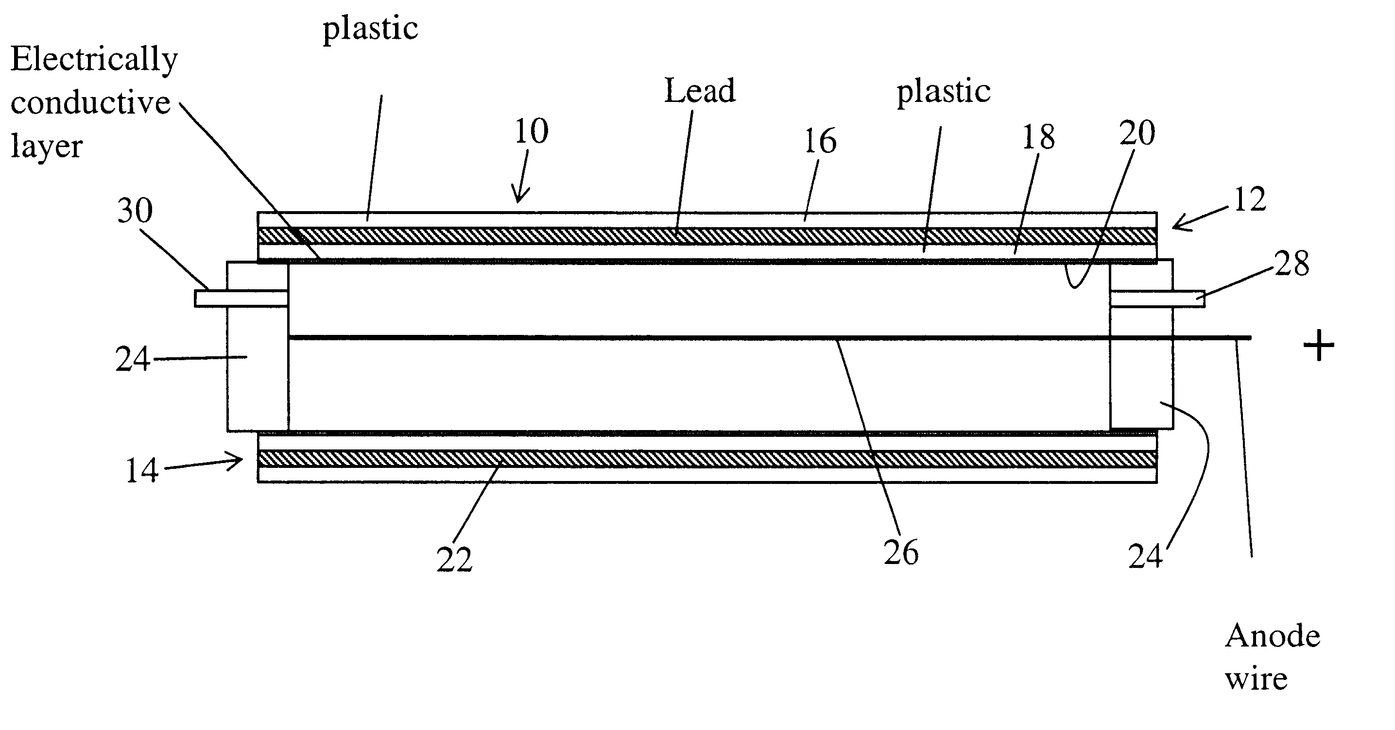
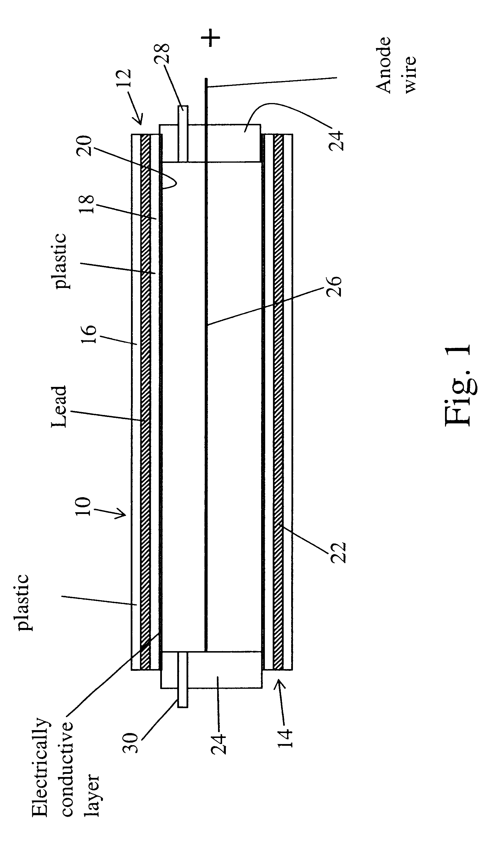
Here it is proposed to use thin Pb converters, which can provide essentially any required degree of reduction of Pb converter thickness, in order to obtain efficient photoelectron escape, while at the same time acceptable time resolution. This is accomplished through use of straw detectors which have become a very well developed inexpensive technology in high energy physics. The radiation detectors of this invention comprise a thin plastic tube, typically 4-5 mm in diameter, coated on the inner wall with a vapor deposited conductive coating, such as copper or aluminum. The plastic wall thickness is 0.002"-0.0005", and the conductive metal layer is of negligible 1,000 angstrom thickness. The basic plastic tube complete with conductive metal coating can be manufactured in mass quantities by utilizing techniques developed in the soda straw industry and since no expensive, consumable materials are required, the cost of such tubing is very small. A central anode wire of diameter 20 micro...


PUM

PropertyMeasurementUnit
pressureaaaaaaaaaa
thicknessaaaaaaaaaa
thicknessaaaaaaaaaa

Abstract

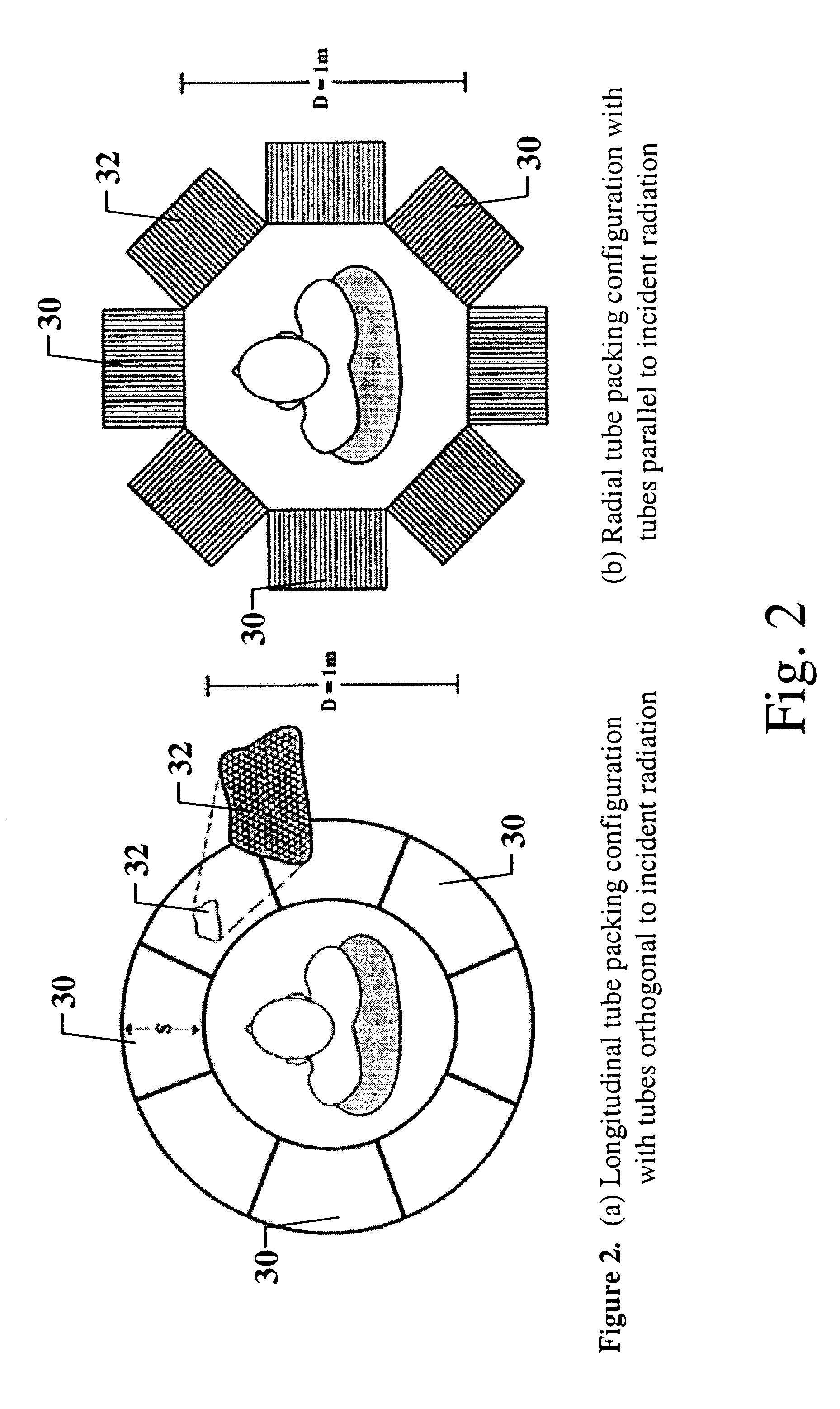
A structure for detection of radiation emitted by positron annihilation comprising a thin wall tube the inner surface of which is covered with an electrically conductive coating which is encompassed and enclosed by a spaced apart thin layer of lead, bismuth or an alloy thereof, an electrically conductive wire element coaxially positioned within said tube and an electrically insulative and pressure retaining element closing each tube end to contain there within from 0.1 to 10 bar of a gaseous atmosphere comprising argon together with a quench gas component or another suitable counting gas; and a PET imaging camera having closely packed arrays of a plurality of such tube structures.

Description

Not applicable.Not applicable.1. Field of the InventionThis invention relates to detector device that may be used to detect radiation emitted by positron annihilations.2. Description of the Related ArtPositron emission tomography (PET) is a powerful imaging technique providing capabilities which no other technology is likely to match. Its most unique feature is its ability to image tissue differentiated by function. In particular, the field is currently dominated by use of the imaging agent 18-FDG, which is a glucose analog. This agent is capable of labeling tissue in proportion to the tissue's ability to metabolize glucose. Such imaging shows great promise in the field of oncology since many tumors have altered metabolic processes relative to surrounding normal tissue. As a result, a large variety of primary and metastatic tumors are well visualized with the technique. No other technique is capable of distinguishing tissue according to its metabolic status.Furthermore, PET produces...

Claims


Application Information

Patent Timeline
Patent Type & Authority Patents(United States)
IPC IPC(8): G01T1/164G01T1/00G01T1/29
CPCA61B6/037G01T1/2985
Inventor LACY, JEFFREY L.
Owner PROPORTIONAL TECH
Learn More