
Implant delivery catheter system and methods for its use

a catheter and implant technology, applied in the field of site-specific delivery of therapeutic agents, can solve the problems of severe chest pain, impaired cardiac function, and damage to the affected tissu

Inactive Publication Date: 2002-11-12
BIOCARDIA
View PDF77 Cites 285 Cited by
  • Summary
  • Abstract
  • Description
  • Claims
  • Application Information

AI Technical Summary

Problems solved by technology

As a result, ischemia, or inadequate blood flow to tissues, can result and damage the affected tissue.
In patients with coronary artery disease, ischemia in the heart can lead to severe chest pain, impaired cardiac function or, if very severe, heart attacks.
The procedure is difficult or impossible to perform on certain patients with multiple vessel disease, diffuse disease, calcified vessels or vessels that are too small to access.
Stent placement has become a good alternative to angioplasty, but the challenges of re-occlusion of the stent have not been completely solved, and stents are not generally used to treat multiple occlusions.
The conventional CABG procedure requires cutting through the sternum of the chest and placing the patient on cardiopulmonary bypass, both of which involve significant risk of morbidity and mortality.
In addition, it is difficult or impossible to perform CABG on certain patients with diffuse atherosclerotic disease or severe small vessel disease or patients, who have previously undergone a CABG procedure.
Often when implantable leads become infected or fail due to fatigue, physicians will extract the entire body of the lead and leave behind the active fixation element which is buried in the myocardium.
However, in ischemic conditions, the growth factor genes often may not produce sufficient amounts of the corresponding proteins to generate an adequate number of new blood vessels.
Current delivery systems however are undesirable for a number of reasons.
However, the extent of collateralization (growth of blood vessels elsewhere in the body, like the brain and lenses of the eye) observed is undesirable, so the dose provided must be less than desired.
Delivery of recombinant growth factors bFGF and VEGF to the coronary arteries has entered Phase II human clinical trials, but the route of administration does not appear to be optimal.
At each of these six steps there are substantial efficiency issues that are difficult to control.
However, currently proposed depots pose difficulties.
Although this is by far the most advanced work done to date, the controlled release depots are too large (0.5 cm to 1.0 cm in diameter) to be delivered percutaneously by a catheter system.
It is also unlikely that the desired target area for these devices is epicardial or even endomyocardial as ischemic zones tend to be localized to the subendocardium.
These issues limit this delivery approach, add risks to the patients who receive it, and increase the procedural costs of this delivery method.
These small controlled release systems have a very large surface-area-to-volume ratio, thus making it difficult to provide optimal release kinetics for many known microsphere systems, such as the Alkermes Prolease system.
It can be difficult to achieve zero order release kinetics in which the dose is delivered at a constant rate over time.
In addition, polymeric microspheres require formulation specific issues to be addressed for each agent that is to be delivered, and these can cause additional problems as already discussed.
If the drug releasing structure is implanted in the left ventricle from the endocardial surface, there is a danger that solid particles can escape into the arterial blood system and be pumped out to the body.
These embolic particles could end up lodged in a vessel and occlude it, causing ischemia or necrosis to tissue elsewhere in the body.
Another danger is that a proliferative agent, such as a growth factor, could embolize and be delivered to an unintended area of the body, such as the brain or the retina, where new uncontrolled blood vessel growth (angiogenesis) could damage healthy tissue.

Method used


Image

  • Implant delivery catheter system and methods for its use
  • Implant delivery catheter system and methods for its use
  • Implant delivery catheter system and methods for its use

Examples


Embodiment Construction

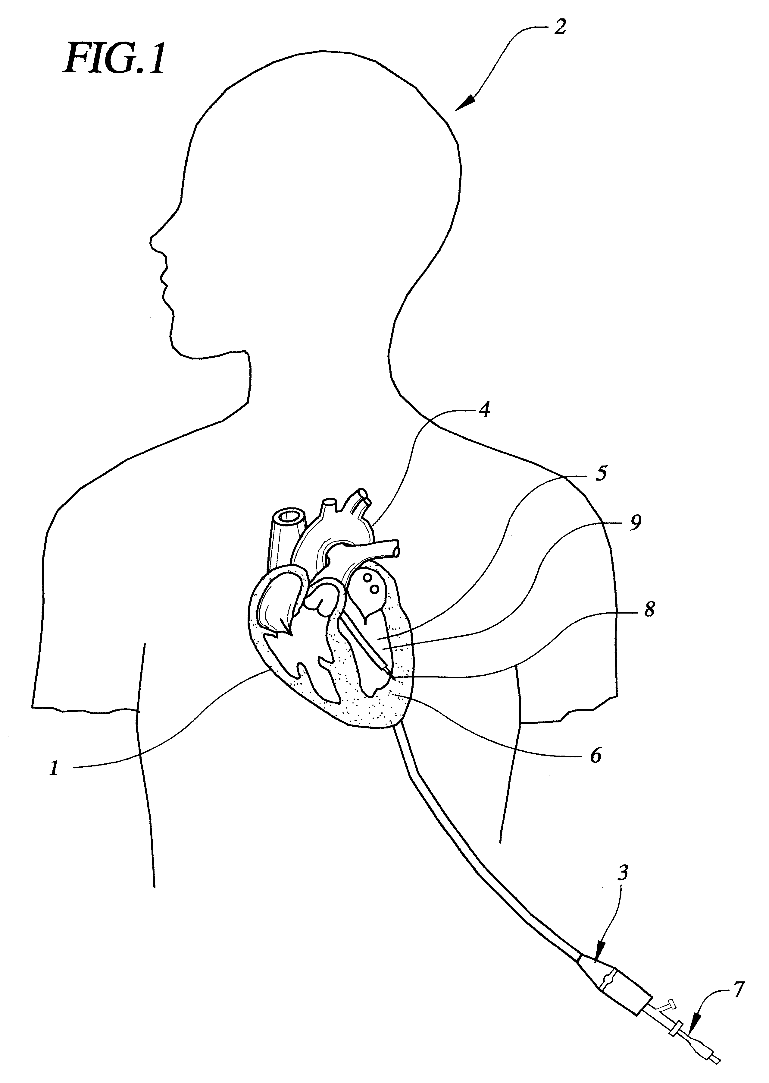
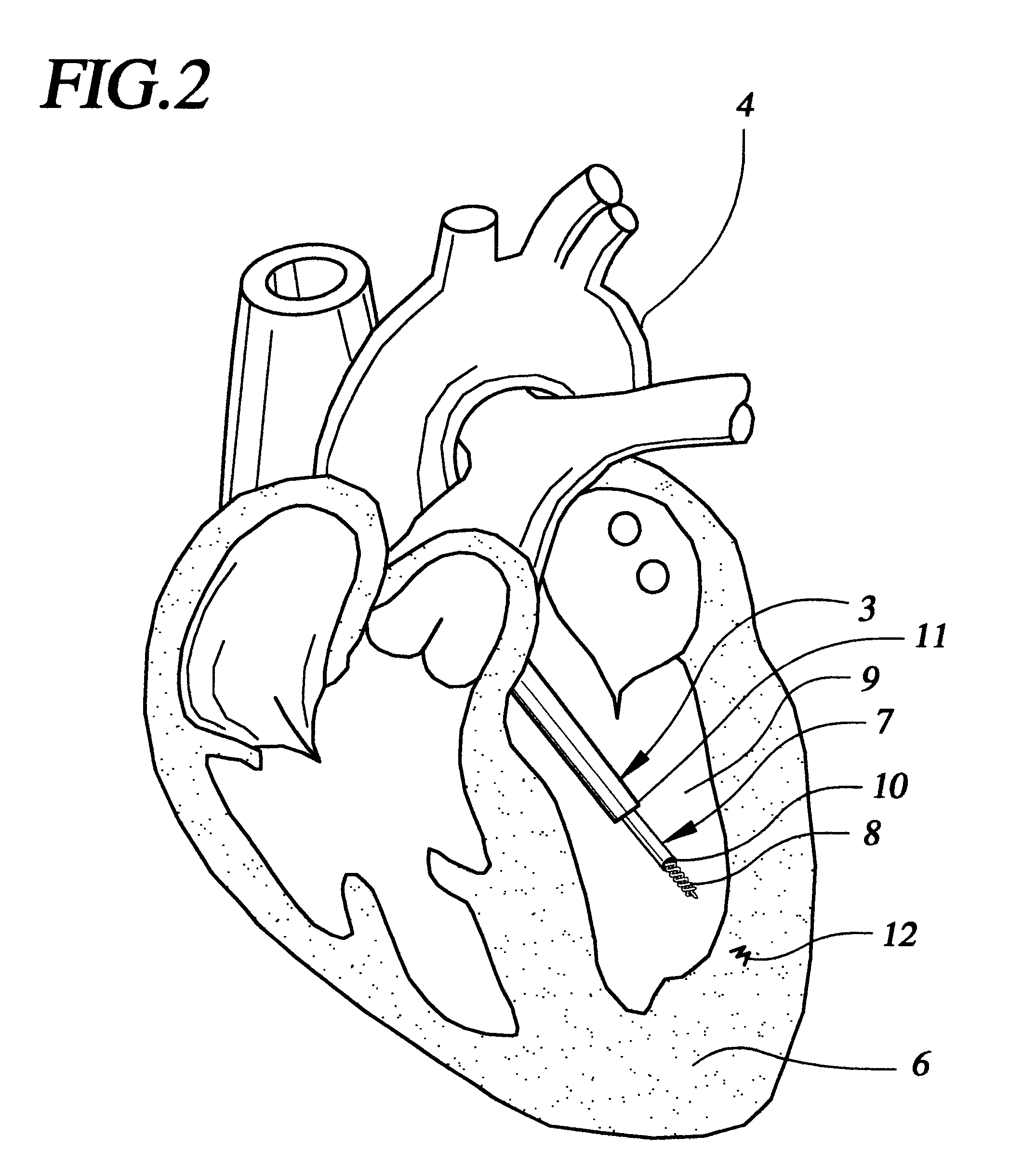
FIG. 1 shows a sectional view of the heart 1 within a patient 2. A steerable guide catheter system 3 is placed within the patient, having been percutaneously inserted into an artery such as the femoral artery, and passed retrograde across the aorta 4 and into the left ventricular chamber 5. Steerable guide catheter 3 is advanced through the patient's vasculature into the left ventricle in order to target a region of the heart wall 6 for delivery. An implant delivery catheter 7 with a fixation element 8 has been inserted through the guide catheter, so that the distal tip of the implant delivery catheter and the fixation element are proximate the target region of the heart. Once oriented toward a region of the heart wall 6 within, for example, the left ventricle wall 9, the centrally located implant delivery catheter 7 is advanced into the heart wall 9 and fixed to the heart tissue by means of the fixation element 8. As described below, the catheter shown in FIG. 1 is different from t...


PUM


Abstract

Catheter systems and methods for implanting helical or dart-like implants into the myocardium or other body tissue. The catheter system includes a helix for fixing the distal end of the catheter to the myocardium, an implant held by the helix, mechanisms for driving the fixation helix into the myocardium, and mechanisms for driving the implant into the myocardium, removing the fixation helix and leaving the implant behind. The implant may be coated, filled, or made of a drug or drug eluting compound, or drug delivery matrix of any composition.

Description

FIELD OF THE INVENTIONSThe inventions described below relate to site-specific delivery of therapeutic agents, devices, structures and catheter systems, means for implanting and using these systems to enable delivery of therapeutic agents to the body, and methods for manufacturing these devices.BACKGROUND OF THE INVENTIONSCardiovascular disease is the leading cause of death in the United States and many other developed countries. A major contributing factor to cardiovascular disease is atherosclerosis, or the hardening of the arteries due to plaque formation. As atherosclerosis progresses, the blood vessels narrow and may close entirely. As a result, ischemia, or inadequate blood flow to tissues, can result and damage the affected tissue. In patients with coronary artery disease, ischemia in the heart can lead to severe chest pain, impaired cardiac function or, if very severe, heart attacks. Approximately 50% of deaths attributable to cardiovascular disease are due to coronary artery...

Claims


Application Information

Patent Timeline
Patent Type & Authority Patents(United States)
IPC IPC(8): A61B17/34A61N1/05A61F2/02A61M25/00A61B17/00A61M5/178A61M31/00A61M37/00
CPCA61B17/3468A61K9/0024A61M37/0069A61B17/3478A61B2017/00247A61B2018/00392A61F2/2493A61M2025/0089A61N1/0568A61N2001/0578
Inventor ROSENMAN, DANIEL C.ALTMAN, PETER A.LOVICH, MARK A.SCHWARTZ, MICHEAL A.MILLER, AARON J.
Owner BIOCARDIA
Learn More