
Biospecific, two photon excitation, fluorescence detection and device

a fluorescence detection and biospecific technology, applied in the field of biospecific assay methods and devices, can solve problems such as complexity, and achieve the effect of reducing the cost of the system by a large amoun

Inactive Publication Date: 2002-03-26
ERKKI SOINI
View PDF16 Cites 26 Cited by
  • Summary
  • Abstract
  • Description
  • Claims
  • Application Information

AI Technical Summary

Benefits of technology

1) Because of the growing interest to avoid centifugal separation of the blood serum, the assay performed in whole blood or other cell suspensions without separation of the cells becomes more potential.
2) Simplification of the liquid handling system reduces strongly the system cost.
The crucial point of the method of this invention is that the assay can simply be performed by focusing the laser beam directly to the reaction suspension and the formation of the complexes on the surface of the microparticles can be monitored without the need of complicated liquid handling and flow systems. This invention leads to a remarkable methodological simplification but it also allows many other useful features which satisfy the assay requirements defined above and which will be discussed later in this text.

Problems solved by technology

The problem with conventional assays and research methods lies in their complexity.
This harmful phenomenon appears only when all assay components are added simultaneously, and is called the "hook effect" in the literature.

Method used


Image

  • Biospecific, two photon excitation, fluorescence detection and device
  • Biospecific, two photon excitation, fluorescence detection and device
  • Biospecific, two photon excitation, fluorescence detection and device

Examples


example

The method described above can be illustrated in more detail using an application example. Reaction charts, reaction equations, standard curves and signal intensity curves presented in FIGS. 2-7 were produced using mathematical modelling.

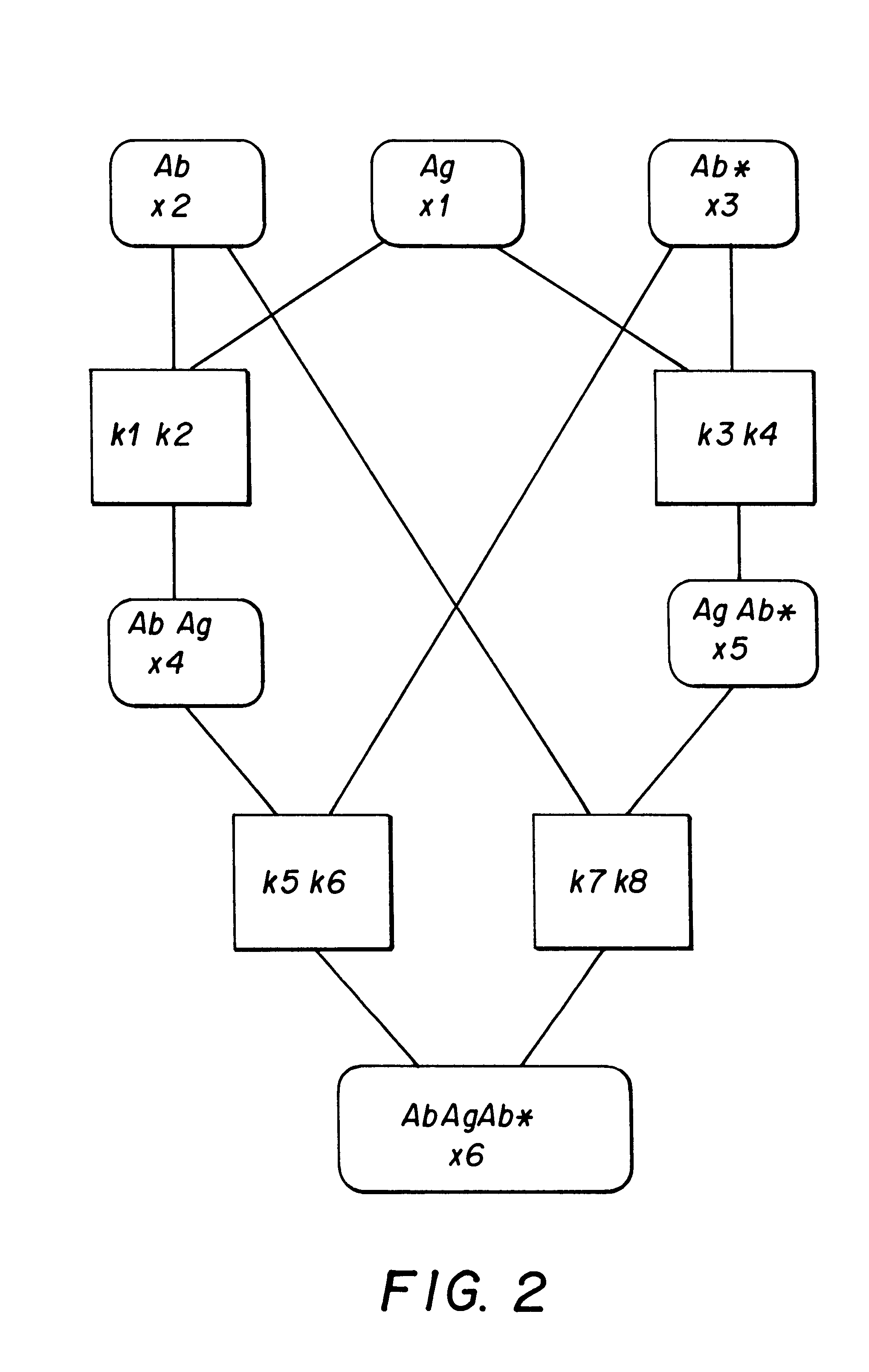
As a practical example of this invention, we present an assay which uses a biospecific reagent bound to the solid phase. FIG. 2 shows the reaction scheme of the assay. The initial components of the reaction include a biospecific reagent Ab bound to the solid phase, analyte Ag and a labelled reagent Ab*. Components Ab, Ag and Ab* are added simultaneously in the reaction solution. The reaction equations (formulas 1-4) presented in FIG. 3 cover all the possible reactions and products. Letters k refer to the reaction association and dissociation rates. Reaction rates naturally differ a lot. Reactions occurring in the liquid phase are rapid, whereas reactions associated with the solid phase are slower. In this example, the selected reaction constant valu...


PUM

PropertyMeasurementUnit
wavelengthaaaaaaaaaa
diameteraaaaaaaaaa
transit timeaaaaaaaaaa

Abstract

A method for measuring the end point and for monitoring the real time kinetics of a bioaffinity reaction in biological fluids and suspensions, employing microparticles as bioaffinity binding solid phase, biospecific reagent labelled with a fluorescent label and a fluorescence detection system which is based on two-photon fluorescence excitation, contacting the analyte, the labelled reagent and the solid phase simultaneously, focusing a two-photon exciting laser beam into the reaction suspension and measuring the fluorescence signal emitted by the microparticles from one particle at a time when they randomly float through the focal volume of the laser beam. In this method the signal is monitored kinetically to obtain information about the analyte concentration before the reaction approaches the highest point of the response. Since the growth rate of the signal intensity is directly proportional to the analyte concentration, the analyte concentration can be predicted in the initial phase of the reaction. The growth rate monitoring also predicts whether the analyte concentration is higher than the binding capacity of the reagent and whether the reaction will be continued over the highest point of the response curve.

Description

Immunoassay is an established biospecific assay method, widely used in routine diagnostics and research laboratories. Another group of biospecific assays is DNA and RNA hybridisation assays. Two biospecific probes, a primary probe and a secondary probe (i.e. an antibody, DNA or RNA probe) are usually used in biospecific assays. They are both connected to the specific determinants of the analyte molecules and form a complex of three molecules (sandwich structure). Normally, one of these two reagents is labelled. Nowadays, commonly used labels are radioisotopes, enzymes, luminescent and fluorescent labels. The most common sample material, which is assayed, is blood serum.The method according to this invention refers to those fluorometric biospecific assays where one biospecific reagent is attached to microparticles serving as a solid matrix. The solid matrix with the attached components is later referred to as the solid phase. The second biospecific reagent is labelled with a fluoresc...

Claims


Application Information

Patent Timeline
Patent Type & Authority Patents(United States)
IPC IPC(8): G01N33/543G01N33/557G01N21/78
CPCG01N33/557G01N33/54326
Inventor HANNINEN, PEKKASOINI, ERKKISOINI, JUHANI
Owner ERKKI SOINI
Learn More