
Optical sensor unit and procedure for the ultrasensitive detection of chemical or biochemical analytes

an analyte detection and ultra-sensitive technology, applied in biochemistry equipment and processes, biochemistry apparatus and processes, material testing goods, etc., can solve the problems of limiting the immunological detection method to procedures including denaturation of samples prior to detection, the overall duration of the test is limited by known analyte detection techniques in terms of sensitivity and quantification, and the overall duration of the test is limited. , to achieve the effect of high sensitivity

Inactive Publication Date: 2002-02-12
CSEM CENT SUISSE DELECTRONIQUE & DE MICROTECHNIQUE SA RECH & DEV
View PDF23 Cites 166 Cited by
  • Summary
  • Abstract
  • Description
  • Claims
  • Application Information

AI Technical Summary

Benefits of technology

It is the object of the present invention to overcome the drawbacks and failures of prior art like complex multistep procedures, limited sensitivity, necessity of label-based analyte detection and necessity of sophisticated equipment. These limitations are addressed in the present invention, which provides an accurate and ultrasensitive detection system for biological ligands, especially prion proteins and proteins which interact with prion proteins, to be used in the diagnosis of prion diseases, or any other biological state involving detection of molecules in biological fluids or tissue homogenates and requiring high sensitivity.
A miniature biosensor chip provides a versatile platform for biomolecule binding, quantitative detection of biomolecules and for selective retention of biomolecules. The disposable biosensor transducer chip (FIG. 4) consists of a high refractive index dielectric waveguiding film (F) deposited on a previously structured substrate (S). The substrate is structured with grating pads (G1, G2) by microstructuring technologies including hot embossing or injection moulding. The grating effects appropriate incoupling of light, signal generation and outcoupling of the light. The size of the sensor pads may vary from tens of mm.sup.2 to submillimeter dimensions. The pads are arranged pairwise or serial, allowing individual and group referencing, respectively. The array sensor platform is essential for multicomponent analysis and on-chip referencing. Preferred shapes of the biosensor chip are rectangular (FIG. 4.d) or circular disc-shaped (FIG. 4.e) forms. Multi-arrays on rectangular platforms can be addressed by fibre guided light, originating from a single source or from multiple light sources. Disc-shaped optical platforms use CD technologies to address individual pads, enabling high repetitive reading frequencies and thus, fast kinetic measurements.
Instead of directly analysing the binding of prions to antibodies immobilized on the chip surface, a competition assay is described. A protein ligand for anti-PrP antibodies is immobilized on the chip surface. Tissue homogenates or body fluids are incubated with anti-PrP antibodies. The mixture is then reacted with the molecules on the chip surface. Residual free antibodies will then bind and elicit a signal. In the case of an animal or human with prion disease. Part or all of the antibody will already be bound to PrP in the sample, thereby preventing the binding of the antibody to the chip surface, effectively reducing the signal observed with a sample from a normal animal or human. Reduction of the signal is therefore indicative of prion disease.
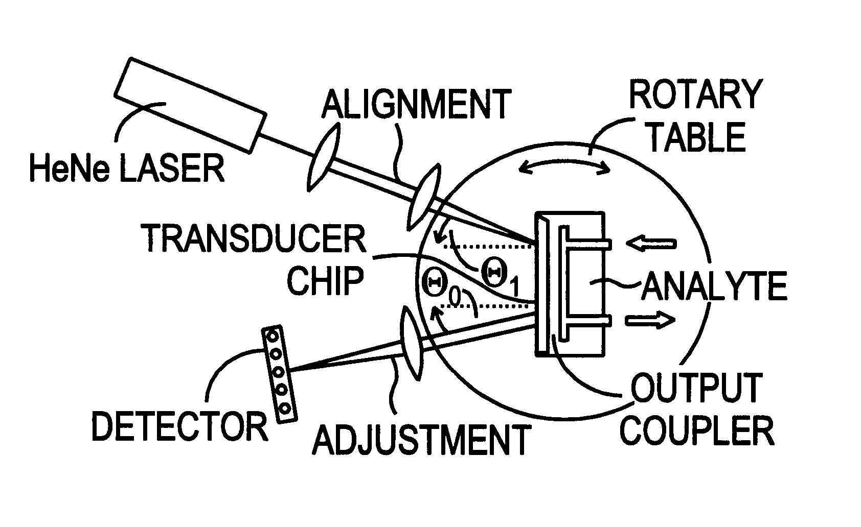
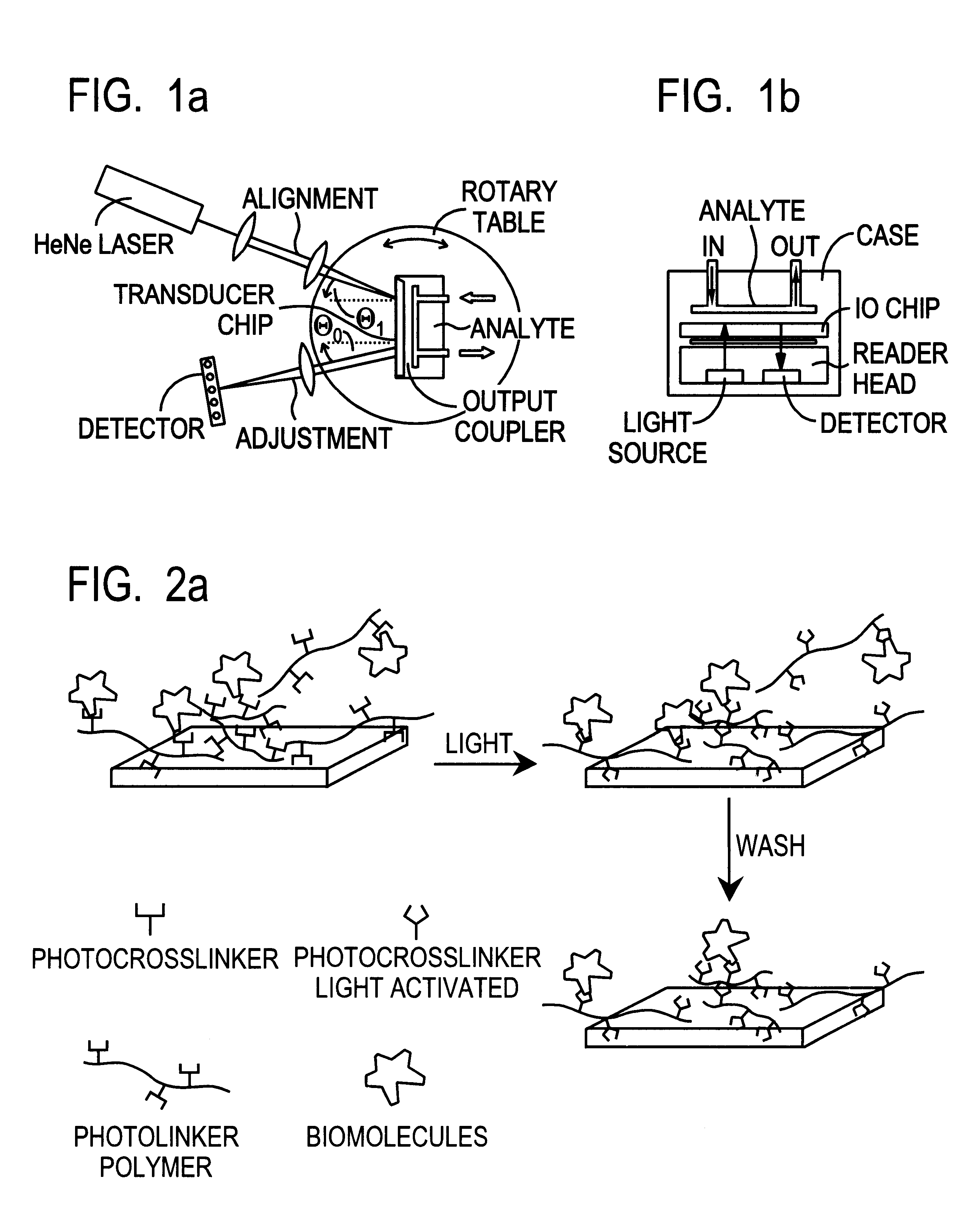
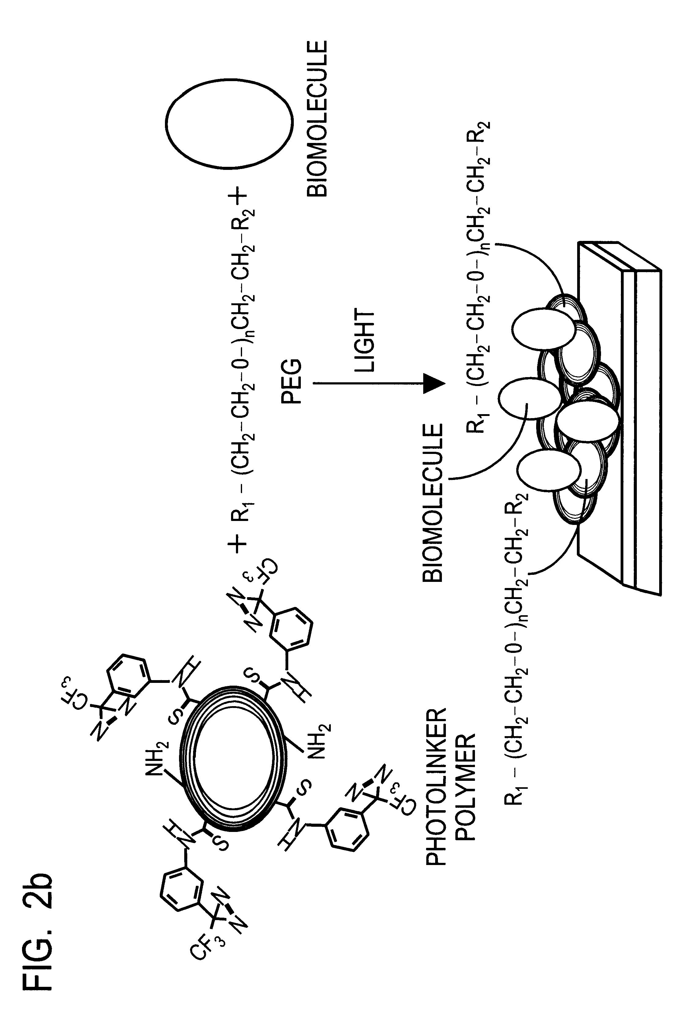
Smart planar optical elements are produced as described. Ligands or particularly monoclonal antibodies recognizing given molecules are bonded as described on these chips on distinct pads. The analyte to be examined is guided onto the chip. A laser or several laser beams are directed to the pads and the system detects the change of the effective refractive index at these pads. This allows performance of highly sensitive quantitation of various molecule concentrations in a complex analyte solution.

Problems solved by technology

Previously, it has been attempted without success to generate antibodies which specifically recognize PrP.sup.Sc, thus limiting the immunological detection methods to procedures including denaturation of samples prior to detection.
Indeed, known analyte detection techniques have their limitations in terms of sensitivity and quantification, the procedure involving several steps, the necessity of detection of labelled components and the overall duration of the test procedure.

Method used


Image

  • Optical sensor unit and procedure for the ultrasensitive detection of chemical or biochemical analytes
  • Optical sensor unit and procedure for the ultrasensitive detection of chemical or biochemical analytes
  • Optical sensor unit and procedure for the ultrasensitive detection of chemical or biochemical analytes

Examples


example 1

Design Fabrication and Testing of Biosensor Chips

Detection of immunological interactions between recombinant bovine PrP and monoclonal anti-PrP antibodies is accomplished by combining optical waveguide detection and light-addressable photobonding. In view of the exceedingly small quantities of analytes in test samples (10.sup.5 to 10.sup.8 molecules), biosensor technologies must meet the requirements of high sensitivity and low non-specific binding. Designed for routine analysis, the detection system preferably consists of few components, fast in response (measuring time in the order of minutes or shorter) and suitable for mass production. The availability of disposable sensor devices, combined with multiple sensing and referencing pads on a single device is advantageous for diagnostic analysis.

I. Optical Waveguide Design and Fabrication

To adapt the resolution and dynamic range of the sensor chip to the needs of diagnostic application, corresponding substrates are fabricated. The gr...

example 2

Building of a Biosensor Module

Since PrP.sup.BSE only occurs in combination with infectious particles, it is important to have a mobile biosensor module which can be used under biosafety conditions P2. A new compact and miniaturized IO sensor module was constructed and designed. A schematic drawing of such a module is shown in FIG. 5.

The complete module consists of three distinct submodules. The tasks of the submodules are illumination (i.e. to provide the optical input), signal transduction into an on-chip measuring variable and signal evaluation. In the sensor module shown in FIG. 5, the electrical input connector (laser driver) powers a laser diode (CD laser diode at 785 mm) for illumination of the sensor chip. A fluid handling system (tubings in the back) is used for analyte application. The sensor output signal is detected using an one-dimensional photodiode array. In this example, the electrical output signal is fed into a laptop computer (RS232 interface) via the connector to ...

example 3

Detection of PrP.sup.BSE

The functional biosensor module as described above was used to test the detection of bovine PrP.sup.Sc. Specifically, the chips carrying either immobilized 6H4 or 15B3 were incubated with proteinase K-digested brain homogenates from normal or BSE-sick cattle. From Western blotting experiments, it is known that about 10-30% of total PrP in a BSE-infected brain correspond to PrP.sup.BSE. Using the above described procedures, antibodies immobilized on the chips were incubated with proteinase K digested brain homogenates. No signal was observed in normal brain while specific binding was detected in homogenates from BSE-sick animals. For antibody 15B3, undigested homogenates were used and a signal was detected only in homogenates from BSE-sick cattle.

This method was further tested for the detection of PrP.sup.BSE in organs other than brain. The exquisite sensitivity of the biosensor system allowed to detect Pre SE in spinal fluid, lymphoid organs, blood, saliva or...


PUM

PropertyMeasurementUnit
Molecular weightaaaaaaaaaa
Areaaaaaaaaaaa
Sensitivityaaaaaaaaaa

Abstract

This document describes an optical sensor unit and a procedure for the specific detection and identification of biomolecules at high sensitivity in real fluids and tissue homogenates. High detection limits are reached by the combination of i) label-free integrated optical detection of molecular interactions, ii) the use of specific bioconstituents for sensitive detection and iii) planar optical transducer surfaces appropriately engineered for suppression of non-specific binding, internal referencing and calibration. Applications include the detection of prion proteins and identification of those biomolecules which non-covalently interact with surface immobilized prion proteins and are intrinsically involved in the cause of prion related disease.

Description

The present invention relates to an optical sensor unit and to a procedure for accurate and ultrasensitive detection of chemical or biochemical analytes. An optical sensor unit is described combining several unique parts inching a laser light source, an optical detection module, and an integrated optical transducer chip. The optical transducer chip consists of a disposable carrier substrate coated with a waveguiding layer onto which sensing biomolecules are bonded in pattern fashion, said sensing biomolecules being capable of interacting with chemicals or bio-molecules of an analyte solution to induce changes in the effective refractive index.More particularly, the invention relates to the detection of prions and prion binding molecules in pico- to femto-molar concentrations with the help of antibodies, preferably monoclonal, allowing specific detection of disease-specific prion proteins on surfaces engineered for low non specific binding of bioconstituents. Conversely, the biosenso...

Claims


Application Information

Patent Timeline
IPC IPC(8): G01N33/543
CPCG01N33/54373Y10S435/80Y10S436/805
Inventor SIGRIST, HANSGAO, HUIKUNZ, RINO E.DUBENDORFER, JURGKORTH, CARSTENMOSER, MARKUSOESCH, BRUNO
Owner CSEM CENT SUISSE DELECTRONIQUE & DE MICROTECHNIQUE SA RECH & DEV
Learn More