Novel methods for the in vitro processing of cancer cells from one individual to accurately preserve the antigenic architecture of multiple surface abnormalities specific to the individual cancer and for rapidly selecting and amplifying anti-cancer molecules highly specific for cancer stem cells and other abnormalities regardless of their rarity while minimizing collateral damage to normal tissue associated with less specific therapies
a cancer cell and in vitro processing technology, applied in the field of in vitro processing of cancer cells from one individual, can solve the problems of inability to accurately preserve the antigenic architecture of multiple surface abnormalities specific to the individual cancer, and achieve the effects of improving the ability to select cancer binding fragments, reducing the risk of cancer survival, and improving safety
- Summary
- Abstract
- Description
- Claims
- Application Information
AI Technical Summary
Benefits of technology
Problems solved by technology
Method used
Image
Examples
embodiment 1 (fig.2)
Embodiment 1 (FIG. 2)
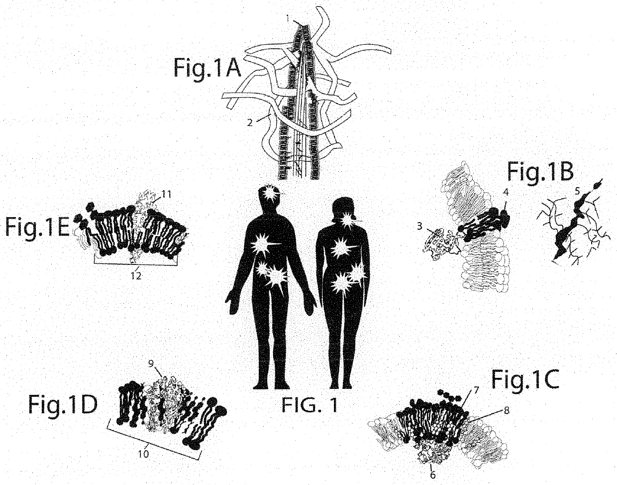
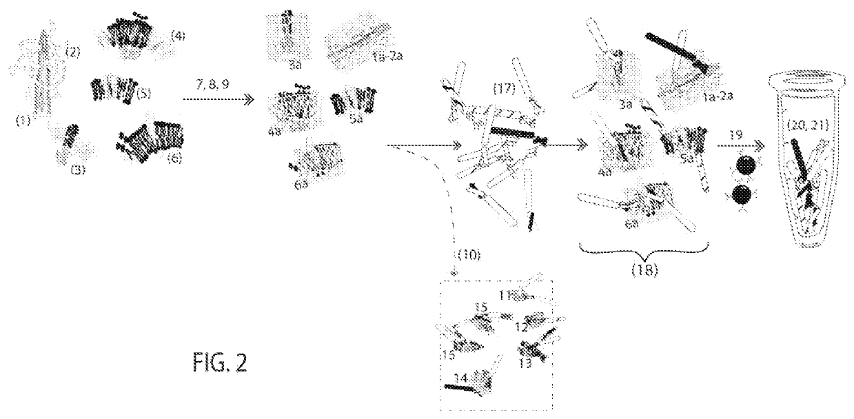
[0104]Embodiment 1 takes advantage of cancer cell surface deformations caused by mutations and their effects to alter membrane-proteins. Embodiment 1 selects and amplifies cancer binding molecules for rapid, routine cancer immunotherapy. The most critical factor in using these cell surface formations as immunotherapy targets is to preserve their 3-dimensional structures and arrangements. If cancer cell surface structures become artificially distorted, protocols will select antibodies that recognize the distorted cancer cell surfaces. These antibodies have a high affinity for the distorted antigen but not for the antigen in tumor samples.[0105]1. Cancer cells comprise structures (1-6) on the same or different cell populations and contributions from the microenvironment (2) such as those from FIG. 1[0106]7. Preserving cancer cell structures begins by enriching for the outer cell membrane surfaces. The Membrane Fraction preparation comprises gentle lysis and differ...
embodiment 2 (fig.5)
Embodiment 2 (FIG. 5)
[0129]To preserve the structure of normal cell and cancer cell antigens, cells are immobilized in microwells on a microplate (“1” in FIG. 5) under conditions that maintain in vivo morphology. Before exposure to a patient derived cancer cell specimen, we generate conditions mimicking cancer cell physiologic environments. First, microwell plates are coated with stromal cells (“2” in FIG. 5.) (such as fibroblasts or smooth muscle cells). Next comes a combination of fibronectin and hyaluron added in sequence (“3” in FIG. 5). Human fibronectin (0.1%) facilitates adhesion through binding to integrin receptors (1-5 mcg / cm2). Hyaluronic acid coating is done by the method of Corradetti et al., 2017. The last addition is animal origin-free recombinant Type 1 human collagen (“4” in FIG. 5) in dilution medium to encourage adhesion.
[0130]After cancer cells are placed on the substrate, the cells are exposed to an edited phage display library. The library has been edited so th...
PUM
| Property | Measurement | Unit |
|---|---|---|
| Surface structure | aaaaa | aaaaa |
| Surface morphology | aaaaa | aaaaa |
| Antigenicity | aaaaa | aaaaa |
Abstract
Description
Claims
Application Information
 - R&D
- Intellectual Property
- Life Sciences
- Materials
- Tech Scout
- Unparalleled Data Quality
- Higher Quality Content
- 60% Fewer Hallucinations
Browse by: Latest US Patents, China's latest patents, Technical Efficacy Thesaurus, Application Domain, Technology Topic, Popular Technical Reports.
© 2025 PatSnap. All rights reserved.Legal|Privacy policy|Modern Slavery Act Transparency Statement|Sitemap|About US| Contact US: help@patsnap.com


