Methods of producing adenovirus
- Summary
- Abstract
- Description
- Claims
- Application Information
AI Technical Summary
Benefits of technology
Problems solved by technology
Method used
Image
Examples
example 1
s Infection Using High and Low MOI
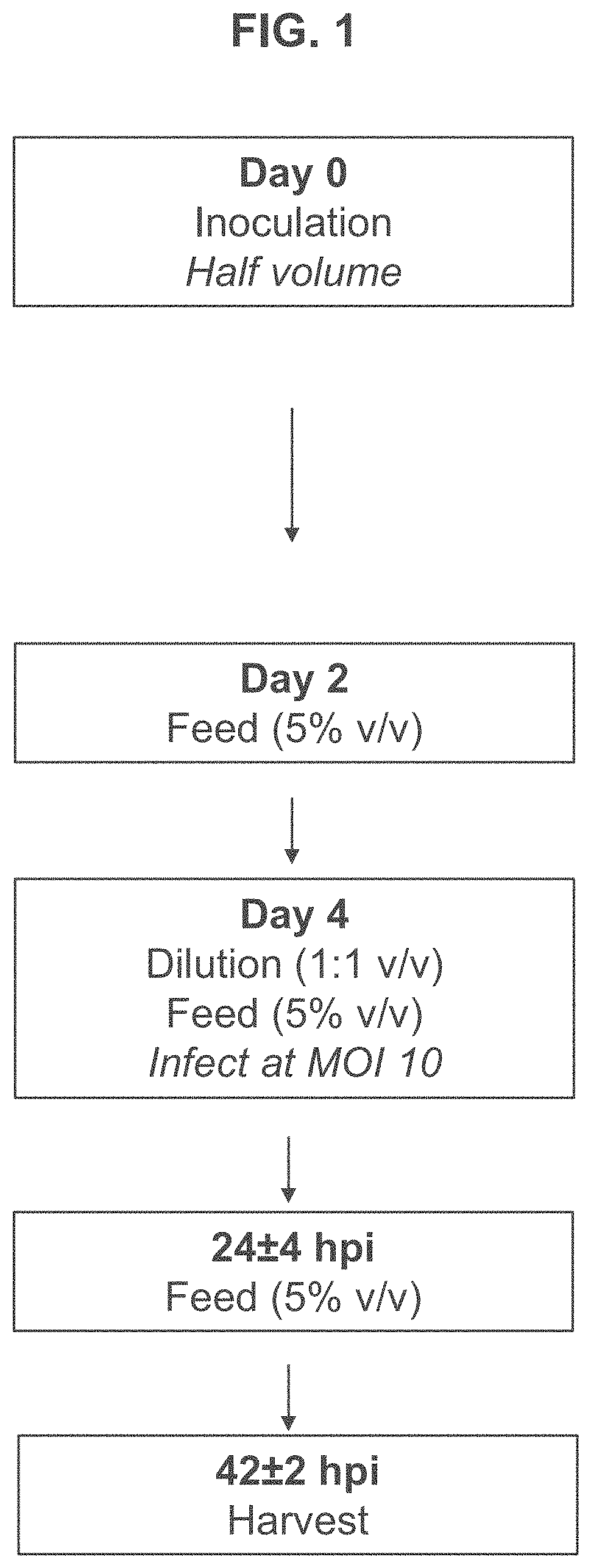
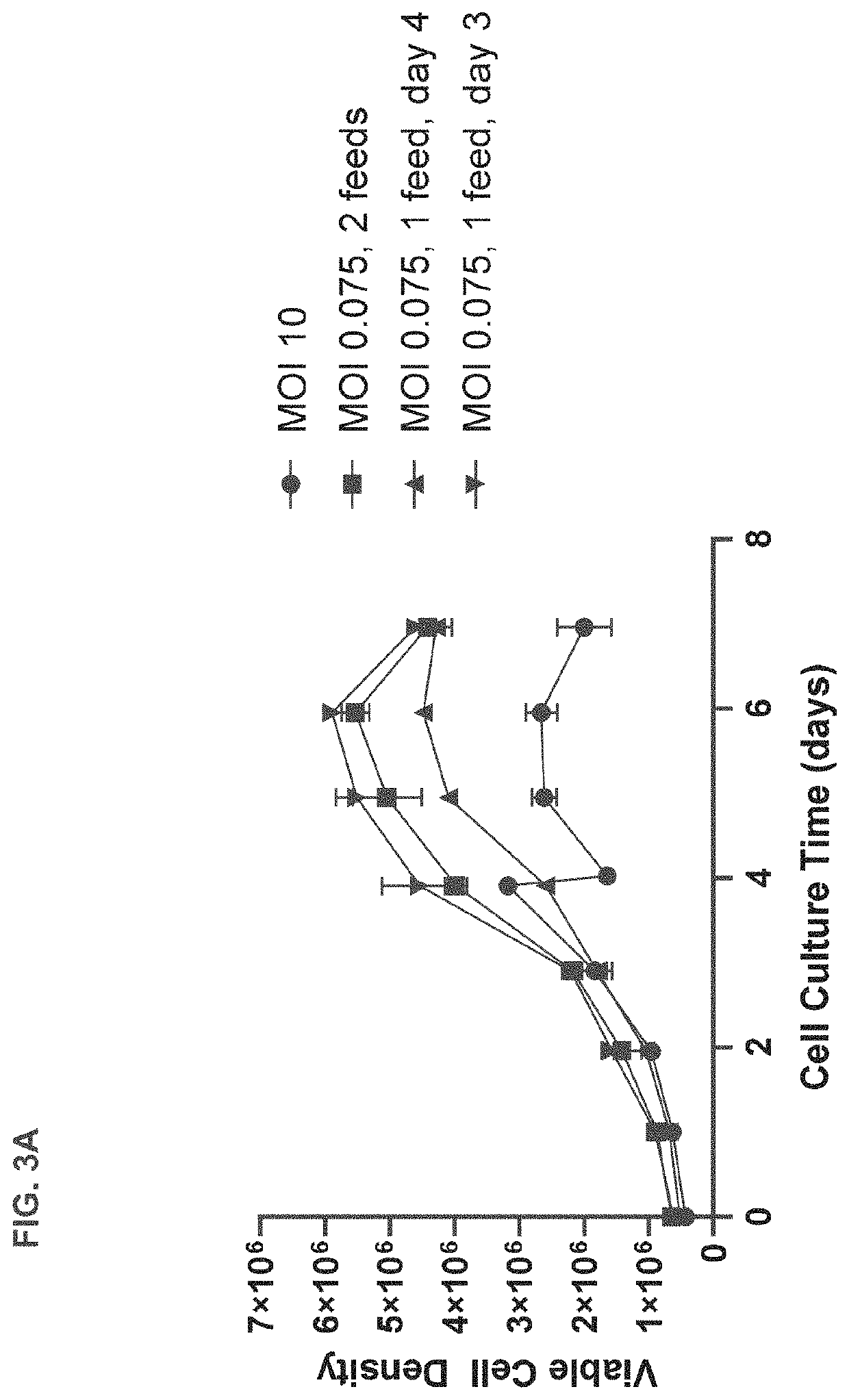
[0189]T-REx™ cells were seeded in 3 L bioreactors at 0.5×10⁶ viable cells per mL and subjected to high or low MOI adenovirus infection regimes based on those shown in FIG. 1 or FIG. 2. Briefly, for the high MOI infection regime, T-REx™ cells were grown until they reached a confluency of approximately 3-5×10⁶ cells / mL at which point they were diluted 1:1 and infected with adenovirus at an MOI of 10. The infected cells were fed with commercially available feed for HEK 293 cells when cell density reached 1×10⁶ viable cell per mL approximately 24 hours after infection. Separate cultures were harvested at 24, 48, and 72 hours after infection for assessment of viral titer.
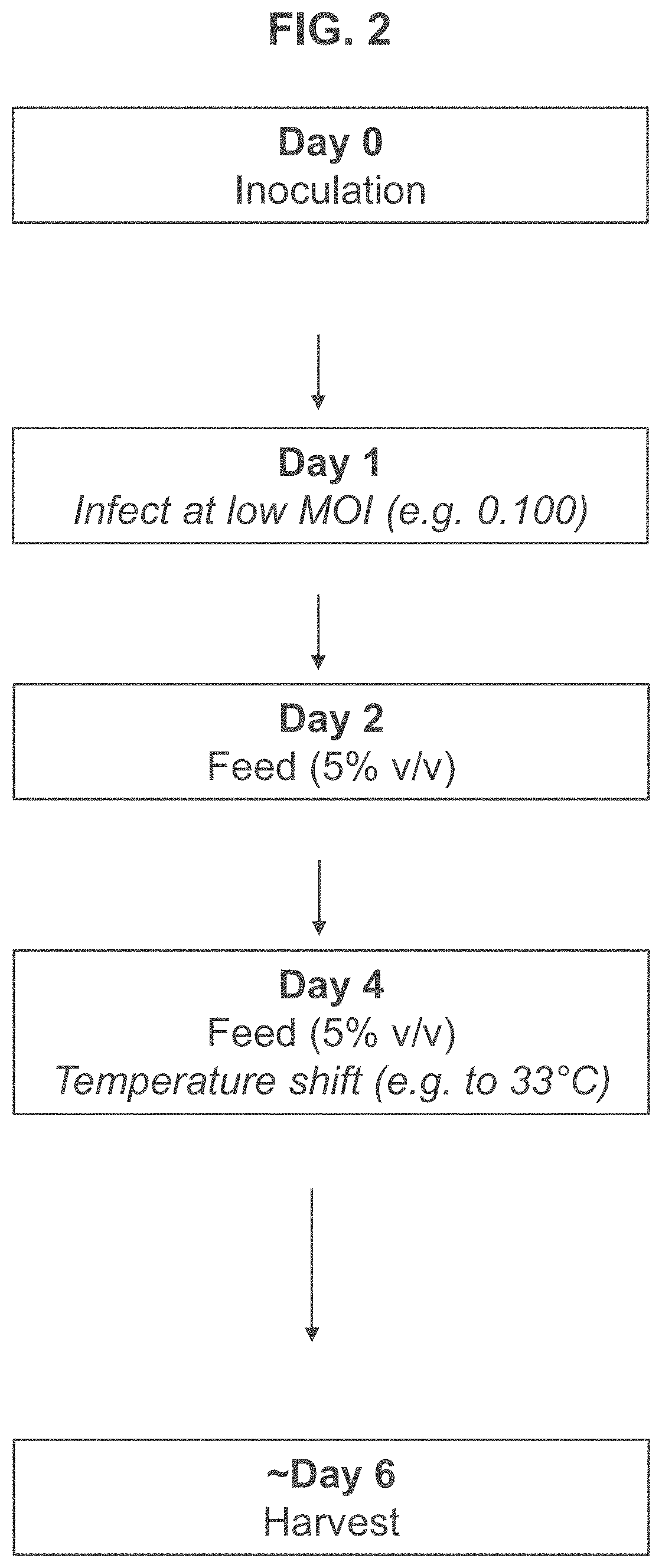
[0190]In contrast, for the low MOI regime, T-REx™ cells were seeded in 3 L bioreactors at 0.7×10⁶ viable cells per mL and infected with adenovirus at an MOI of 0.075 approximately 24 hours after seeding. The infected cells were fed on day 4 and separate cultures were harvested at 72, 96,...
example 2
f Infection at Low MOIs on Viral Titer
[0198]Next, the inventors tested the effect of a range of low MOIs on peak viable cell density, cell viability and virus titer. Briefly, T-REx™ cells were seeded in 3 L bioreactors at 0.7×10⁶ viable cells per mL and infected with adenovirus at an MOI of 0.026-0.270 approximately 24 hours after seeding. The infected cells were fed on day 2 and day 4 and separate cultures were harvested approximately 5, 6, and 7 days after seeding. As described previously, viable cell density and viability was measured for each of the cultures daily.
[0199]As shown in FIG. 5A, cells infected at a lower MOI had a higher peak viable cell density. Specifically, cells infected at an MOI of 0.026-0.030 had a peak cell density of about 7-8×10⁶ cells / mL, whereas cells infected at an MOI of 0.232-0.270 had a peak cell density of about 3×10⁶ cell / mL.
[0200]As shown in FIG. 5B, cell viability tended to decrease with increasing MOI. Thus, cells infected at an MOI of 0.026-0.03...
example 3
f Cell Seeding Density on Viral Titer
[0202]The inventors next assessed viral titer at different initial cell seeding densities with infection on either day 0 or day 1. In brief, T-REx™ cells were seeded in ambr 250 vessels at 0.5-1.2×10⁶ cells / mL and infected with adenovirus at target MOIs of 0.025 or 0.075 on day 0 or day 1 after seeding. Cells were cultured for up to 7 days post infection and cell culture was harvested for assessment of viral titer.
[0203]As shown in FIG. 6A, increasing cell density surprisingly increases viral titer for cultures infected at day 0 after cell seeding. Specifically, a cell seeding density of 0.5×10⁶ cells / mL resulted in a viral titer of 11 VG / mL when cultures were infected at an MOI of 0.025, whereas a cell seeding density of 1.2×10⁶ cells / mL resulted in a dramatically higher viral titer of approximately 4.5×10¹¹ VG / mL when cultures were infected at the same MOI. A similar effect was observed for cultures infected with an MOI of 0.075. FIG. 6B shows ...
PUM
| Property | Measurement | Unit |
|---|---|---|
| Time | aaaaa | aaaaa |
| Volume | aaaaa | aaaaa |
| Volume | aaaaa | aaaaa |
Abstract
Description
Claims
Application Information
 - R&D
- Intellectual Property
- Life Sciences
- Materials
- Tech Scout
- Unparalleled Data Quality
- Higher Quality Content
- 60% Fewer Hallucinations
Browse by: Latest US Patents, China's latest patents, Technical Efficacy Thesaurus, Application Domain, Technology Topic, Popular Technical Reports.
© 2025 PatSnap. All rights reserved.Legal|Privacy policy|Modern Slavery Act Transparency Statement|Sitemap|About US| Contact US: help@patsnap.com


