Drp1-FILAMIN COMPLEX FORMATION INHIBITORS
a technology of dynamin-related protein and complex, which is applied in the field of dynamin-related protein 1 (drp1)filamin complex formation inhibitors, can solve the problems of energy metabolic disorder, chronic heart failure,
- Summary
- Abstract
- Description
- Claims
- Application Information
AI Technical Summary
Benefits of technology
Problems solved by technology
Method used
Image

Examples
first embodiment
[0125]Regarding the Drp1-filamin complex formation inhibitor of a first embodiment, a compound represented by formula (1) may be such that R1 represents a phenyl group, 2-pyridyl group, 3-pyridyl group or 4-pyridyl group, R2 represents a hydrogen atom, and R3 represents a nitro group. Alternatively, regarding the Drp1-filamin complex formation inhibitor of the first embodiment, a compound represented by formula (1) may be such that R1 represents a 2-pyridyl group, 3-pyridyl group or 4-pyridyl group, R2 represents an amino group, and R3 represents a hydrogen atom.
[0126]As described later in Examples, the present inventors clarified that the cilnidipine derivative of the first embodiment has the Drp1-filamin complex formation inhibitor. The present inventors also clarified that the cilnidipine derivative of the first embodiment does not act to block the calcium channel, unlike cilnidipine that blocks the calcium channel. That is, the cilnidipine derivative of the first embodiment does...
second embodiment
[0131]Regarding the Drp1-filamin complex formation inhibitor of the second embodiment, a compound represented by formula (1) may be such that R1 represents a phenyl group, 2-pyridyl group, 3-pyridyl group or 4-pyridyl group, R2 represents an amino group, and R3 represents a hydrogen atom.
[0132]As described later in Examples, the present inventors clarified that the cilnidipine derivative of the second embodiment exhibits suppressive ability over mitochondrial division, approximately three times stronger than that of cilnidipine.
[0133]Specific examples of the cilnidipine derivative of the second embodiment include a compound represented by chemical formula (5) below (referred to as “NS4-700”, hereinafter), and a compound represented by chemical formula (6) below (referred to as “NS4-238”, hereinafter).
[0134]As described later in Examples, NS4-700 exhibits suppressive ability over mitochondrial division, to a high degree comparative to that of NS4-238, but does not block the calcium c...
examples
[0156]The present invention will further be detailed below referring to Examples. The present invention is, however, not limited to Examples below.
[0157](Exemplary Experiment 1)
[0158]
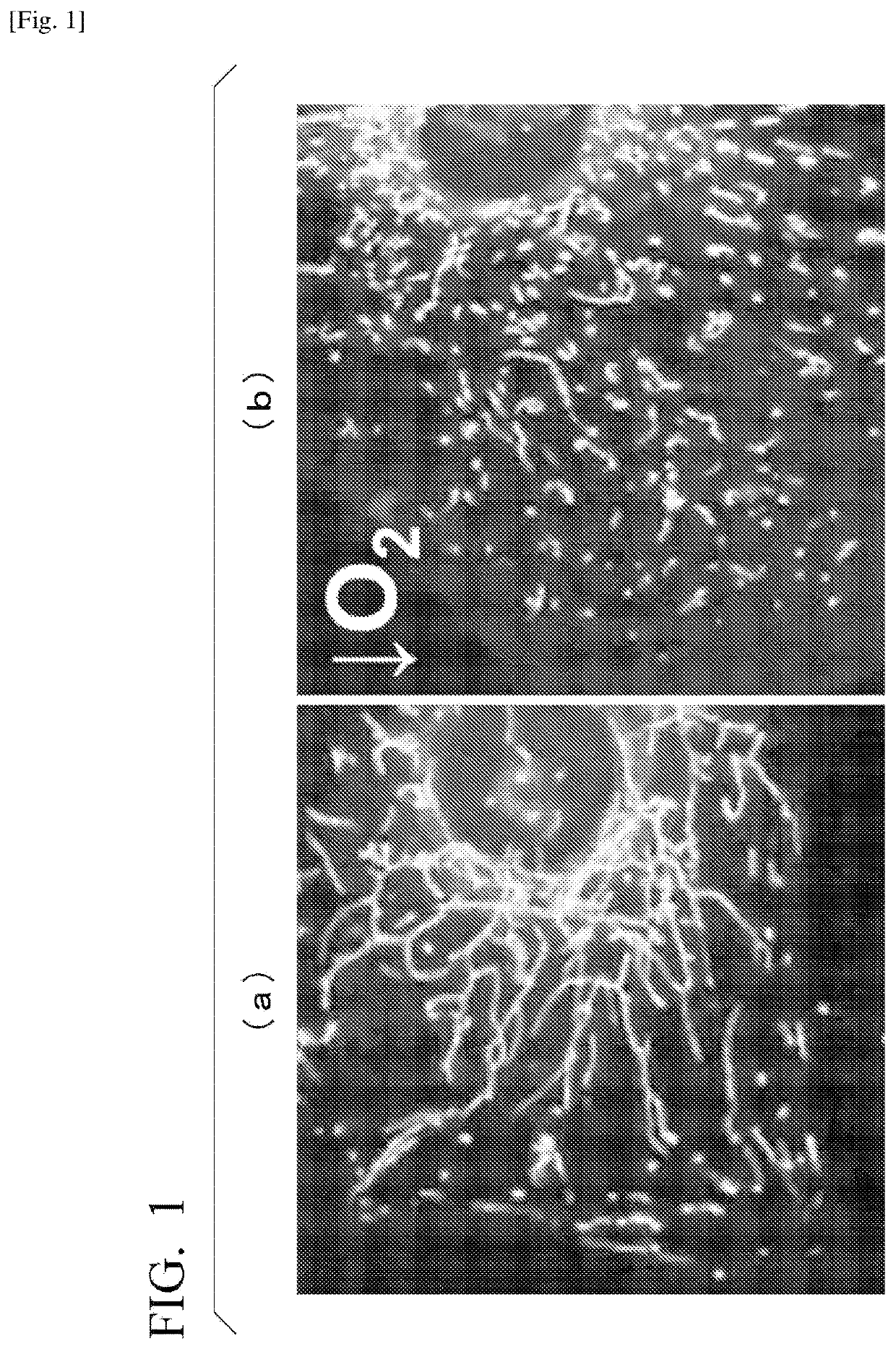
[0159]A system for quantitatively evaluating degree of intracellular mitochondrial division was built. More specifically, myocardial cells obtained from a neonatal rat were cultured under normoxia (20%) or under hypoxia (1%) for 16 hours. The cells were then stained with a fluorescent dye (MitoTracker Green FM, from Thermo Fisher Scientific Inc.) that selectively stains mitochondria. The cells were observed under a fluorescent microscope, and the shape of mitochondria was quantified by image analysis.
[0160]FIGS. 1(a) and 1(b) are representative fluorescent microphotographs of mitochondria. FIG. 1(a) is a fluorescent microphotograph of a cell cultured under normoxia, and FIG. 1(b) is a fluorescent microphotograph of a cell cultured under hypoxia.
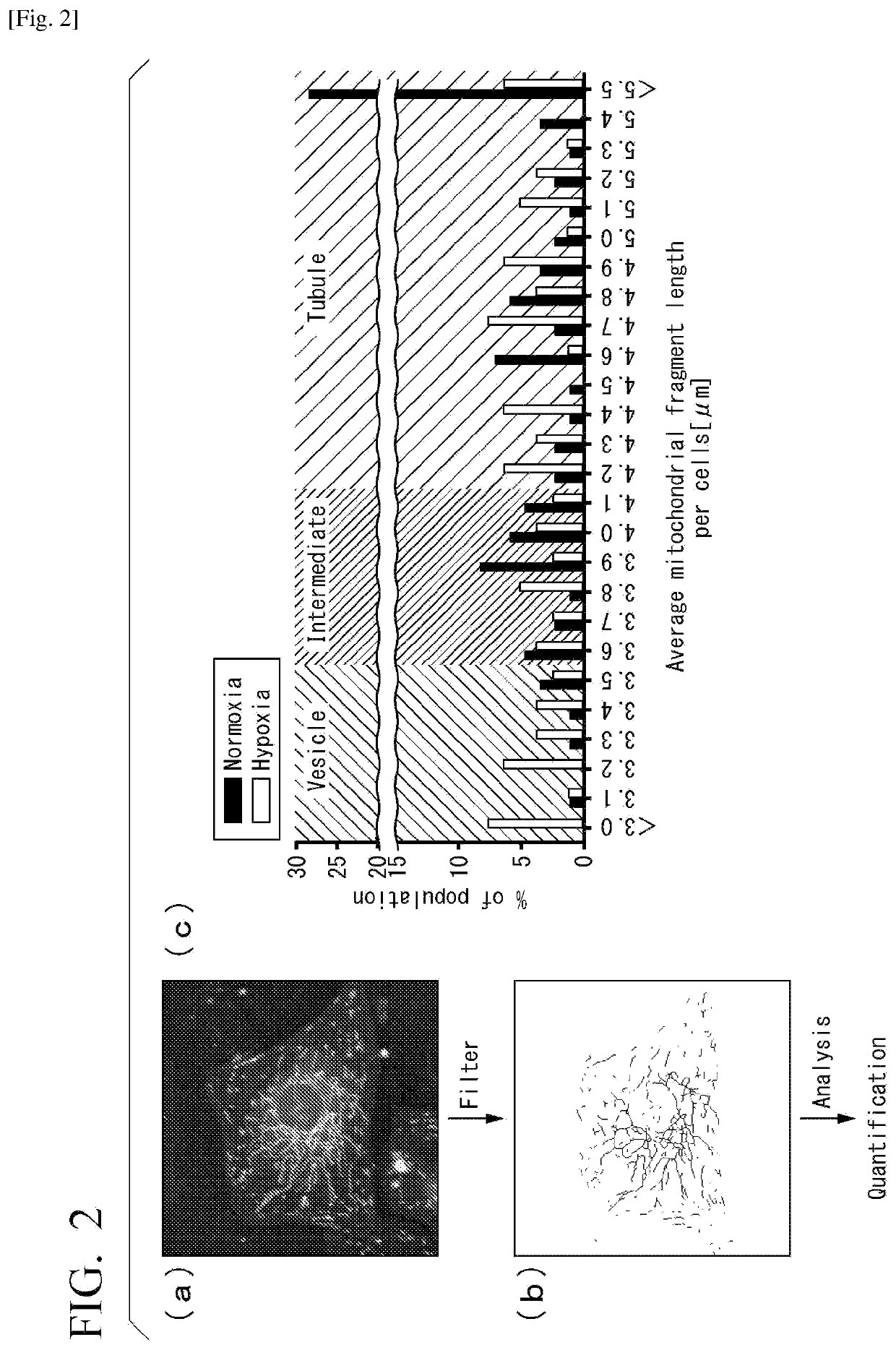
[0161]FIGS. 2(a) and 2(b) are photographs and images explai...
PUM
| Property | Measurement | Unit |
|---|---|---|
| time | aaaaa | aaaaa |
| size | aaaaa | aaaaa |
| size | aaaaa | aaaaa |
Abstract
Description
Claims
Application Information
 - R&D
- Intellectual Property
- Life Sciences
- Materials
- Tech Scout
- Unparalleled Data Quality
- Higher Quality Content
- 60% Fewer Hallucinations
Browse by: Latest US Patents, China's latest patents, Technical Efficacy Thesaurus, Application Domain, Technology Topic, Popular Technical Reports.
© 2025 PatSnap. All rights reserved.Legal|Privacy policy|Modern Slavery Act Transparency Statement|Sitemap|About US| Contact US: help@patsnap.com