Devices, systems and methods for biomarker analysis

a biomarker and device technology, applied in the field of devices, systems and methods for biomarker analysis, can solve problems such as cell-free circulating nucleic acids analysis

Pending Publication Date: 2022-04-21
JUNO DIAGNOSTICS INC
View PDF5 Cites 1 Cited by
  • Summary
  • Abstract
  • Description
  • Claims
  • Application Information

AI Technical Summary

Benefits of technology

[0006]Analysis of cell-free circulating nucleic acids is met with a number of technical challenges. For instance, amplification of circulating nucleic acids in blood may be inhibited by some of the components in whole blood (e.g., hemoglobin and associated iron). The devices, systems, kits and methods disclosed herein aim to overcome many of these technical challenges. In addition, the devices, systems, kits and methods offer the advantage of being (1) minimally invasive, (2) applicable in home with little or no technical training (e.g., do not require complex equipment); and (3) informative at early stages of a condition (e.g., pregnancy, infection). Furthermore, the avoidance of repeated doctor / hospital visits for the purpose of blood draws and centralized testing may improve patient compliance and allow more frequent monitoring, ultimately leading to improved health outcomes at lower cost to the healthcare system.
[0009]By way of non-limiting example, devices, systems, kits and methods are disclosed herein may be used for determining the gender of a fetus. Devices, systems, kits and methods disclosed herein allow for gender determination in the privacy of a home, without the need for laboratory equipment and without the risk of sample swapping. These devices, systems, kits and methods generally analyze cell free fetal DNA and / or cell free fetal RNA. Devices, systems, kits and methods disclosed herein may advantageously determine the gender of the fetus at early stages of gestation because they require very little fetal nucleic acid material. Devices, systems, kits and methods disclosed herein provide gender status from a very low volume of sample because the devices, systems, kits and methods are capable of detecting fragments of the Y chromosome including genes or any amplifiable regions which can uniquely identify the presence or absence of the Y chromosome in a biological sample, often present in multiple copies on the Y chromosome. The effective concentration of these fragments is higher than those fragments of genes that are not present in multiple copies in most cases.

Problems solved by technology

Analysis of cell-free circulating nucleic acids is met with a number of technical challenges.

Method used


Image

  • Devices, systems and methods for biomarker analysis
  • Devices, systems and methods for biomarker analysis
  • Devices, systems and methods for biomarker analysis

Examples


example 1

r Analysis of Cell-Free Nucleic Acids from Whole Blood

[0315]A device for purifying separating plasma from maternal whole blood for the purpose of analyzing cell-free fetal nucleic acids was constructed. The device consists of 6 layers. From bottom to top these are:

[0316](1) Lower Adhesive Sheet

[0317](2) Lower Separation Disc: 16 mm diameter disc of adhesive sheet material (polymer material that is inert to DNA or Plasma) with glue on the side facing the Lower Adhesive Sheet

[0318](3) Polyethersulfone (PES) membrane, various sizes, typically between 6 and 16 mm, preferred design features 10 mm PES membrane. The membrane serves as wicking material which attracts the plasma from the filter through capillary force.

[0319](4) Filter Disc (e.g., Pall Vivid™ Membrane), 16 mm diameter, rough side facing up, shiny side facing the PES membrane.

[0320](5) Upper Separation Disc: same material as Lower Separation Disc, size 12 or 14 mm diameter, containing a 4 mm hole in the center. When using adhe...

example 2

r Analysis of Fetal Cell-Free Nucleic Acids from Maternal Blood

[0325]The device consists of multiple layers as exemplified in Example 1.

[0326]Application of blood and filtration to the device occurs as follows:

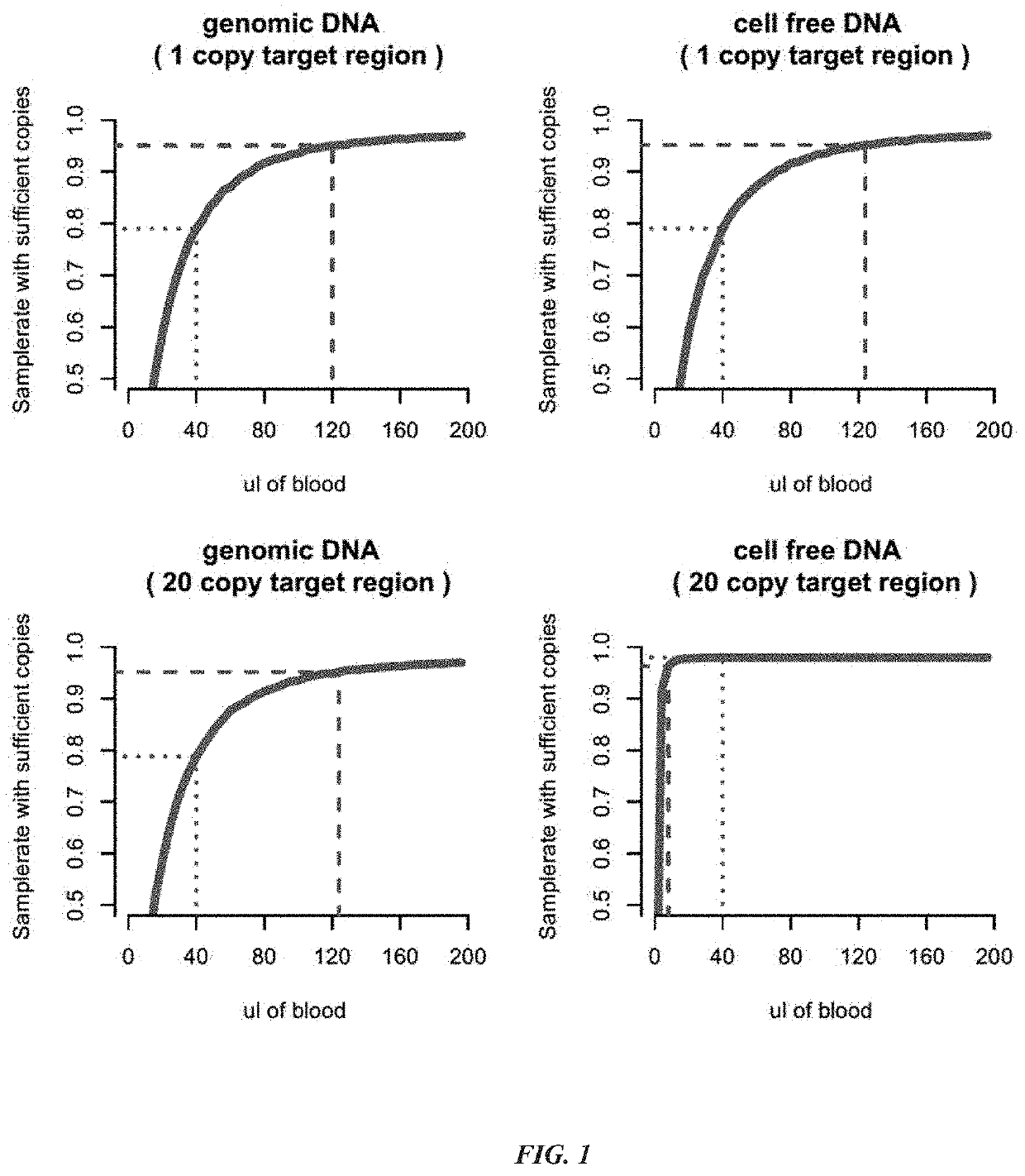
[0327]40 μl to 60 μl of whole blood is applied to the center of the device through the hole in the Upper Adhesive Sheet and the hole in the Upper Separation Disc. The blood distributes centripetally throughout the Filter Disc by capillary forces. Plasma is also wicked through the Filter Disc into the PES membrane by capillary forces. After about two minutes, the maximum amount of plasma has been transferred into the PES membrane.

[0328]The PES membrane containing cell-free nucleic acids is recovered as follows:

[0329]The device is cut out around the edges of the PES membrane. The membrane separates easily from the Filter and the Lower Disc.

[0330]DNA is eluted from the membrane as follows:

[0331]The PES membrane containing the plasma is transferred into an Eppendorf tube (0.5 ml) ...

example 3

of Human Y Chromosome DNA Using Recombinase Polymerase Amplification

[0333]Amplification and detection of human Y chromosome DNA in plasma samples were carried out by the following various methods:

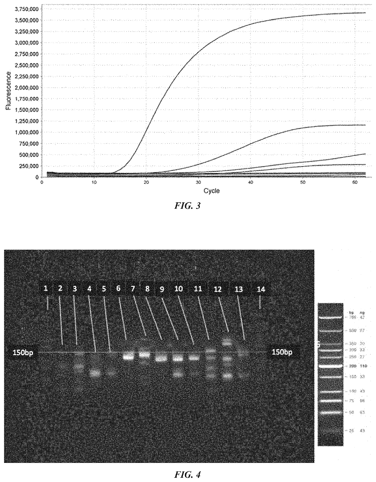
Detection of Y Chromosome Targets Using RPA and Polyacrylamide Gel Electrophoresis (PAGE):

[0334]Recombinase polymerase amplification of 50 ng (15151 copies) of male genomic DNA was conducted using the TwistAmp Basic Kit (TwistDx, Cambridge, UK) following the standard protocol. Briefly, 29.5 μl of Rehydration Buffer was combined with 3 μl of each amplification primer (10 μM) (IDT, Coralville, Iowa), 2 μl of water and 10 μl of DNA template. 47.5 μl of this mixture was mixed with the lyophilized RPA enzymes as provided (uvsX, uvsY, gp32, Bsu). Following resuspension of the lyophilized RPA enzymes the reaction mixture was added to 2.5 μl of 280 mM magnesium acetate and mixed thoroughly to activate the RPA reaction. The reaction was incubated at 37 degrees Celsius for 20 minutes with agitation e...


PUM

PropertyMeasurementUnit
pore sizeaaaaaaaaaa
volumeaaaaaaaaaa
volumeaaaaaaaaaa

Abstract

Provided herein are devices, systems, kits and methods for predicting or determining the gender of a fetus using cell free fetal nucleic acids in a small amount of maternal biological sample. Devices can be used at point of need during early stages of pregnancy and are compatible with communication devices.

Description

CROSS-REFERENCE[0001]This application is a continuation of U.S. patent application Ser. No. 16 / 648,819, filed on Mar. 19, 2020, which is a U.S. National Phase application of International Application No. PCT / US2018 / 052891, filed on Sep. 26, 2018, which application claims priority to U.S. Provisional Patent Application No. 62 / 563,314, filed on Sep. 26, 2017, and U.S. Provisional Patent Application No. 62 / 609,086, filed on Dec. 21, 2017. Priority is claimed pursuant to 35 U.S.C. § 119. The above noted patent applications are incorporated by reference as if set forth fully herein.SEQUENCE LISTING[0002]The instant application contains a Sequence Listing which has been submitted electronically in ASCII format and is hereby incorporated by reference in its entirety. Said ASCII copy, created on Dec. 10, 2021, is named 52781-701_301_SL.txt and is 49,173 bytes in size.BACKGROUND OF THE INVENTION[0003]Genetic testing is a means for obtaining information about a subject's DNA and / or expression...

Claims


Application Information

Patent Timeline
Patent Type & Authority Applications(United States)
IPC IPC(8): C12N15/10C12Q1/686
CPCC12N15/1017C12Q2565/625C12Q1/686C12N15/10B01L3/5023B01L2300/0681B01L2400/0406B01L2300/0825C12Q2565/629
Inventor VAN DEN BOOM, DIRKEHRICH, MATHIASOETH, PAULCHAUVAPUN, JIM
Owner JUNO DIAGNOSTICS INC
Why Eureka
Try Eureka