System and method for rapid and accurate histologic analysis of tumor margins using machine learning

a tumor margin and machine learning technology, applied in the field of systems and methods for analyzing images of tissue samples, can solve the problems of local tumor recurrence, increased risk of distant metastasis and decreased survival, and potential loss of information, so as to improve patient outcomes, rapid and accurate tumor margin analysis, and rapid and accurate tumor margin readout

Pending Publication Date: 2021-12-02
DARTMOUTH HITCHCOCK CLINIC
View PDF3 Cites 0 Cited by
  • Summary
  • Abstract
  • Description
  • Claims
  • Application Information

AI Technical Summary

Benefits of technology

This invention has a system and method for quickly and accurately detecting tumors and their borders using machine learning algorithms. This can be used in both clinical and research settings. If a tumor is detected, the system can help map it and plan for further tissue resection. This automation process makes the process more efficient and improves patient outcomes. Additionally, the system can analyze incomplete tissue sections to determine if they are clear of tumor.

Problems solved by technology

Incomplete excision of tumors results in both local tumor recurrence and increased risk of distant metastasis and decreased survival.
A caveat to en face margins is that the peripheral margins are separated from the deep margin resulting in the potential for lost information at this point.
Both en face margins and MMS result in very low local recurrence rates while also limiting the amount of healthy surrounding tissue removed.
Note that the use of the breadloafing technique in its current form, and permanent section histology, results in taking significantly larger margins to increase the likelihood of complete tumor resection as it is not performed in real-time.
Increasing the number of pieces generated and sectioned in a breadloafed model can increase the percentage of margin analyzed though this is time consuming both at the generation step and the pathologic reading step.
Acquiring complete tissue sections can be challenging depending on the type and size of the tissue.
This issue also arises in the sectioning of permanent paraffin sections, but less frequently.
Challenges with en face margin and MMS margin analysis is that, as the size of the tissue increases, the surface area or total margin to be analyzed increases exponentially.
Depending on the size of section analyzed and the number of tissue sections this can be challenging to accurately and efficiently map the tumor.
Subsequent resection is reliant on precise mapping and failure to accurately map the tissue results in “clear” tumor margins when, in reality, it is an incomplete tumor resection.
Communication via telephone requires staff or the surgeon to recreate the tumor map either mentally or physically presenting the opportunity for mistakes.
Not surprisingly, the risk of adverse events increases the longer a patient is under general anesthesia.

Method used


Image

  • System and method for rapid and accurate histologic analysis of tumor margins using machine learning
  • System and method for rapid and accurate histologic analysis of tumor margins using machine learning
  • System and method for rapid and accurate histologic analysis of tumor margins using machine learning

Examples


Embodiment Construction

I. Background Considerations

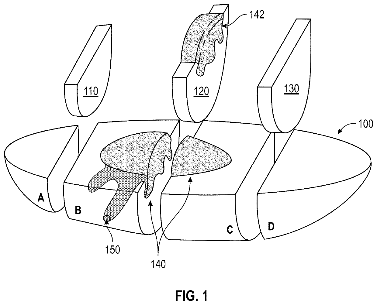
[0019]FIG. 1 shows a generalized diagram of an exemplary excised tumor tissue sample 100—for example a skin lesion—that has been subjected to the breadloafing technique, by way of non-limiting example. In this technique, the excised tissue sample 100 is sliced laterally, as shown, into sections (A-D), from which are taken thin slices 110, 120 and 130 at various points along the length of the tissue sample 100. The distribution of slices 100-130 through the tissue is chosen to intercept the likely extent of the cancerous growth 140 therein. Hence in slice 120, there resides a piece 142 of the overall cancerous growth 140. Based upon one or more slides (described below) that are created from the slices, the pathologist should be able to determine the boundaries of the cancerous tissue, thereby ensuring that it is completely removed by the excision process.

[0020]However, using conventional techniques, it is possible that irregularly or unpredictably shaped t...


PUM


Abstract

This invention provides a histologic system and method for rapidly and accurately assessing tumor margins for the presence or absence of tumor using machine learning algorithms. This affords a rapid and accurate histologic tumor readout and increase process efficiency and decreases the chance for human error. Advantageously and uniquely, the system and method allows for analyzing the tissue section as complete or incomplete as the first criteria to determine whether a tissue section is clear of tumor. A machine learning process receives whole slide images (WSI) of tissue and determines (a) if each image of the WSI contains complete / incomplete tissue samples and (b) if each image of the WSI contains tumorous tissue or an absence thereof. A reconstruction process generates a model of the tissue that maps types of tissue therein, and a display process provides results of the model or report for use and manipulation by a user.

Description

FIELD OF THE INVENTION[0001]This invention relates to systems and method for analyzing images of tissue samples for medical and research diagnostic purposes, and more particularly to those employing machine learning and / or neural networks operating on computing systems.BACKGROUND OF THE INVENTION[0002]Successful treatment of solid cancers relies on complete surgical excision of the tumor for either definitive treatment or prior to adjuvant therapy. Incomplete excision of tumors results in both local tumor recurrence and increased risk of distant metastasis and decreased survival. Recurrence rate of tumors following surgical excision range from as high as 50% for subtypes of brain cancer to as low as 0.5% for skin cancers. Tissue preparation / grossing, inking, grossing, and analysis may contribute to this wide range in local recurrence rates. Varied methodologies can be used to gross and ink the tissue prior to tissue sectioning and then analysis by the pathologist. The “breadloafing”...

Claims


Application Information

Patent Timeline
Patent Type & Authority Applications(United States)
IPC IPC(8): G16H50/20A61B34/10G16H50/50G16H30/40G16H10/40G16H15/00G16H70/60G16H50/70G06Q30/00G06Q20/10G06N3/08H04L29/06G06T7/00
CPCG16H50/20G06T2207/20081G16H50/50G16H30/40G16H10/40G16H15/00G16H70/60G16H50/70G06Q30/0185G06Q20/102G06N3/08H04L63/08G06T7/0012G06T2207/30096G06T2207/30024A61B34/10G06T2207/20084G06T2207/10056G06N3/045
Inventor LEBOEUF, MATTHEWVAICKUS, LOUIS J.
Owner DARTMOUTH HITCHCOCK CLINIC
Why Eureka
Try Eureka