
Heart valve prosthesis anchored to interventricular septum and conveying and releasing method thereof

a technology of heart valve and interventricular septum, which is applied in the field of medical equipment, to achieve the effect of avoiding the traction of the valve annulus and reducing the influence of the outflow tra

Active Publication Date: 2021-09-16
JENSCARE SCI CO LTD
View PDF18 Cites 0 Cited by
  • Summary
  • Abstract
  • Description
  • Claims
  • Application Information

AI Technical Summary

Benefits of technology

The present application provides a heart valve prosthesis that can be anchored to the interventricular septum to replace a damaged valve. It solves the problems of traditional anchoring technology that can affect the outflow tract after the supporting frame is released and avoids traction for the native valve. This innovation helps to improve the surgical procedure and patient outcomes.

Problems solved by technology

As a result, the right ventricle becomes fat and thick, and right heart failure easily happens.
In the surgical methods, the typical thoracotomy and open heart surgeries have too large invassiveness, and an extracorporeal circulation needs to be established, therefore, there will be a high complication incidence and a risk of infection.
Many patients cannot bear the huge surgical risk and have to helplessly wait for death.
However, as for the intervention treatment of atrioventricular valve, a relatively large blank still exists in the art.
A few products for the intervention treatment for atrioventricular valve are applied in transcatheter intervention valve shaping and repairment, but in the aspect of transcatheter intervention valve replacement, no mature products are available in the world.
After long-term implantation, disorders such as heart failure will arise.
Especially when the native valve leaflet of the patient has severe calcification, the valve clamping mechanism of the native valve leaflet increases the difficulty of the surgery operation.
In the clinical application of this product of Edwards, the native valve leaflet of the patient were not able to be correctly grabbed, which causes multiple cases of surgery failure, instrument displacement and urgent transferring the patients to surgical thoracotomy operation native valve, and the patients finally died or were faced with the risk of death.
Finally, the technique of clamping the native valve leaflet inevitably affects the function of the supporting frame of returning to the sheath, and once released, it cannot be withdrawn, which brings a large operation risk.
The valve with huge diameter severely blocks the blood supply from the left ventricular outflow tract, and in the aortic valve orifice, the flow rate is increased and the pressure is boosted, which, for a long time, easily causes heart failure.
Although the patent states that the so-called D-shaped net-like tube may match with the shape of the receiving space that is enclosed by the protogenetic body wall of the in-situ mitral valve, and compared with a supporting frame whose cross-section is circular, the D-shaped net-like tube may avoid the narrowing of the outflow tract of the heart caused by the excessive stress exerted on the non-round contour of the mitral valve by the supporting frame, the problems of the technique are that, even if the cross-section of the supporting frame is modified to be D-shape, the direct contact of the whole lattice structure with the mitral valve annulus still exerts stress on its surrounding tissue; moreover, in the technical solution, the maximum tube diameter of the first sub-supporting frame is the same as the tube diameter of the second sub-supporting frame, which means that the diameter of the cross-section of the supporting frame is still at least equal to the diameter of the mitral valve annulus, therefore, the influence exerted on the outflow tract by the huge supporting frame itself still exists; and finally, in the technical solution, the second sub-supporting frame is configured to be D-shaped, and the valve leaflet is stitched to the supporting frame, and the non-round region definitely affects the clinging state after the valve leaflet is stitched.
Although the technique does not particularly disclose the stitching mode of the valve, it can still be seen from the drawings that the valve leaflet is a three-leaf valve.
Therefore, the non-round suture will be harmful to the closing property of the valve leaflet and long-term valve leaflet fatigue.
The problems of the solution are that, the expanded external supporting frame still supports and radially expands the original valve annulus, and the relatively large cross-sectional area of the supporting frame definitely takes effects on the outflow tract.
In addition, the portion of the external supporting frame that is disposed on the mitral valve annulus cannot conform to the non-uniform contour of the atrial wall or of the native valve annulus of the patient, pressing the aorta or other heart tissues, and moreover, the leak resistance effect is poor.
The current clinical results indicate that, there are no ideal products for valve intervention and replacement of atrioventricular valve.
The major reason is that, both of the mitral valve and the tricuspid valve have particular physiological structures, and the physiological environments under the valve annulus are complicated, which makes it difficult to accurately position and fix the products.
The problems of the prior art are summarized as follows: (1) the conventional anchoring techniques mostly rely on the supporting force exerted on the atrioventricular valve annulus by the supporting frame; and doctors usually select a valve specification larger than the valve annulus of the autogenous atrioventricular valve of the patient, so as to conform to the contour of the mitral valve tissue, thus the huge supporting frame itself not only affects the outflow tract, but also easily presses the surrounding tissues, and further blocks the blood flow in the left ventricular outflow tract; (2) in the prior art, the supporting frame portion disposed in the atrium mostly configured to be a lattice, the huge supporting force of which easily presses the heart tissue; and (3) as for the mitral valve replacement, the supporting frame specification is too large, and the anterior valve of the mitral valve is easily pushed toward the left ventricular outflow tract; and the design of clamping the valve leaflet, which is introduced to fix the anterior valve of the mitral valve, makes the releasing step extremely complicated and be influenced by the calcification degree of the valve leaflet, which affects the success rate of the operation.
In conclusion, although the above described techniques individually have certain effects on the atrioventricular valve replacement, they still have defects.

Method used


Image

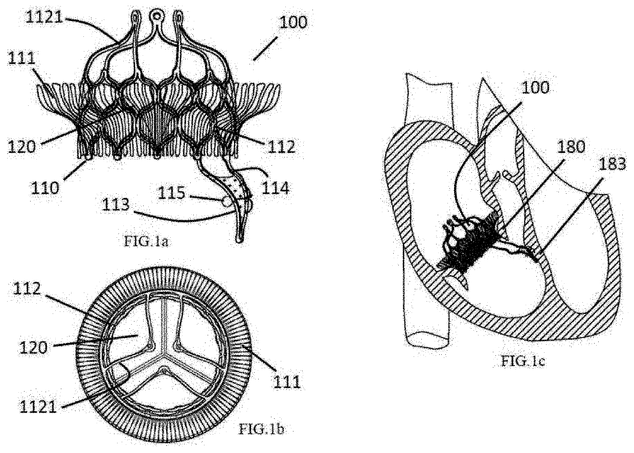
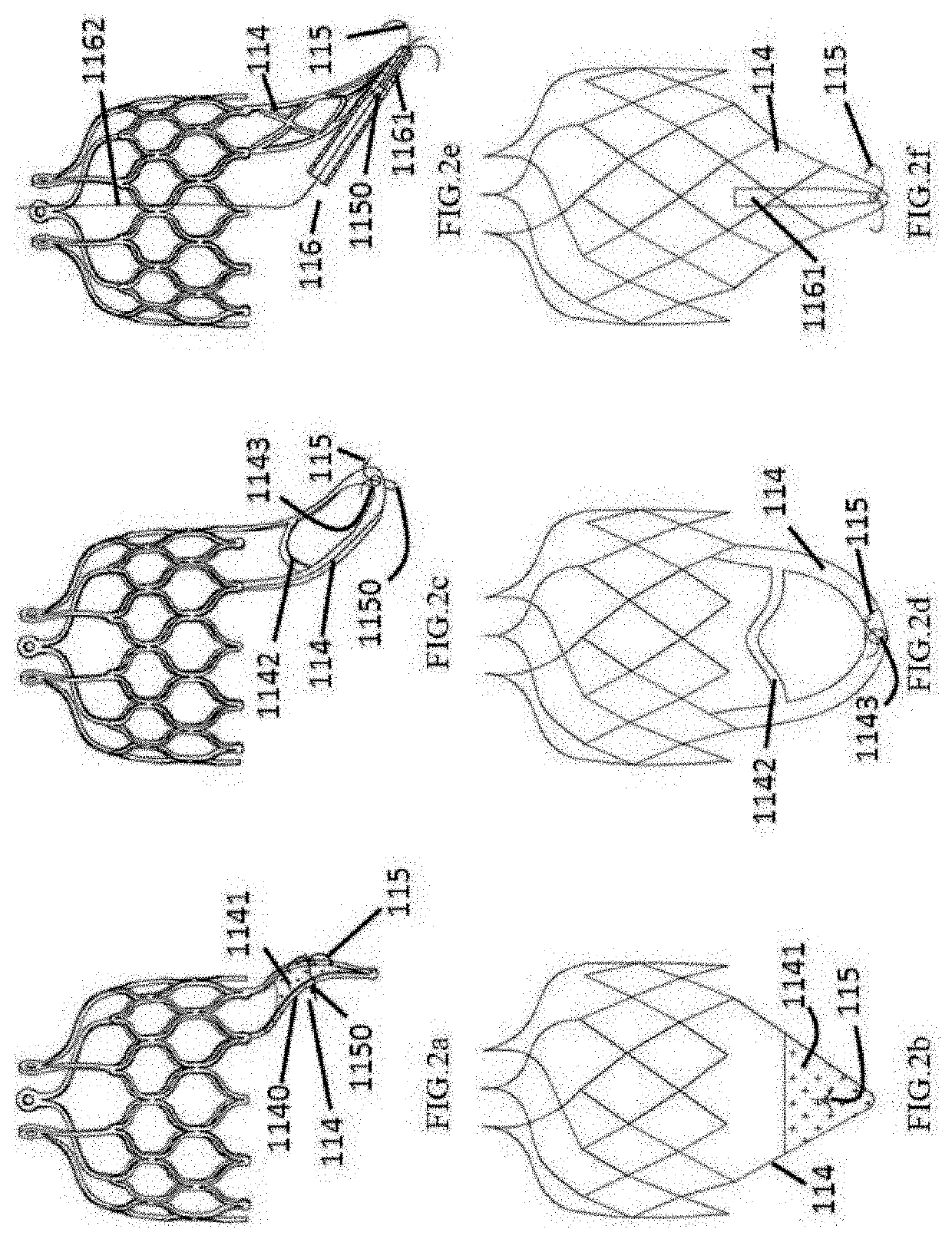
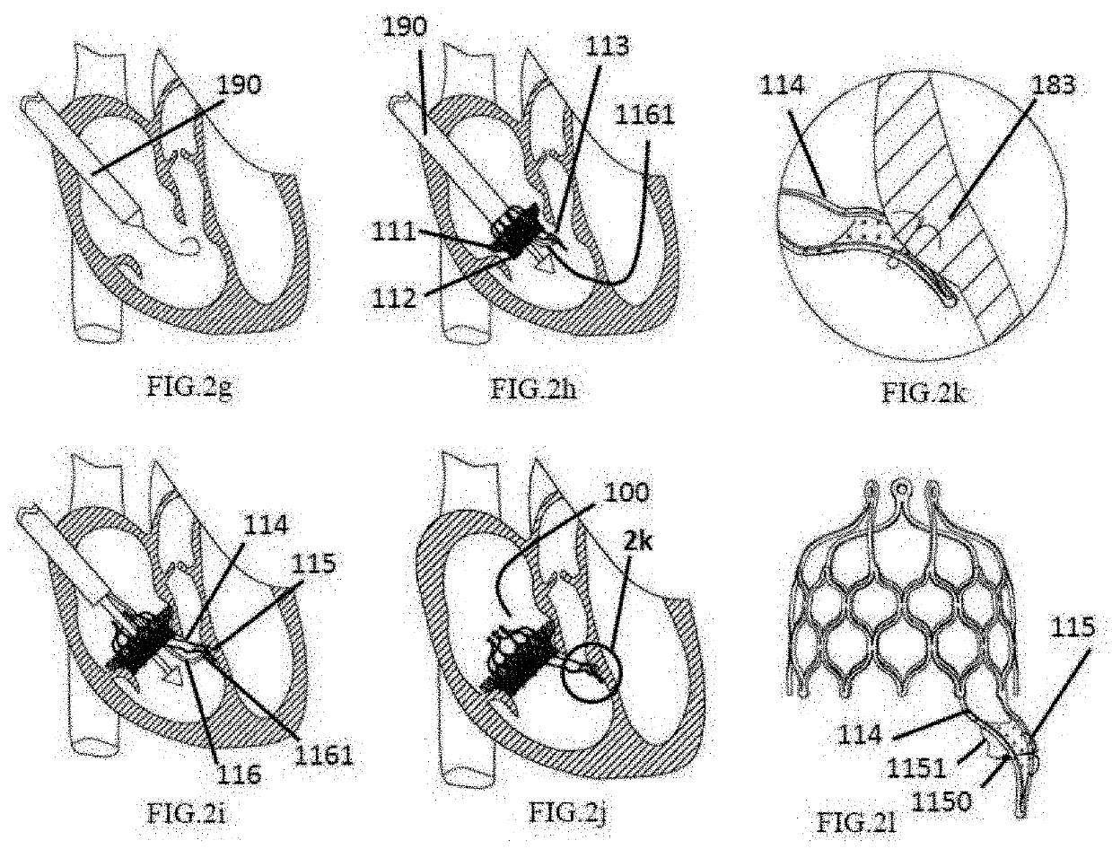
  • Heart valve prosthesis anchored to interventricular septum and conveying and releasing method thereof
  • Heart valve prosthesis anchored to interventricular septum and conveying and releasing method thereof
  • Heart valve prosthesis anchored to interventricular septum and conveying and releasing method thereof

Examples


embodiment 1

[0093]For a long time, large valve manufacturers, whether Edwards corporation or Medtronic corporation, all achieve a sufficient anchoring force of the supporting frame by increasing the radial expansion ratio of the supporting frame to the valve annulus, which has already been widely applied and become a common view in the field of aortic valve intervention and replacement, and in the field of pulmonary valve intervention and replacement (generally, 10% -15% is the ideal perimeter expansion ratio). Moreover, subsequently, both Jenavalve corporation and Symetic corporation applied the valve leaflet clamping mechanism to the products, which still has a certain expansion ratio for the valve of the patient. However, because the physiological structure and the pathological mechanism of the atrioventricular valve (including the mitral valve and the tricuspid valve) are complicated, it is quite difficult to accurately position and fix the products. Currently, for the technique of atrioven...

embodiment 2

[0100]In an embodiment, as shown in FIGS. 3a and 3b, a heart valve prosthesis 200 anchored to an interventricular septum is provided for mitral valve intervention and replacement treatment. The heart valve prosthesis comprises a valve supporting frame 210 and a fixing device 213; the valve supporting frame 210 comprises a valve stitching section 212 and an artificial valve (not shown); the artificial valve is fixedly connected to the valve stitching section 212; the fixing device 213 comprises a fixing and supporting section 214 and a fixing member 215; one end of the fixing and supporting section 214 is connected to a proximal portion of the valve stitching section 212, and the other end of the fixing and supporting section 214 is connected to the interventricular septum of the patient by the fixing member 215, to support the heart valve prosthesis 200 and limit the axial movement of the heart valve prosthesis 200. The heart valve prosthesis 200 further comprises a positioning ring...

embodiment 3

[0105]In an embodiment, as shown in FIGS. 5a-5c, a heart valve prosthesis 300 anchored to an interventricular septum is provided for tricuspid valve intervention and replacement treatment, wherein the heart valve prosthesis comprises a valve supporting frame 310 and a fixing device 313; the valve supporting frame 310 comprises a valve stitching section 312 and an artificial valve (not shown); the artificial valve is fixedly connected to the valve stitching section 312; the valve stitching section 312 is a tube-like wave-shaped structure; the fixing device 313 comprises a fixing and supporting section 314 and a fixing member 315; the fixing and supporting section 314 is an inverted cone-shaped structure; the fixing and supporting section 314 includes a plurality of rods or wires; one end of the fixing and supporting section 314, which has a larger diameter, is connected to the proximal end of the valve stitching section 312 through well-known techniques such as stitching, clipping or...


PUM


Abstract

A heart valve prosthesis anchored to an interventricular septum. The heart valve prosthesis includes a valve supporting frame and a fixing device. The valve supporting frame includes a valve stitching section and an artificial valve. The artificial valve is fixedly connected to the valve stitching section. The fixing device includes a fixing and supporting section and a fixing member. One end of the fixing and supporting section is connected to a proximal end of the valve stitching section, and another end of the fixing and supporting section is connected to the interventricular septum of a patient by the fixing member, to support the heart valve prosthesis and prevent the heart valve prosthesis from axially moving.

Description

CROSS-REFERENCE TO RELATED APPLICATIONS[0001]The present application claims the priority of the Chinese patent application entitled “Heart Valve Prosthesis Anchored to Interventricular Septum and Conveying and Releasing Method Thereof”, and filed on Oct. 24, 2016 with the application number 201610921114.9, which is hereby incorporated herein in its entirety by reference.TECHNICAL FIELD[0002]The present application relates to the field of medical equipment, and particularly relates to a heart valve prosthesis anchored to an interventricular septum and a conveying and releasing method thereof.BACKGROUND[0003]Mitral valve is located at the left atrioventricular orifice, and includes five parts: valve annulus, valve leaflet, chordae tendineae, papillary muscle and commissure, and the mitral valve's accurate name in anatomia is mitral apparatus or mitral complex. Mitral valve annulus is a fibrous tissue strip attached to the edge of the left atrioventricular orifice, and is in an irregul...

Claims


Application Information

Patent Timeline
Patent Type & Authority Applications(United States)
IPC IPC(8): A61F2/24
CPCA61F2/2418A61F2220/0075A61F2220/0008A61F2210/0014A61F2/2466A61F2220/0025
Inventor LV, SHIWENLI, YIBINXU, ZHIYUNCHEN, ZHILU, FANGLINLI, JIANAN
Owner JENSCARE SCI CO LTD
Learn More