
Use of the tas1r3 protein as a marker for therapeutic, diagnostic, and/or prognostic purposes for tumors that express said protein

a technology of tas1r3 and tas1r3, which is applied in the field of tas1r3 protein, can solve the problems of difficult to achieve total cell destruction in the case of solid tumors, the most prevalent form of human cancer is still resistant, and the difficulty of most chemotherapeutic agents to achieve the effect of total cell destruction

Pending Publication Date: 2021-09-09
FUNDACION INST DE INVESTIGACION SANITARIA DE SANTIAGO DE COMPOSTELA FIDIS +1
View PDF0 Cites 0 Cited by
  • Summary
  • Abstract
  • Description
  • Claims
  • Application Information

AI Technical Summary

Benefits of technology

The patent describes the use of a receptor called TAS1R3 as a biomarker for cancer diagnosis, monitoring, and therapy. TAS1R3 is found in tumor cells and can be used to identify therapies and direct drug-release systems. It is also present in circulating tumor cells, which can help detect metastasis and monitor disease progression. The use of TAS1R3 as a biomarker could lead to improved diagnosis and treatment for cancer.

Problems solved by technology

However, despite the progress made so far in the field of oncology, and in particular in chemotherapy, many of the most prevalent forms of human cancer are still resistant to the current chemotherapeutic treatments.
Simply put, it is much more difficult for most chemotherapeutic agents to reach all the cells of a solid tumor mass, than to reach the soft tumors, and therefore, it is much more difficult to achieve total cell destruction in the case of solid tumors.
In this regard, to treat such tumors, solid tumors, the doses of chemotherapeutic agents are significantly increased resulting in side effects, which generally limits the overall effectiveness of the therapy.
This objective has been difficult to achieve, because there are few qualitative differences between neoplastic and normal tissues.

Method used


Image

  • Use of the tas1r3 protein as a marker for therapeutic, diagnostic, and/or prognostic purposes for tumors that express said protein
  • Use of the tas1r3 protein as a marker for therapeutic, diagnostic, and/or prognostic purposes for tumors that express said protein
  • Use of the tas1r3 protein as a marker for therapeutic, diagnostic, and/or prognostic purposes for tumors that express said protein

Examples


example 1

Expression of TAS1R3 Receptor in Tumor Cells

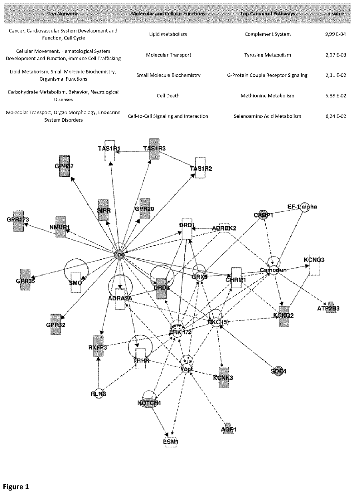
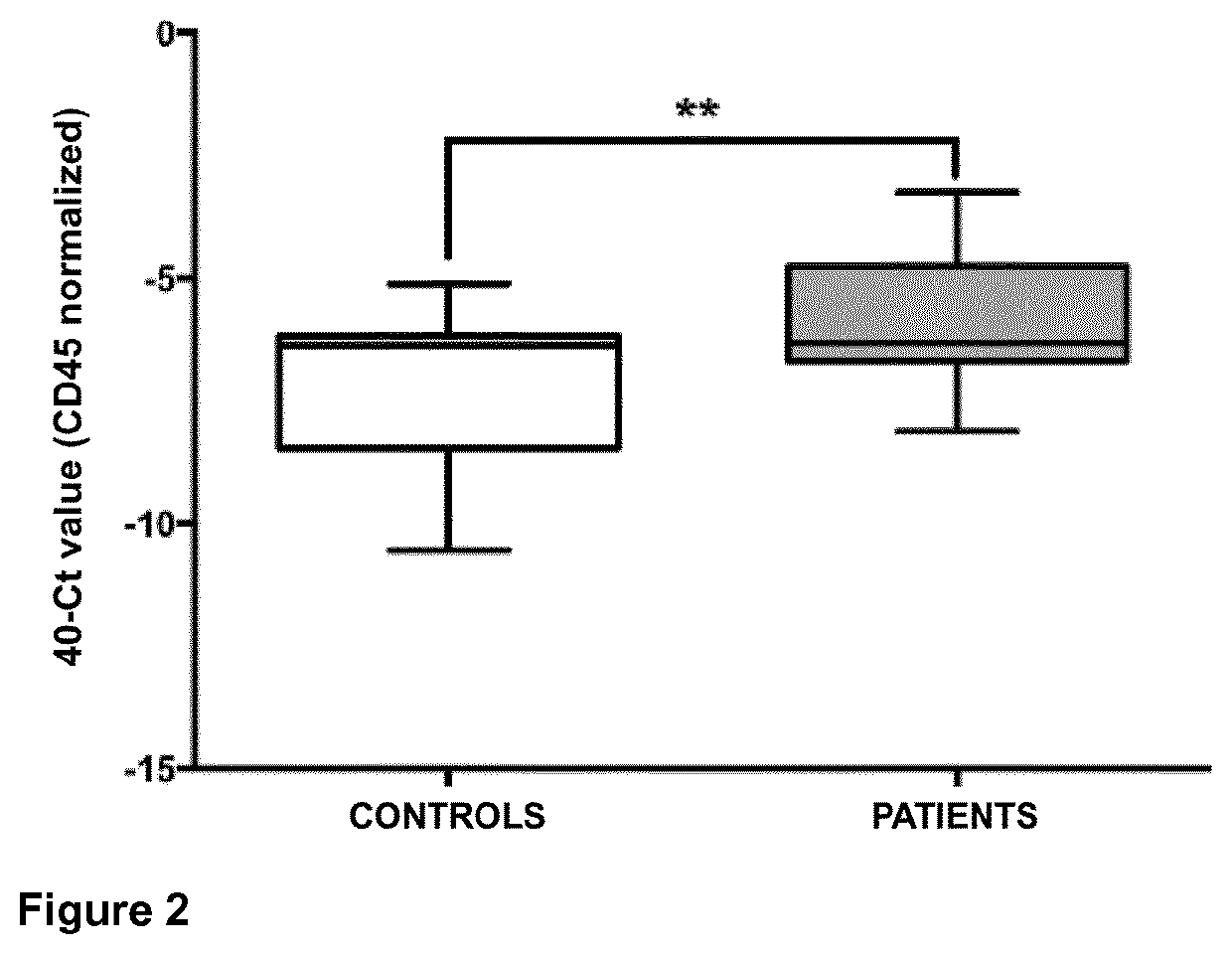
[0088]The analysis of the gene expression of the TAS1R3 receptor in CTCs (circulating tumor cells) of patients with metastatic lung cancer was performed from peripheral blood samples (patients=10, controls n=4), using magnetic particles for their isolation (EpCAM-based CELLection™ Epithelial Enrich Dynabeads® kit), following the diagram shown in FIG. 13. After extracting the RNA from the samples, they were hybridized in gene expression arrays. The signal was captured and processed in order to obtain a list of differentially expressed genes in patients with respect to the controls, among which were the TAS1R3 receptor. In summary, the total RNA of CTCs was amplified, the complementary DNA was hybridized in gene expression microarrays (Agilent) and the raw data were processed giving rise to 2,392 points, (7.01%) that met the criteria of quality. The average signal was 60,889 units, with 76,597 and 16,979 units for the patient and control gro...

example 2

Expression of TAS1R3 in Culture Media with Different Levels of Glucose Concentration

[0095]The expression of TAS1R3 in the colon line SW620 varies depending on the glucose content in the culture medium (high levels of glucose give rise to a lower expression, in relation to that observed in FIG. 6A for cells cultured in low content of glucose). This result, additionally, we have confirmed by western blot (FIG. 6B). Moreover, as seen in FIG. 6, receptor expression increases when SW620 colon cells are grown in the absence of glucose, which relates the expression of this receptor to cellular metabolism.

[0096]Cell proliferation studies were carried out in SW620 cells, FIG. 7A, cultured in medium with high and low glucose concentration. The cells were trypsinized and plated the same number of cells in order to count them at times 0, 24, 72, 96 and 168 hours, using for this the Neubauer chamber, after trypsinization thereof. In an analogous manner, cell proliferation studies were carried ou...

example 3

TAS1R3 as a Target for the Development of New Anti-Tumor Therapies

[0097]The presence of lactisole in the culture medium, a ligand of the TAS1R3 receptor, causes an increase in the expression of the receptor under study (FIG. 8A). Although at higher concentrations, the presence of ligand interferes with cell proliferation (FIG. 8B), a fact that we have related to the induction of cellular senescence (FIG. 8D). In addition, we have also seen by western blot that the presence of ligands of TAS1R3 such as lactisole or glucose (HG) in the culture medium of SW620, activates pathways related to tumor progression, such as p-ERK, p-AKT or IGF-IR (FIG. 8E). It was also determined how other ligands for TAS1R3, a peptide derived from brazzein and cyclamate, also interfere with proliferation, to a greater or lesser extent depending on the concentration and each molecule (FIG. 8F).

[0098]To determine whether after cell incubation with the ligand cell senescence is induced, 500,000 cells of the SW6...


PUM

PropertyMeasurementUnit
concentrationsaaaaaaaaaa
concentrationaaaaaaaaaa
concentrationsaaaaaaaaaa

Abstract

In the present invention, the use of the TAS1R3 receptor as a biomarker for application in cancer diagnosis, monitoring, and therapy is described for the first time. In this sense, the authors of the present invention have demonstrated that TAS1R3 is a biomarker of interest in oncology, useful for the diagnosis of the disease, and capable of providing relevant information thereupon, to monitor the evolution, select the treatment, and selectively direct therapeutic molecules. It has been determined that it is possible to identify therapies against this receptor, and that it is also possible to direct conjugates and controlled release systems of drugs, such as nanoparticles, observing a very effective intracellular accumulation thereof in primary, disseminated and metastatic tumor cells. On the other hand, the presence of TAS1R3 in circulating tumor cells (CTCs) has also been demonstrated. These are tumor cells released into the bloodstream by the primary tumor and are considered key factors in the creation of metastases, so that their detection in early stages will serve as an early detector of metastasis, and are also useful in monitoring the disease and evaluating of the response to drugs.

Description

BACKGROUNDTechnical Field[0001]The invention presents the use of the TAS1R3 protein as a new marker and molecular target in epithelial tumors and other human pathologies. In particular, the use of the detection of the TAS1R3 protein as well as its expression products, as a marker for therapeutic, diagnostic or prognostic purposes, for tumors that normally express said protein is part of the present invention.Description of the Related Art[0002]Significant progress has been made in the chemotherapy of neoplastic diseases over the past 30 years. This includes some progress in the development of new chemotherapeutic agents, and more particularly, in the development of regimens for the concurrent administration of drugs. A significant understanding of the neoplastic processes at the cellular and tissue level, and the mechanism of action of the basic antineoplastic agents, has also made it possible to obtain advances in the chemotherapy of a number of neoplastic diseases, including chori...

Claims


Application Information

Patent Timeline
Patent Type & Authority Applications(United States)
IPC IPC(8): A61K47/64G01N33/574A61K47/69C07K16/28A61K47/68
CPCA61K47/64G01N33/57492G01N33/57407A61K47/6907G01N2333/726A61K47/6851A61K47/6849A61K47/6803C07K16/28G01N33/57488A61K47/62A61P35/00G01N2800/52G01N33/57484
Inventor DE LA FUENTE FREIRE, MARÍALÓPEZ LÓPEZ, RAFAELALONSO NOCELO, MARTAVÁZQUEZ RÍOS, ABI JUDIT
Owner FUNDACION INST DE INVESTIGACION SANITARIA DE SANTIAGO DE COMPOSTELA FIDIS
Learn More