
Method and apparatus for quantitative ultrasound imaging using single-ultrasound probe

a technology of quantitative ultrasound and probe, applied in the field of image reconstruction technology using ultrasound, can solve the problems of long measurement time, high cost, and high radiation exposure risk of x-ray, mri, etc., and achieve the effect of improving contrast and accuracy, simplifying imaging, and small performance difference according to user proficiency

Pending Publication Date: 2021-06-17
KOREA ADVANCED INST OF SCI & TECH
View PDF5 Cites 0 Cited by
  • Summary
  • Abstract
  • Description
  • Claims
  • Application Information

AI Technical Summary

Benefits of technology

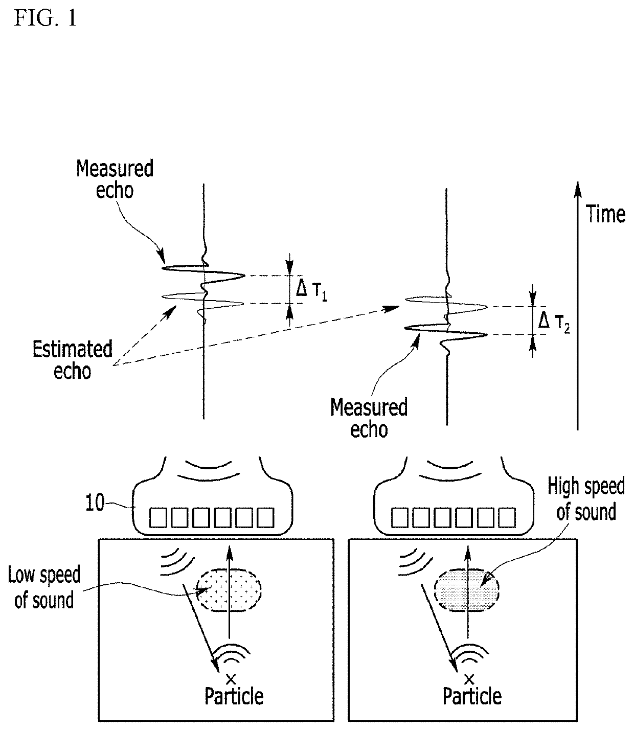
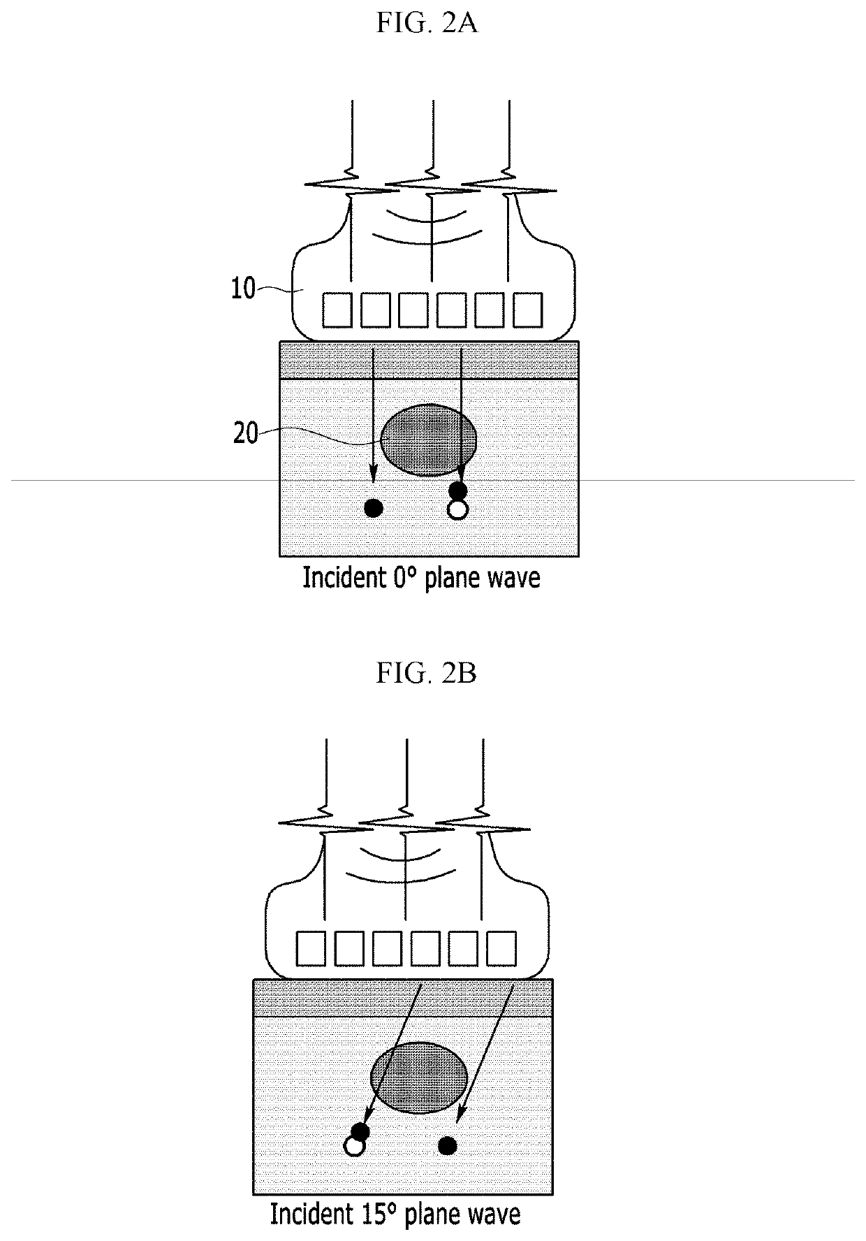
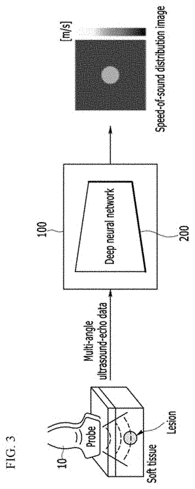
The patent describes a method for using a deep neural network to improve the accuracy and speed of ultrasound imaging. The method involves generating beamformed RF-mode images with phase shift information and training the deep neural network to minimize reconstruction loss by comparing the phase shift maps with a speed-of-sound distribution image of the tissue. This allows for the imaging of various organs and tissues using existing ultrasound imaging devices, simplifying the imaging process and reducing the need for expensive equipment. The method can also improve the contrast and accuracy of quantitative images and securely reconstruct speed-of-sound characteristics from the ultrasound-echo data obtained in noise environment. Overall, the patent provides a technical solution for improving ultrasound imaging capabilities in a cost-effective and efficient manner.

Problems solved by technology

X-ray, MRI, and CT have a risk of radiation exposure and a drawback in that the measurement time is long and the cost is too high.
However, the B-mode imaging system has drawbacks in that the quality of the obtained images may vary according to the proficiency of a user and quantitative characteristics cannot be imaged.
That is, since the B-mode technique provides only geometric information of a tissue, sensitivity and specificity may be low in a differential diagnosis that distinguishes benign tumor and malignant tumor, which are classified by histological characteristics.
However, the Elastography requires an additional dedicated device and consumes a lot of energy.
Therefore, the Elastography can be applied only to expensive ultrasound equipment and is not suitable for imaging dynamically moving tissues due to a low frame rate.
However, since ultrasonic sensors should be placed to enclose an object, the ultrasound computed tomography is applied to imaging breast only and has a limitation in measuring various organs.
In addition, imaging using the ultrasound computed tomography takes over one minute, so that to observe real time movement is impossible.
Further, it is impossible to move the system due to the very large size thereof.

Method used


Image

  • Method and apparatus for quantitative ultrasound imaging using single-ultrasound probe
  • Method and apparatus for quantitative ultrasound imaging using single-ultrasound probe
  • Method and apparatus for quantitative ultrasound imaging using single-ultrasound probe

Examples


Embodiment Construction

[0045]In the following detailed description, exemplary embodiments of the present disclosure will be described in detail with reference to the accompanying drawings so that those of ordinary skill in the art may easily implement the present disclosure. However, the present disclosure may be implemented in various different forms and is not limited to the embodiments described herein. Accordingly, the drawings and description are to be regarded as illustrative in nature and not restrictive. Like reference numerals designate like elements throughout the specification.

[0046]As used herein, unless explicitly described to the contrary, the word “comprise”, “include” or “have”, and variations such as “comprises”, “comprising”, “includes”, “including”, “has” or “having” will be understood to imply the inclusion of stated elements but not the exclusion of any other elements. In addition, the term “unit”, “-er”, “-or” or “module” described in the specification mean a unit for processing at l...


PUM


Abstract

A method of operating an image device operated by at least one processor is provided. The method of operating an image device comprises generating beamformed RF-mode images including phase shift information for each incident angle by using multi-angle ultrasound-echo data, generating phase shift maps representing a displacement of speckle pattern between adjacent beamformed RF-mode images, and obtaining a speed-of-sound distribution image corresponding to the phase shift maps by using a deep neural network which is trained to reconstruct speed-of-sound distribution of a tissue from training phase shift maps.

Description

CROSS-REFERENCE TO RELATED APPLICATION[0001]This application claims priority to and the benefit of Korean Patent Application No. 10-2019-0166617 filed in the Korean Intellectual Property Office on Dec. 13, 2019, and Korean Patent Application No. 10-2020-0086059 filed in the Korean Intellectual Property Office on Jul. 13, 2020, the entire contents of which are incorporated herein by reference.BACKGROUND(a) Field[0002]The present disclosure relates to an image reconstruction technology using ultrasounds.(b) Description of the Related Art[0003]Since it is difficult to detect a cancer early, periodic examinations are required and the size and characteristics of a lesion should be continuously monitored. Representative imaging equipment for this includes an X-ray, a magnetic resonance imaging (MRI), a computed tomography (CT), and an ultrasound. X-ray, MRI, and CT have a risk of radiation exposure and a drawback in that the measurement time is long and the cost is too high. In contrast, ...

Claims


Application Information

Patent Timeline
Patent Type & Authority Applications(United States)
IPC IPC(8): A61B8/08G06T11/00A61B8/14A61B8/00G16H30/40G16H50/50G06N3/08
CPCA61B8/5207G06T11/005A61B8/145G06T2210/41G16H30/40G16H50/50G06N3/08A61B8/587G16H30/20G16H40/63G06N3/048G06N3/045
Inventor BAE, HYEON-MINKIM, MYEONG GEELEE, GIBBEUMOH, SEOKHWANKIM, YOUNGMIN
Owner KOREA ADVANCED INST OF SCI & TECH
Learn More