
Three-Dimensional Dynamic Contrast Enhanced Ultrasound and Real-Time Intensity Curve Steady-State Verification during Ultrasound-Contrast Infusion

a three-dimensional dynamic contrast and ultrasound technology, applied in the field of three-dimensional dynamic contrast enhanced ultrasound and steady-state verification during ultrasound-contrast infusion, can solve the problems of erroneous quantitative measurements, high heterogeneity, and difficulty in navigating a transducer to different, and achieve the effect of contrast-enhanced ultrasound imaging

Inactive Publication Date: 2020-10-15
THE BOARD OF TRUSTEES OF THE LELAND STANFORD JUNIOR UNIV
View PDF0 Cites 0 Cited by
  • Summary
  • Abstract
  • Description
  • Claims
  • Application Information

AI Technical Summary

Benefits of technology

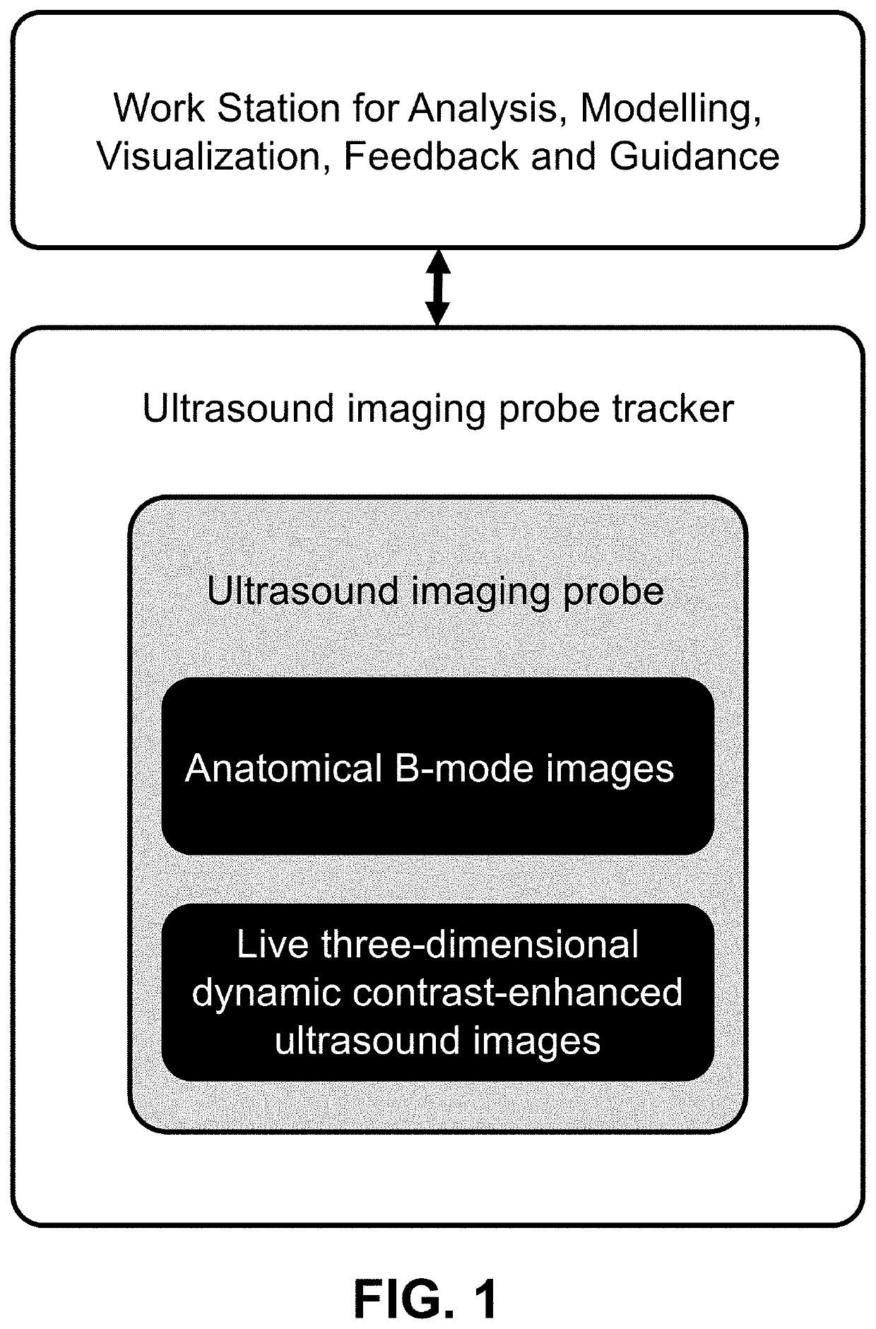
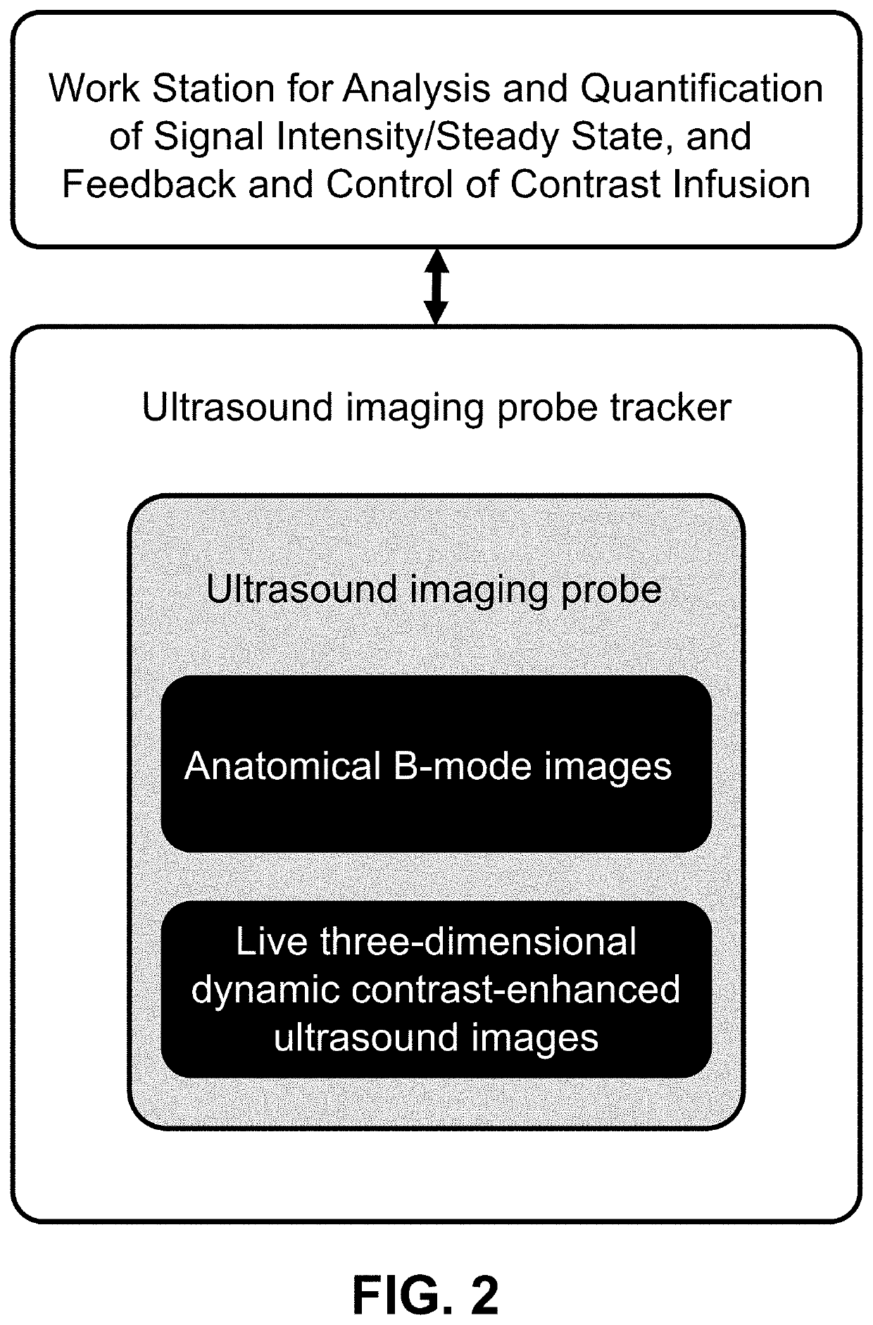
The patent describes a computer code that can measure the intensity of signals in an anatomical area and show it on a screen. This allows users to adjust the rate at which they infuse a substance. The code can also help users determine the steady-state of the infusion and trigger microbubble destruction pulses during a three-dimensional dynamic contrast-enhanced ultrasound imaging.

Problems solved by technology

An important limitation in longitudinal applications of ultrasound is positioning the transducer to obtain the same imaging plane for day-to-day comparison.
This is especially problematic in cancer imaging, where tumors tend to be highly heterogeneous, on a plane-to-plane basis.
As a result, the operator-dependent nature of ultrasound can result in erroneous quantitative measurements and difficulties in navigating a transducer to different anatomical sites during time-sensitive scan sessions to capture different 2D planes.
However, several major challenges remain in the use of 3D DCE-US.
However, unlike conventional 2D contrast modes in clinical systems, current commercially available 3D DCE-US modes do not display a low mechanical index B-mode and contrast-mode image side-by-side, thus leaving the operator with no positioning feedback during lengthy acquisitions to capture the dynamic contrast signal.
In addition, without stable anatomical features from B-mode images, post-acquisition motion correction can be a significant challenge.

Method used


Image

  • Three-Dimensional Dynamic Contrast Enhanced Ultrasound and Real-Time Intensity Curve Steady-State Verification during Ultrasound-Contrast Infusion
  • Three-Dimensional Dynamic Contrast Enhanced Ultrasound and Real-Time Intensity Curve Steady-State Verification during Ultrasound-Contrast Infusion

Examples


Embodiment Construction

[0013]Continuous positioning feedback such as available in 2D, would potentially improve the quality of the data for post-processing and quantification, by ensuring that the operator maintains a steady position throughout a scan. The inventors developed an interventional acquisition system for 3D DCE-US imaging that aims to provide users with navigation feedback and temporal transducer coordinates to re-align the imaging planes in 4D for post-processing and quantification. The interventional system was also designed with disruption-replenishment (DR) imaging in mind, which has been shown to be more repeatable and quantitative over conventional bolus-based acquisitions.

[0014]In brief, DR uses short high-power ultrasound pulses (disruptions within diagnostic range) to momentarily burst microbubbles flowing at steady state. The rate of replenishment of microbubbles is then modeled to extract quantitative parameters. The advantages of DR obviate the need to estimate the indicator input ...


PUM


Abstract

Guidance and visualization for three-dimensional dynamic contrast-enhanced ultrasound imaging is provided. Anatomical B-mode images and live three-dimensional dynamic contrast-enhanced ultrasound images (3D-DCE-US) of an anatomical region of interest are acquired. An ultrasound imaging probe tracker tracks six degrees of freedom position and orientation data of the ultrasound imaging probe. With reference to the common three-dimensional coordinate frame, and for each of the acquired images, the anatomical B-mode images, the live 3D-DCE-US images, and a three-dimensional computer-generated model are visualized and overlaid with each other. The visualization provides guidance and feedback to a user of the ultrasound imaging probe during three-dimensional dynamic contrast-enhanced ultrasound imaging.

Description

CROSS-REFERENCE TO RELATED APPLICATIONS[0001]This application claims priority from U.S. Provisional Patent Application 62 / 831505 filed Apr. 9, 2019, which is incorporated herein by reference.FIELD OF THE INVENTION[0002]This invention relates to methods for three-dimensional dynamic contrast-enhanced ultrasound imaging and interventions.BACKGROUND OF THE INVENTION[0003]Ultrasound (US) is amongst the most widely accessible medical imaging modalities. In comparison to MRI or CT, it offers inexpensive bedside diagnostics without radiation and / or contrast restrictions related to kidney failure. As a result, functional ultrasound using 1D array transducers has been proposed as an ideal tool for longitudinal imaging applications such as cancer treatment monitoring to assess early tumor perfusion changes as surrogate for response.[0004]An important limitation in longitudinal applications of ultrasound is positioning the transducer to obtain the same imaging plane for day-to-day comparison. ...

Claims


Application Information

Patent Timeline
IPC IPC(8): A61B8/08A61B8/00A61M5/00A61M5/172
CPCA61B8/5246A61B8/4245A61B2090/365A61B8/46A61B8/463A61M5/172A61M5/007A61B8/481A61B8/483A61M2005/14208A61B2034/2063
Inventor EL KAFFAS, AHMEDHRISTOV, DIMITRE H.WILLMANN, JUERGEN K.
Owner THE BOARD OF TRUSTEES OF THE LELAND STANFORD JUNIOR UNIV
Learn More