
Microbubble and nanobubble expansion using perfluorocarbon nanodroplets for enhanced ultrasound imaging and therapy

Active Publication Date: 2020-10-08
BOARD OF RGT THE UNIV OF TEXAS SYST
View PDF0 Cites 0 Cited by
  • Summary
  • Abstract
  • Description
  • Claims
  • Application Information

AI Technical Summary

Benefits of technology

The present patent relates to the field of medical diagnostics and therapeutics. Specifically, it describes a new strategy for improved detection and treatment of diseased tissue using a combination of perfluorocarbon (PFC) gas filled microbubbles and nanobubbles. The PFC gas can inflate the microbubbles and nanobubbles, making them easier to detect and treat. The patent also describes a method for in vitro rare cell isolation using PFC gas filled nanobubbles and PFC liquid nanodroplets. The PFC gas can transfer to the nanobubbles, increasing their buoyancy and making them easier to recover. The patent also describes a method for safely occluding tumor microvessels non-invasively and without the need for ultrasound. Overall, the patent presents a novel approach for improving medical diagnosis and treatment.

Problems solved by technology

This is most likely due to the larger ND dose needed compared to microbubbles (MBs), the inability to vaporize all NDs, the amount of phospholipid surfactant that is insufficient to properly stabilize newly formed MBs that are five times larger, and the spontaneous uncontrolled vaporization of ND that could lead to potential toxicity.
However, the key impediment of NDs is their lesser echogenicity and thus lesser US contrast compared to MBs due to their limited reflectivity and compressibility (10), requiring large doses that potentially increase side-effects (11).
However, using micron-size PFP-filled droplets potentially leads to spontaneous droplet vaporization, particularly during injection due to shear stress and cavitation, which is a concern for clinical translation (13).
In addition, occlusion was accomplished exclusively in the larger arteries, resulting in undesired non-discriminant downstream embolization and side effects (14-16) or incomplete embolization for tumors still fed from smaller arteries.
While a few groups have successfully vaporized liquid low boiling point droplets of PFP (17), or perfluorobutane (PFB) (18-20) to MBs in animals, their clinical translation and widespread use has been limited by critical efficacy and safety challenges.
Secondly, the required dose of droplets or NDs is high, which potentially leads to side-effects (11).
Thirdly, the inability to vaporize all NDs due to tissue attenuation, and uncontrolled spontaneous vaporization of sub-micron droplets at body temperature (13) raises safety concerns as it could lead to unwanted microvascular occlusion.
In addition to these safety concerns, MBs formed by ADV might not be stable enough for effective gas embolotherapy as they originate from NDs that are 5 times smaller (23), and thus do not have enough phospholipid surfactants to be properly stabilized.
Although the presence of MBs near NDs decreases the ADV threshold (24), the same challenges remain for the clinical translation of ADV.
Interestingly, while both NBs and NDs individually exhibit very limited ultrasound signal on B-mode imaging when exposed to low acoustic power, their combination results in a rapid and dramatic increase in signal visible on standard ultrasound imaging.

Method used


Image

  • Microbubble and nanobubble expansion using perfluorocarbon nanodroplets for enhanced ultrasound imaging and therapy
  • Microbubble and nanobubble expansion using perfluorocarbon nanodroplets for enhanced ultrasound imaging and therapy
  • Microbubble and nanobubble expansion using perfluorocarbon nanodroplets for enhanced ultrasound imaging and therapy

Examples


Embodiment Construction

lass="d_n">[0055]An overview of exemplary embodiments of the present disclosure will be presented initially, followed by further discussion of specific aspects.

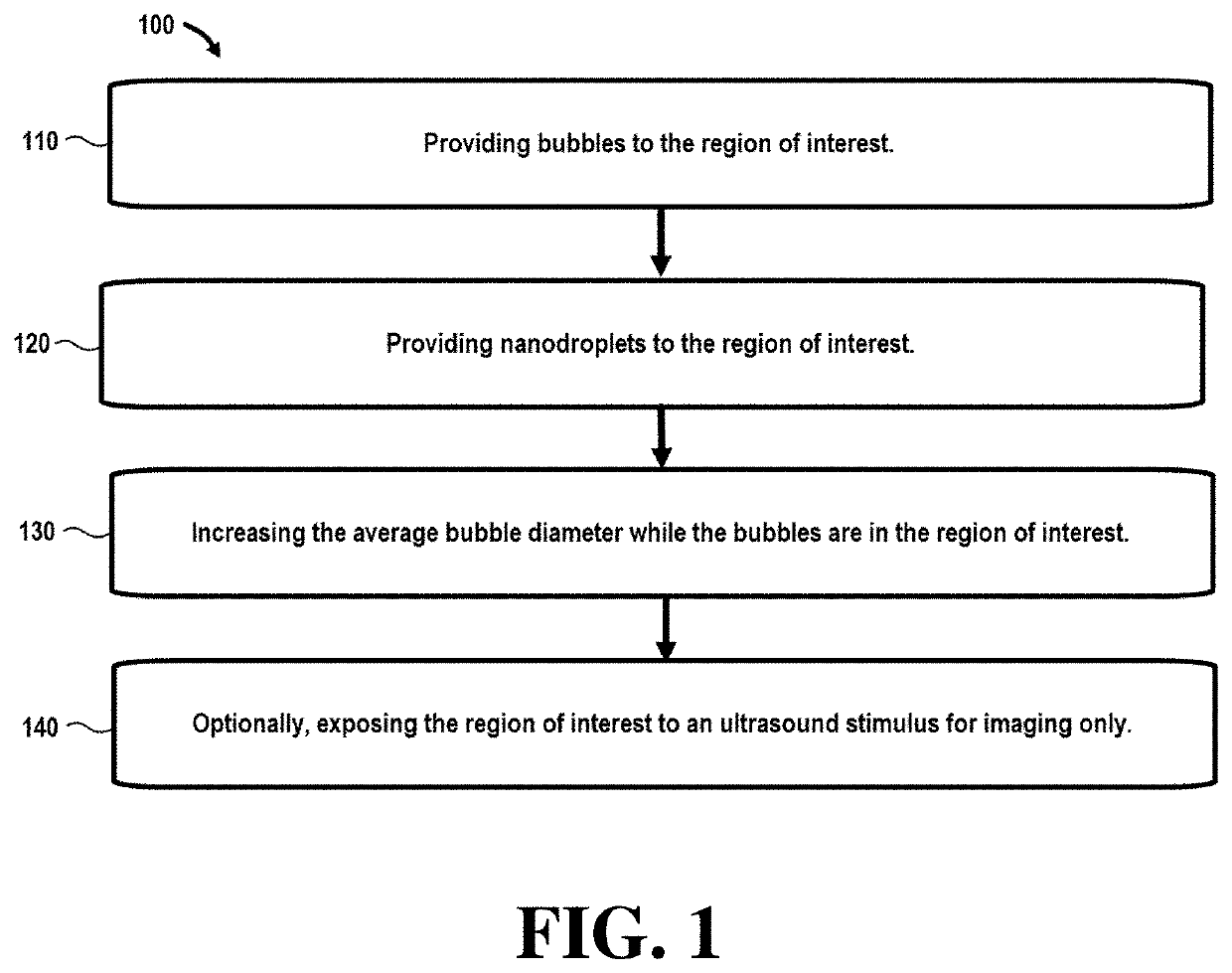
[0056]Referring initially to FIG. 1, a flowchart 100 illustrates steps in a method that can be used in ultrasound imaging techniques. In this embodiment, step 110 comprises providing bubbles to the region of interest. In certain embodiments, the bubbles may be nanobubbles or microbubbles with an average bubble diameter between around 200-600 nm (nanobubbles) and 1-10 μm (microbubbles).

[0057]To circumvent the challenges of ADV for effective gas embolotherapy, the inventors took inspiration from a biological process and the second law of thermodynamics: the process by which PFC is eliminated from the body by exhalation (25). When PFC liquid emulsions or PFC gas-filled MBs traverse the alveolar capillaries, PFC transfers to the air-filled alveolar space because of the large partial pressure gradient (1). In addition, since liqui...


PUM


Abstract

The disclosure describes imaging and therapy techniques comprising nanodroplets. More particularly, aspects of the disclosure relate to the use of nanodroplets to modify nanobubbles or microbubbles to provide improved imaging and / or therapeutic techniques and compositions.

Description

CROSS-REFERENCE TO RELATED APPLICATIONS[0001]This application claims priority to U.S. Provisional Patent Application Ser. No. 62 / 809,280 filed Feb. 22, 2019, the entire contents of which are incorporated herein by reference, including the appendices attached thereto.BACKGROUND[0002]The invention was made with government support under and Cancer Prevention Research Institute of Texas (CPRIT) RR150010, DOD Idea Award 14W81XWH-17-1-0401 and HCC Development Project Pilot Award (Internal—pre SPORE). The government has certain rights in the invention.A. Field[0003]This disclosure relates to imaging and therapy techniques comprising nanodroplets. More particularly, embodiments of the disclosure relate to the use of nanodroplets to inflate nanobubbles or microbubbles to provide improved imaging and / or therapeutic aspects.B. Related Art[0004]Various techniques have been developed to image and provide therapy to conditions that require medical treatment, including for example, cancerous tumor...

Claims


Application Information

Patent Timeline
IPC IPC(8): G06T7/00G06T7/62A61N7/02
CPCG06T2207/20104G06T2207/10132A61N2007/0039G06T2207/30096A61N7/02A61N2007/0004G06T7/0012G06T7/62A61N7/00A61B8/481
Inventor MATTREY, ROBERT F.DE GRACIA LUX, CAROLINELUX, JACQUESGAO, ZHENGHONGBRAMBILA, CARLOS J.
Owner BOARD OF RGT THE UNIV OF TEXAS SYST
Learn More