
Mechanically locking adjustable cardiac tether

Pending Publication Date: 2020-08-27
ARGO CARDIOVASCULAR INC
View PDF2 Cites 0 Cited by
  • Summary
  • Abstract
  • Description
  • Claims
  • Application Information

AI Technical Summary

Benefits of technology

The invention is a method and device for improving heart function by positioning an elongate member across a heart chamber and fixing it in place using anchor members. This reduces the size of the ventricle, changes the shape of the ventricle, and helps to keep valves in place, resulting in improved cardiac function. The device can be adjusted during real-time monitoring of cardiac function using echocardiographic and / or fluoroscopic imaging. Overall, the invention provides a minimally invasive way to improve heart function in patients with various heart conditions.

Problems solved by technology

Ischemic and Non Ischemic Dilated Cardiomyopathy causes the heart to become enlarged and to function poorly.
As a result, the muscle of the heart becomes weak, thin or floppy and is unable to pump blood efficiently around the body.
This inability to efficiently pump blood typically causes fluid to build up in the lungs, which then become congested, resulting in a feeling of breathlessness.
Often there is also right heart failure which causes fluid to accumulate in the tissues and organs of the body, usually the legs and ankles, and the liver and abdomen.
Left ventricular dilation can also lead to secondary Mitral valvular regurgitation, further worsening cardiac performance, referred to as functional Mitral Regurgitation.
This may lead to an area of less coaptation of the valve leaflets, and, as a result, eventually to valve leakage.
In heart failure, there is poor ventricular function and elevated wall stress.
These effects tend to reduce annular contraction and distort annular size, often exacerbating mitral valve regurgitation.
During this movement of the papillary muscles, however, the various chordae lengths remain substantially constant, which limits the full closure ability of the leaflets by exerting tension prematurely on the leaflets.
This condition is commonly referred to as “chordal tethering.” The combination of annular changes and papillary changes results in a poorly functioning valve.
The tether and the local deformation they impart may further alter the positions of the papillary muscles in such a way that the chordae do not allow as complete of a closure of the mitral valve, or that rotation of portions of the ventricular wall (to which additional chordae may be attached) may “tighten” one valve leaflet and “loosen” the other.
In this manner, the leaflets may not close at the same level relative to the annulus, causing increased retrograde leakage through the valve.
This type of access necessitates the use of cardiopulmonary bypass or open heart access, which can introduce additional complications to the surgical procedure.

Method used


Image

  • Mechanically locking adjustable cardiac tether
  • Mechanically locking adjustable cardiac tether
  • Mechanically locking adjustable cardiac tether

Examples


Embodiment Construction

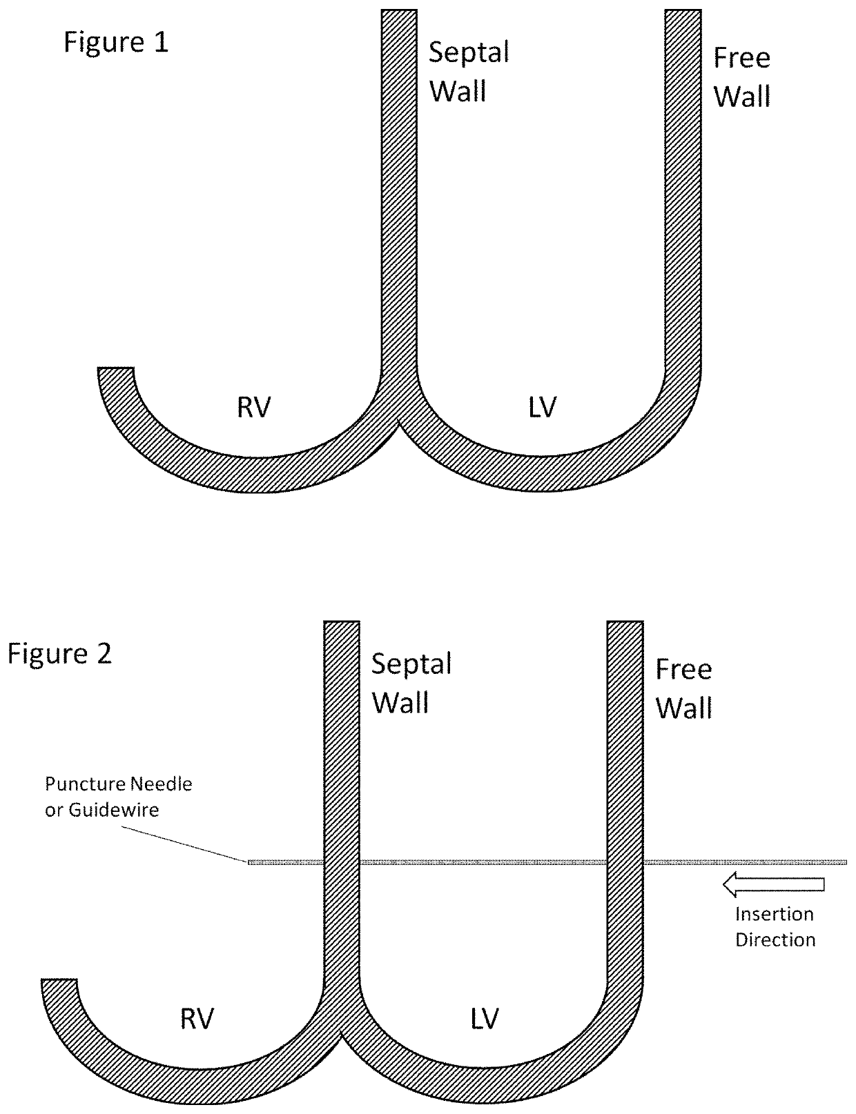
[0033]The various aspects of the invention to be discussed herein generally pertain to devices and methods for treating heart conditions, including, for example, dilatation, valve incompetencies, including mitral valve leakage, and other similar heart failure conditions. Each device of the present invention preferably operates passively in that, once placed in the heart, it does not require an active stimulus, either mechanical, electrical, or otherwise, to function. Implanting one or more of these devices alters the shape or geometry of the heart, both locally and globally, and thereby increases the heart's efficiency. That is, the heart experiences an increased pumping efficiency through an alteration in its shape or geometry and concomitant reduction in stress on the heart walls. In addition, the devices of the present invention may operate to assist in the apposition of heart valve leaflets to improve valve function.

[0034]The inventive devices and related methods offer numerous ...


PUM


Abstract

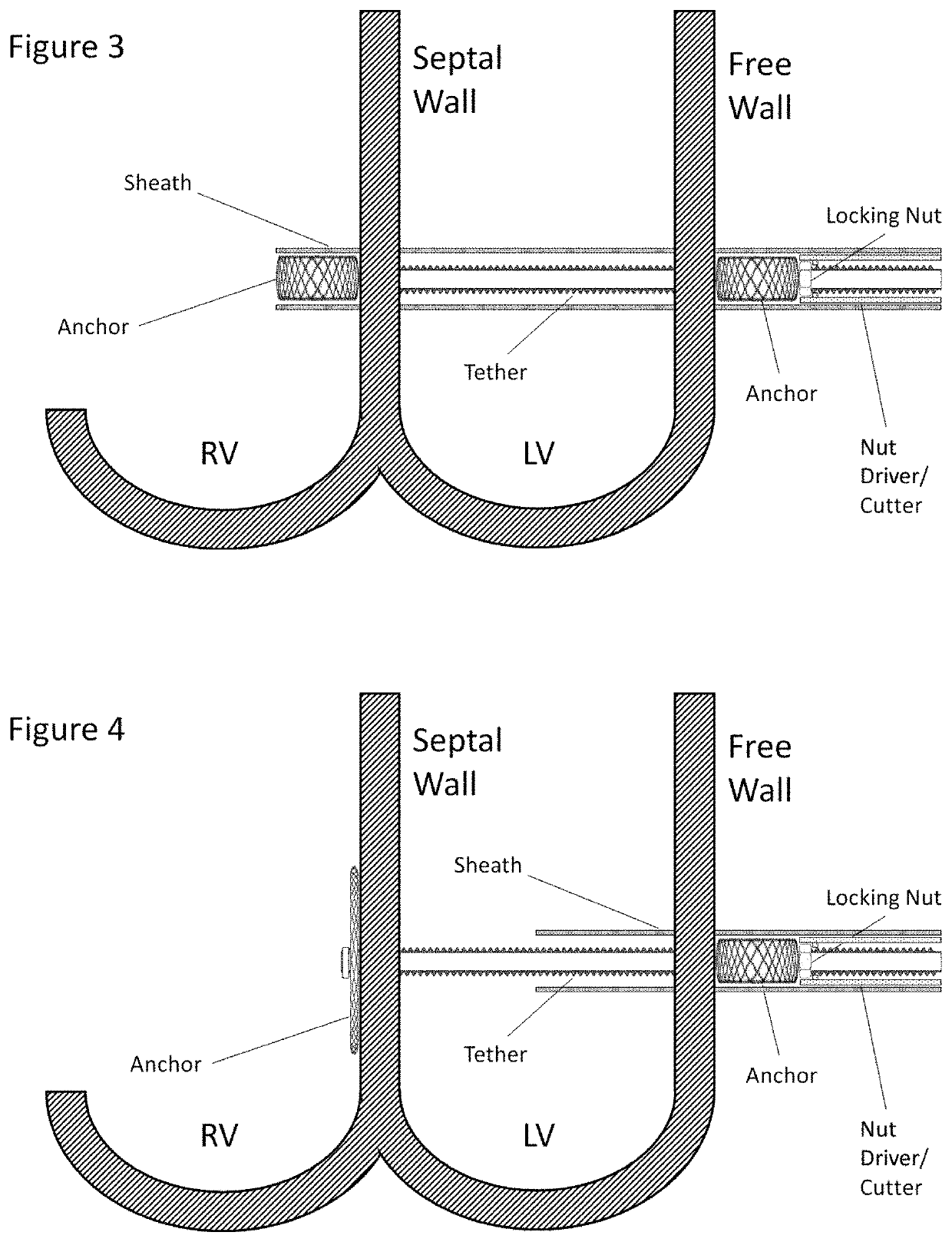
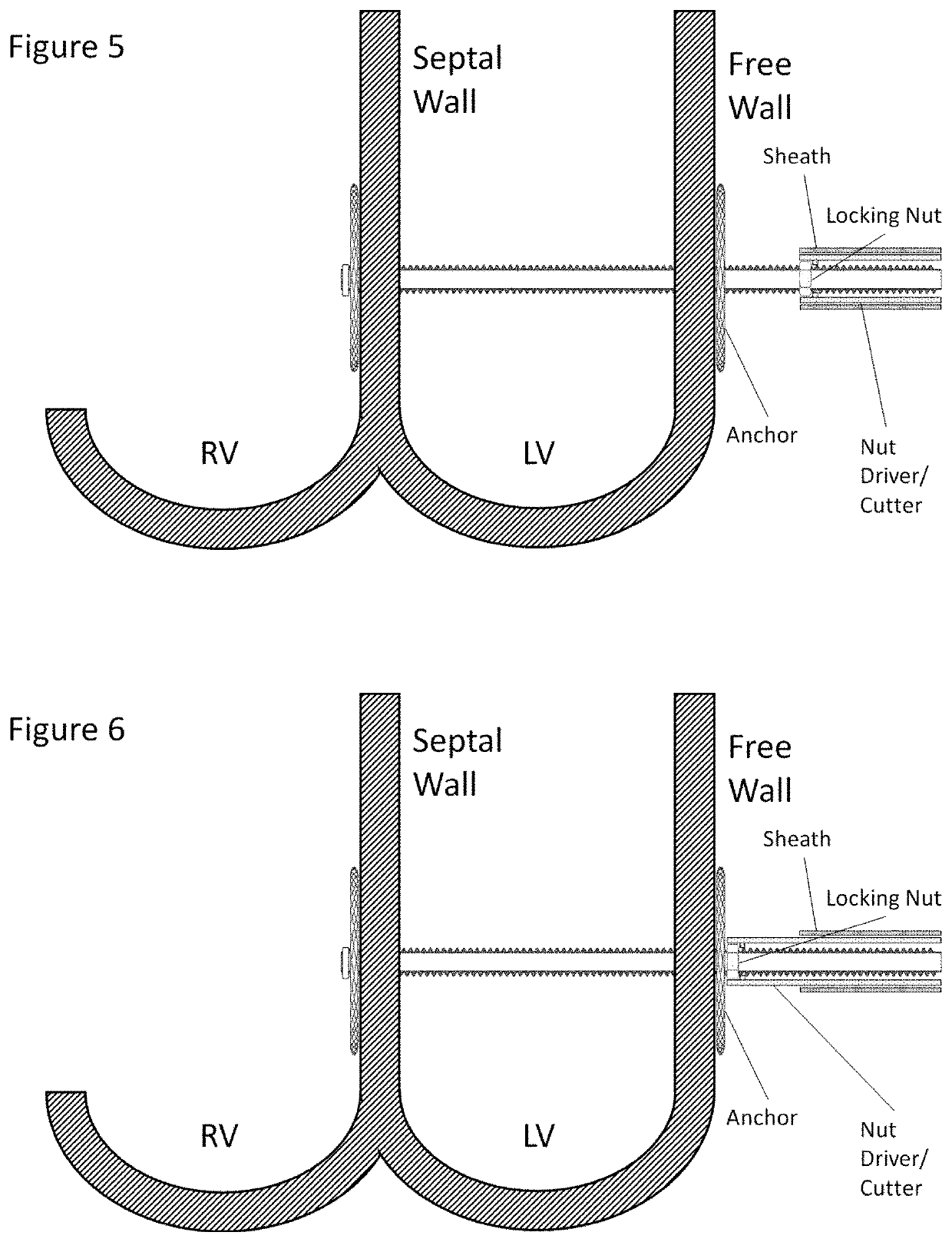
A method of modifying a shape of a patient's heart by placing a first anchor against a surface of a first wall of the heart; extending a tether proximally from the first anchor to a second wall of the heart; placing a second anchor against a surface of the second wall; rotating a nut about the tether to move the second anchor distally along the tether, thereby moving the second wall toward the first wall; and permitting the nut to remain in place after the rotating step. The invention also includes a device for practicing the method.

Description

CROSS REFERENCE TO RELATED APPLICATIONS[0001]This application claims the benefit of priority to U.S. Provisional Application No. 62 / 809,116, filed Feb. 22, 2019, which application is incorporated by reference herein.INCORPORATION BY REFERENCE[0002]All publications and patent applications mentioned in this specification are herein incorporated by reference in their entirety to the same extent as if each individual publication or patent application was specifically and individually indicated to be incorporated by reference.BACKGROUND[0003]Ischemic and Non Ischemic Dilated Cardiomyopathy causes the heart to become enlarged and to function poorly. Some people have stable disease and there is little worsening of their condition. Others have progressive disease. As a result, the muscle of the heart becomes weak, thin or floppy and is unable to pump blood efficiently around the body. This inability to efficiently pump blood typically causes fluid to build up in the lungs, which then become...

Claims


Application Information

Patent Timeline
IPC IPC(8): A61F2/24
CPCA61F2/2487A61F2220/0016A61F2002/249A61F2220/0041A61F2/2481A61F2002/2484
Inventor KAO, STEPHEN
Owner ARGO CARDIOVASCULAR INC
Learn More