Patient risk stratification based on body composition derived from computed tomography images using machine learning

a computed tomography and patient technology, applied in tomography, image enhancement, instruments, etc., can solve the problems of inability to generalize approaches, inability to automatically analyze body composition on large datasets, and manual correction of segmentation errors, etc., to achieve more accurate analysis

Active Publication Date: 2020-07-02
THE GENERAL HOSPITAL CORP
View PDF0 Cites 5 Cited by
  • Summary
  • Abstract
  • Description
  • Claims
  • Application Information

AI Technical Summary

Benefits of technology

[0007]The present disclosure addresses the aforementioned drawbacks by providing a system and method for determining patient risk stratification based on body composition derived from computed tomography images using machine learning. The system may enable real-time segmentation of muscle and fat tissue, for example, facilitating clinical application of body morphological analysis sets. In one configuration, a fully-automated deep learning system for the segmentation of skeletal muscle cross sectional area (CSA) is proposed. In an example configuration, the system may use an axial computed tomography (CT) image taken at the third lumbar vertebra (L3) for the CSA. A fully-automated deep segmentation model may be derived from an extended implementation of a Fully Convolutional Network with weight initialization of a pre-trained model, such as an ImageNet model, followed by post processing to eliminate intramuscular fat for a more accurate analysis.

Problems solved by technology

However, segmentation errors require manual correction based on visual analysis by highly-skilled radiologists.
As a result, semi-automated body composition analysis on large datasets is impractical due to the expense and time required.
As a result, these approaches cannot be generalized.
While many physicians rely on an “eyeball test” to assess whether patients will tolerate major surgery or chemotherapy, “eyeballing” is inherently subjective and difficult to quantify.
However, the determination of morphometric age is time intensive and requires highly trained experts.

Method used


Image

  • Patient risk stratification based on body composition derived from computed tomography images using machine learning
  • Patient risk stratification based on body composition derived from computed tomography images using machine learning
  • Patient risk stratification based on body composition derived from computed tomography images using machine learning

Examples


Embodiment Construction

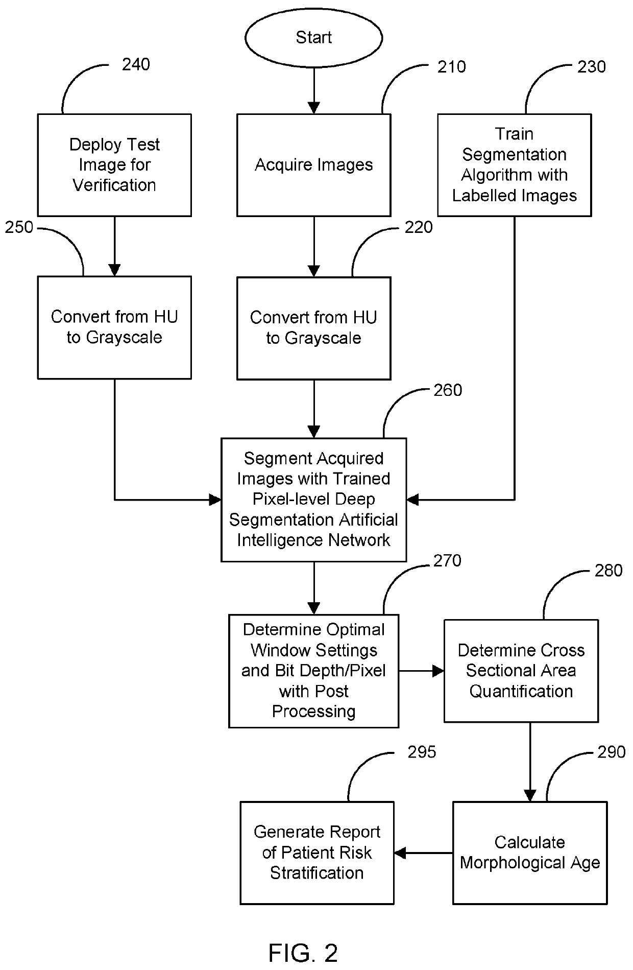
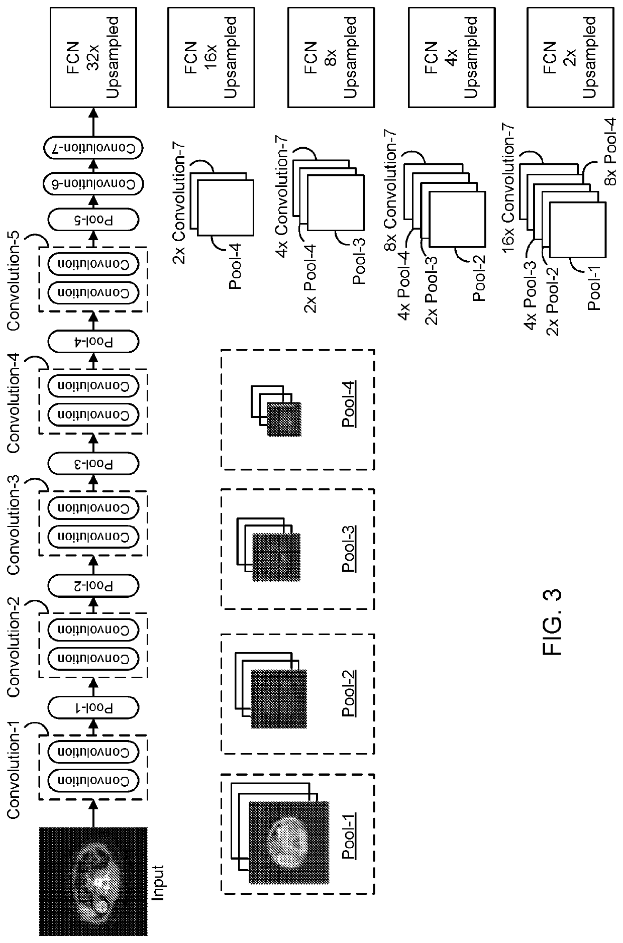
[0021]Muscle cross sectional area has been shown to correlate with a wide-range of posttreatment outcomes. However, integration of muscle CSA measurements in clinical practice have been limited by the time required to generate this data. By dropping calculation time from 1800 seconds to less than one second, research into new applications for morphometric analysis may be drastically sped up. CT is an essential tool in the modern healthcare arena with approximately 82 million CT examinations performed in the US in 2016. In lung cancer in particular, the current clinical paradigm has been on lesion detection and disease staging with an eye toward treatment selection. However, accumulating evidence suggests that CT body composition data could provide objective biological markers to help lay the foundation for the future of personalized medicine. Aside from preoperative risk stratification for surgeons, morphometric data may be used to predict death in radiation oncology and medical onc...


PUM


Abstract

A system and method for determining patient risk stratification is provided based on body composition derived from computed tomography images using segmentation with machine learning. The system may enable real-time segmentation for facilitating clinical application of body morphological analysis sets. A fully-automated deep learning system may be used for the segmentation of skeletal muscle cross sectional area (CSA). Whole-body volumetric analysis may also be performed. The fully-automated deep segmentation model may be derived from an extended implementation of a Fully Convolutional Network with weight initialization of a pre-trained model, followed by post processing to eliminate intra-muscular fat for a more accurate analysis.

Description

CROSS-REFERENCE TO RELATED APPLICATIONS[0001]This application claims the benefit of U.S. Provisional Patent Application Ser. No. 62 / 555,771 filed on Sep. 8, 2017, and entitled “Patient risk stratification based on body composition derived from computed tomography images using machine learning.”STATEMENT REGARDING FEDERALLY SPONSORED RESEARCH[0002]N / ABACKGROUND[0003]Deep learning has demonstrated enormous success in improving diagnostic accuracy, speed of image interpretation, and clinical efficiency for a wide range of medical tasks, ranging from the interstitial pattern detection on chest CT to bone age classification on hand radiographs. Particularly, a data-driven approach with deep neural networks has been actively utilized for several medical image segmentation applications, ranging from segmenting brain tumors on magnetic resonance images, organs of interest on CT to segmenting the vascular network of the human eye on fundus photography. This success is attributed to its capab...

Claims


Application Information

Patent Timeline
Patent Type & Authority Applications(United States)
IPC IPC(8): G16H50/30G06T7/11G06T7/155G06T7/00G16H30/40G16H15/00A61B34/10A61B6/03A61B6/00
CPCA61B6/5223G06T2207/30048G16H50/30G16H30/40G06T2207/20084G06T2207/30061G06T2207/30016G06T7/0016A61B6/5217A61B34/10G06T7/11G06T2207/30041G06T2207/30008A61B6/032G06T2207/10081G06T7/155G06T2207/30096A61B2034/107G16H15/00G06N3/08G06N3/045
Inventor DO, SYNHOFINTELMANN, FLORIANLEE, HYUNKWANG
Owner THE GENERAL HOSPITAL CORP
Why Eureka
Try Eureka