


Quick Research
Generate reliable direction feasibility study reports for your R&D in just a few steps.

Technical Q&A
Discover and master advanced knowledge NOW. Basics, ideas, possibilities, all at once.

Find Solutions
As an expert in R&D theories, this can generate solutions to your technical problems instantly.

Evaluate Feasibility
Analyze your overall solution with one click, know your potential R&D risks in advance.

Monitor Landscape
Get weekly tech updates, stay abreast of the latest tech innovations and key insights.
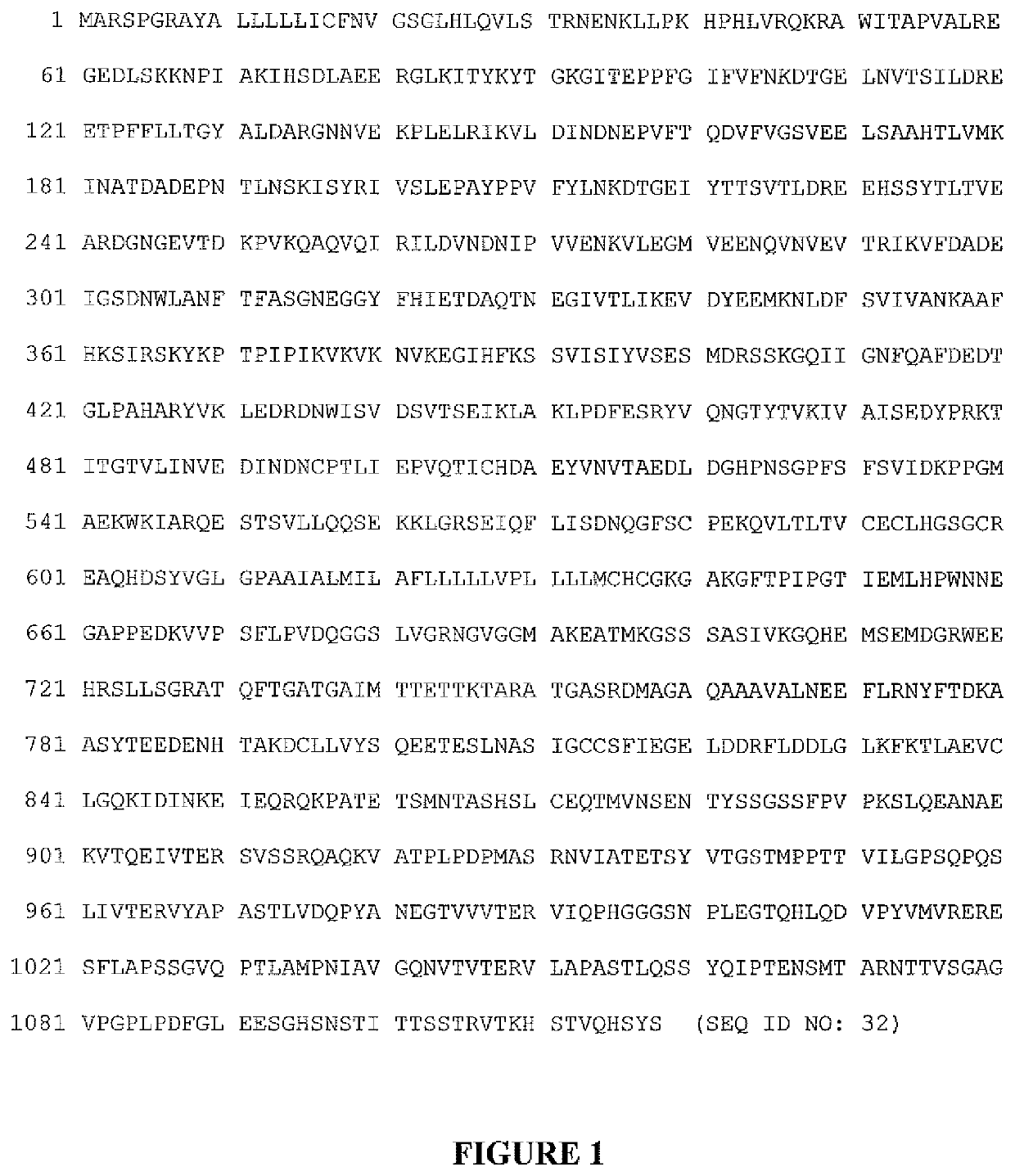
Method for Diagnosis of Arrhythmogenic Right Ventricular Cardiomyopathy
a right ventricular cardiomyopathy and arrhythmogenic technology, applied in disease diagnosis, measurement devices, instruments, etc., can solve the problems of many lost years of life, arvc remains clinically and genetically difficult to diagnose, and multiple tests performed to determine task force criteria are expensiv
- Summary
- Abstract
- Description
- Claims
- Application Information
AI Technical Summary
Benefits of technology
Problems solved by technology
Method used
Image

Examples
example 1
body Determination Using Western Blot Analysis
[0043]Sera from ARVC cases were analyzed for specific anti-desmosomal and anti-adherens junction autoantibodies. Since the extracellular components of desmosomal and adherence junctions are cadherins (Desmoglein-2 and desmocollin-2 for desmosomes and N-cadherin for adherence junctions), these three cadherin proteins were used to detect their respective serum autoantibodies on Western blots exposed to patient and control sera. Sera from any consenting patient with ARVC, whether sporadic or familial, gene-identified or elusive, was analyzed.
[0044]The Western Blot analysis was adopted from Chatterjee-Chakraborty & Chatterjee (Brain Res. 2010. 1348:10-20). The analysis of serum from control and patient samples was carried out using recombinant human DSG2, DSC and Cadherin proteins. Recombinant human DSG2 (Creative Biomart / cat #DSG2-1601H), DSC (Creative Biomart / cat # DSC2-3856H) and Cadherin (Creative Biomart / cat # HEK293) were reconstituted...
example 2
body ELISA Protocol
[0053]Direct ELISA from “Abeam” online protocol was used after some modifications. Recombinant human DSG2 protein (Creative Biomart / cat #DSG2-1601H) was diluted in 100 mM bicarbonate buffer (pH 9.6) to a final concentration of 2 microgram per ml. ELISA microtitre plate (Thermo, Cat # M9410-1CS) was coated with the diluted antigen (100 μl / well). The plate was covered with adhesive plastic and incubated at room temperature for 2 hours. The coating solution was removed and the wells were washed 2× with phosphate-buffered saline containing 0.05% Tween-20 (PBS-T), pH 7.4, and the wells were incubated with blocking buffer (PBS containing 2% bovine serum albumin) for 2 hours at room temperature. After incubation, the wells were washed 2× with PBS-T.
[0054]One hundred (100) μl of diluted human serum (usually 1:100 dilution in blocking buffer) were added to each well, covered with adhesive plastic and incubated for 2 hours at room temperature. After incubation, the wells we...
example 3
on with Disease Severity
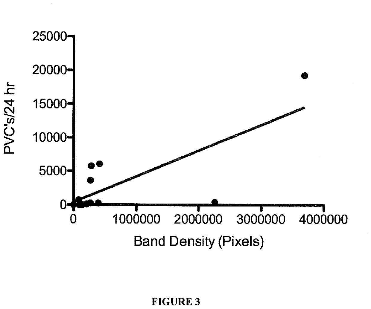
[0056]It was assessed whether or not antibody density as measured by pixel count of the Western blot or ELISA O.D. correlated with disease severity as measured by 24-hour burden of premature ventricular contractions (PVCs). PVCs burden during 24-hours was measured by 24-hour ambulatory ECG recordings. The R-squared value was 0.59 (p=0.0021) for PVC's versus pixel count of the Western band, indicating that 59% of the variation in PVC count could be accounted for by its linear relationship with antibody density (FIG. 3). Similarly, R-squared was 0.38 (p=0.026) for PVC's versus the O.D. measure of antibody concentration from the ELISA (FIG. 4).
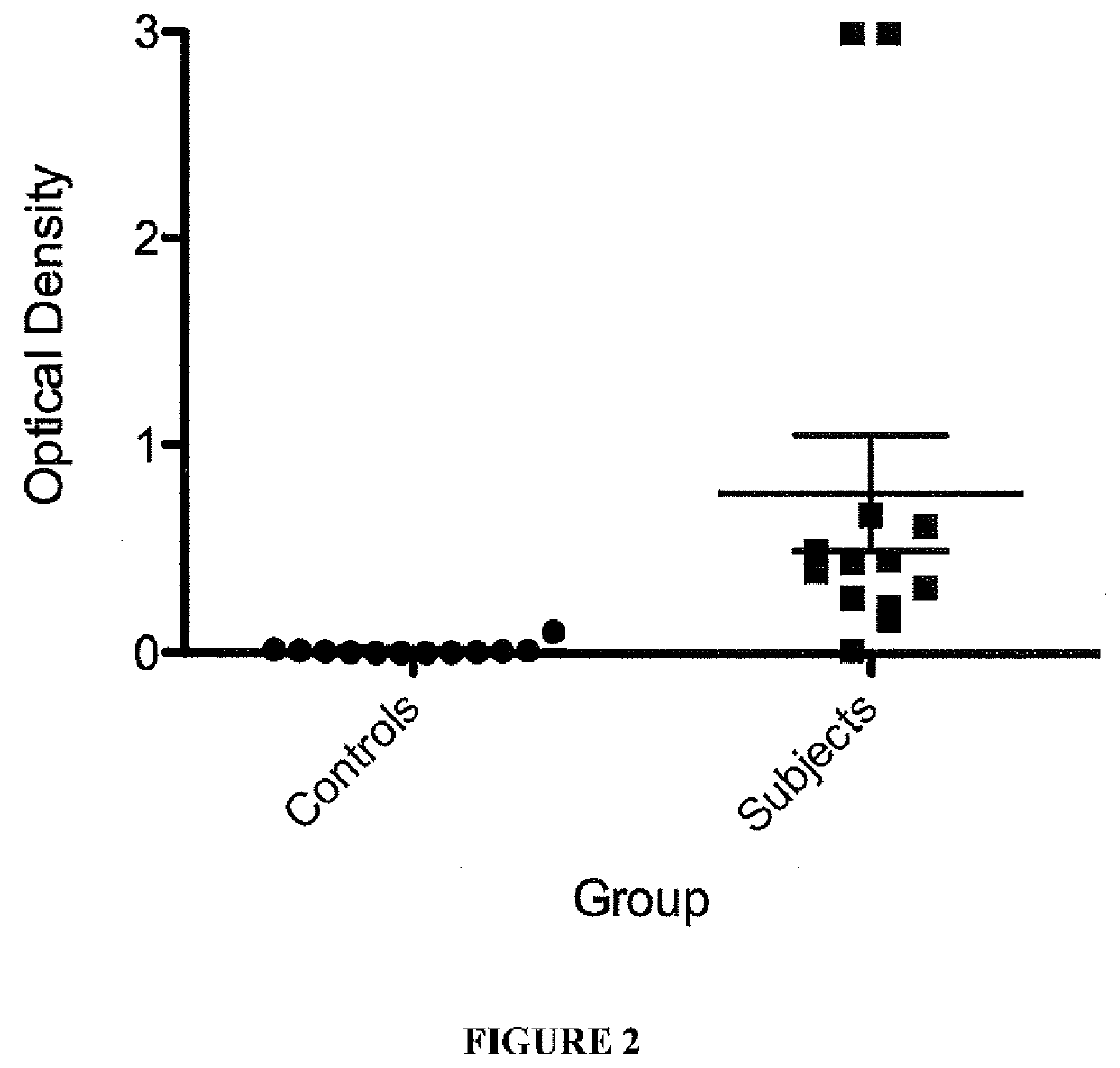
[0057]Assessment of additional subjects, including 32 controls as well as patients assessed in clinic and considered to have no ARVC, shows that the biomarker level (as measured by enzyme-linked immune-sorbent assay, ELISA) is very low or absent. In comparison, patients with possible ARVC, borderline ARVC and definite ARVC...
PUM
| Property | Measurement | Unit |
|---|---|---|
| optical density | aaaaa | aaaaa |
| concentration | aaaaa | aaaaa |
| pH | aaaaa | aaaaa |
Abstract
Description
Claims
Application Information
 - R&D Engineer
- R&D Manager
- IP Professional
- Industry Leading Data Capabilities
- Powerful AI technology
- Patent DNA Extraction
Browse by: Latest US Patents, China's latest patents, Technical Efficacy Thesaurus, Application Domain, Technology Topic, Popular Technical Reports.
© 2024 PatSnap. All rights reserved.Legal|Privacy policy|Modern Slavery Act Transparency Statement|Sitemap|About US| Contact US: help@patsnap.com