
Non-invasive Systems and Methods to Detect Cortical Spreading Depression for the Detection and Assessment of Brain Injury and Concussion

a cortical spreading depression and cortical surface technology, applied in the field of brain injury detection and diagnosis, can solve the problems of high cost of mtbi, concussion, indirect cost, low detection efficiency, etc., and achieve the effect of increasing brain damag

Active Publication Date: 2019-02-07
SCIENCEPLUSPLEASE LLC
View PDF0 Cites 3 Cited by
  • Summary
  • Abstract
  • Description
  • Claims
  • Application Information

AI Technical Summary

Benefits of technology

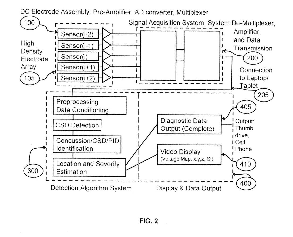
The present invention is a non-invasive system that uses the unique characteristics of DC properties of CSDs (cerebral spinal discharges) to detect them. The system includes a High Density Electrode Array that is placed on the test subject's head, DC-coupled electroencephalography amplifiers, post-processing algorithms, and software. It also includes a user interface that provides a solution for non-invasive CSD detection. The system can help reduce the complex and often inaccurate process of concussion diagnosis and treatment based on symptomology and can also allow for the non-invasive observation of brain-damaging CSDs in acute neurological injury.

Problems solved by technology

Available data indicate that the total costs for mTBI, or concussion, are high, and that the indirect costs, such as for sick leave, early retirement and loss of productivity, are the main expense.
Currently, CSDs and their neurophysiological and other properties are not detectable from the surface of the scalp by existing commercial methods and devices.
The detection and diagnosis of concussion has been problematic for several reasons.
None of these methods is able to directly detect or quantify the neurological basis of concussion, and instead are performed to detect secondary indicators that can be ambiguous and inconsistent in nature.
Imaging via MRI, CT, PET, ultrasound, or near-infrared spectroscopy also is not compatible with observing CSD in the setting of concussion.
Although it has become the de facto standard, it is also acknowledged to be imprecise and ineffectual.

Method used


Image

  • Non-invasive Systems and Methods to Detect Cortical Spreading Depression for the Detection and Assessment of Brain Injury and Concussion
  • Non-invasive Systems and Methods to Detect Cortical Spreading Depression for the Detection and Assessment of Brain Injury and Concussion
  • Non-invasive Systems and Methods to Detect Cortical Spreading Depression for the Detection and Assessment of Brain Injury and Concussion

Examples


example 1

ted Electrode Helmet Transmitting Data to a Sideline Laptop for Analysis

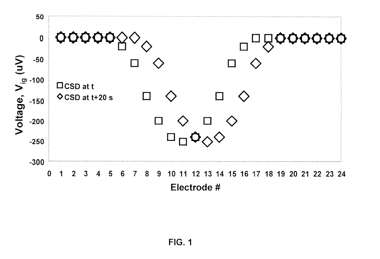
[0079]In this example, a football player wearing a helmet instrumented with an electrode array cap that includes a High Density Electrode Array that transmits DC-coupled-EEG data to the sidelines is hit by another player on the right upper temporal aspect of the player's head. The player falls to the ground and is unable to get up. Data transmitted from the helmet cap to the sideline analysis system composed of the portable processing unit with visual display then is assessed. The system begins the analysis and identification procedure, while at the same time a doctor logs the standard concussion assessment questions and patient history into system. The system continues its performance for about thirty minutes, at which time the system provides information that the player has suffered a concussion by displaying a region of depressed voltage of about 200 V on a diagrammatic representation of the player's head in ...

example 2

Cap at the Sidelines Associated with a Laptop for Analysis

[0080]In this example, a football player is hit by another player on the right upper temporal aspect of the player's head. The player falls to the ground and is unable to get up. The player is assisted off the field and placed on the sidelines. A portable headband / electrode net and processing unit with visual display is fastened onto the player's head and a button is clicked to start the software for the analysis and identification procedure of the condition of the player. At the same time, a doctor logs the standard concussion assessment questions and patient history into system. The system continues its performance for about thirty minutes, at which time the system provides information that the player has suffered a concussion by displaying a region of depressed voltage of about 200 V on a diagrammatic representation of the player's head in the form of an expanding ring with a width of about 3.0 mm that originates at the ri...

example 3

Cap in a Neuro-Intensive Care Unit for Traumatic Brain Injury, Acute Ischemic Stroke, Intracerebral Hemorrhage, and Subarachnoid Hemorrhage Associated with a Laptop for Analysis

[0081]In this example, a patient in the neuro-intensive care unit is monitored with an electrode head unit which includes a High Density Electrode Array for the presence of CSDs and PIDs using DC-coupled-EEG data and the processing scheme as described above in Examples 1 and 2. A health care practitioner places the electrode head unit on the patient's head over the border between normal undamaged brain and the suspected region of brain damage as deduced from imaging or a neurological exam. The electrode unit is connected to the processing and display unit via wireless or wired connections. Over a period of up to seven days, the system collects data consisting of globular regions of about 3.0 mm diameter of depressed voltage of about 200 μV which travel along the border of the suspected region of damaged brain...


PUM


Abstract

The present invention provides systems and methods for detection and diagnosis of concussion and / or acute neurologic injury comprising a portable headwear-based electrode array and computerized control system to automatically and accurately detect cortical spreading depression and acute neurological injury-based peri-infarct depolarization (CSD / PID). The portable headwear-based electrode system is applied to a patient or athlete, and is capable of performing an assessment automatically and with minimal user input. The user display indicates the presence of CSD / PID, gauges its severity and location, and stores the information for future use by medical professionals. The systems and methods of the invention use an instrumented DC-coupled electrode / amplifier array which performs real-time data analysis using unique algorithms to produce a voltage intensity-map revealing the temporally propagating wave depressed voltage across the scalp that originates from a CSD / PID on the brain surface.

Description

CROSS REFERENCE TO RELATED APPLICATIONS[0001]The present application claims priority to U.S. patent application Ser. No. 14 / 948,995, filed Nov. 23, 2015, which claims priority to U.S. Provisional Application No. 62 / 084,247, filed Nov. 25, 2014, both of which are incorporated in their entirety herein by reference.FIELD OF THE INVENTION[0002]The present invention relates to the field of detection and diagnosis of concussion and / or acute neurologic injury; to neurologic triage for acute ischemic stroke, traumatic brain injury, intracerebral hemorrhage, and subarachnoid hemorrhage; and to the evaluation of athletic-induced head injury; specifically, to an athletic field sideline-placed or portable apparatus using a computer-based method for performing emergency neurological triage on a subject who has recently suffered a blow to the head to determine if the subject is suffering from episodes of cortical spreading depression (CSD) associated with concussion; and specifically, to a neuro-...

Claims


Application Information

Patent Timeline
Patent Type & Authority Applications(United States)
IPC IPC(8): A61B5/0478A61B5/0482A61B5/04A61B5/00A61B5/375
CPCA61B5/0478A61B5/0482A61B5/04004A61B5/04002A61B5/6803A61B5/04012A61B5/4076A61B2562/046A61B2562/0215A61B5/2415A61B5/30A61B5/316A61B5/291A61B5/375
Inventor JONES, STEPHEN CARTERGOPALAKRISHNA, PRAHLAD MENONKHARLAMOV, ALEXANDERSAUTER, PHILIP M.ALBA, NICOLAS A.HUND, SAMUEL J.SINGER, ERIC
Owner SCIENCEPLUSPLEASE LLC
Learn More