Astrocyte traumatome and neurotrauma biomarkers
a neurotrauma and astrocyte technology, applied in the field of astrocyte traumatome and neurotrauma biomarkers, can solve the problems of difficult repeated administration of neuroimaging tools, especially advanced modalities, difficult diagnosis, and lack of standardization of readout values, so as to improve the diagnostic accuracy of mild tbi
- Summary
- Abstract
- Description
- Claims
- Application Information
AI Technical Summary
Benefits of technology
Problems solved by technology
Method used
Image
Examples
example 1
cortical Astrocyte Cell Fates after Mechanical Trauma
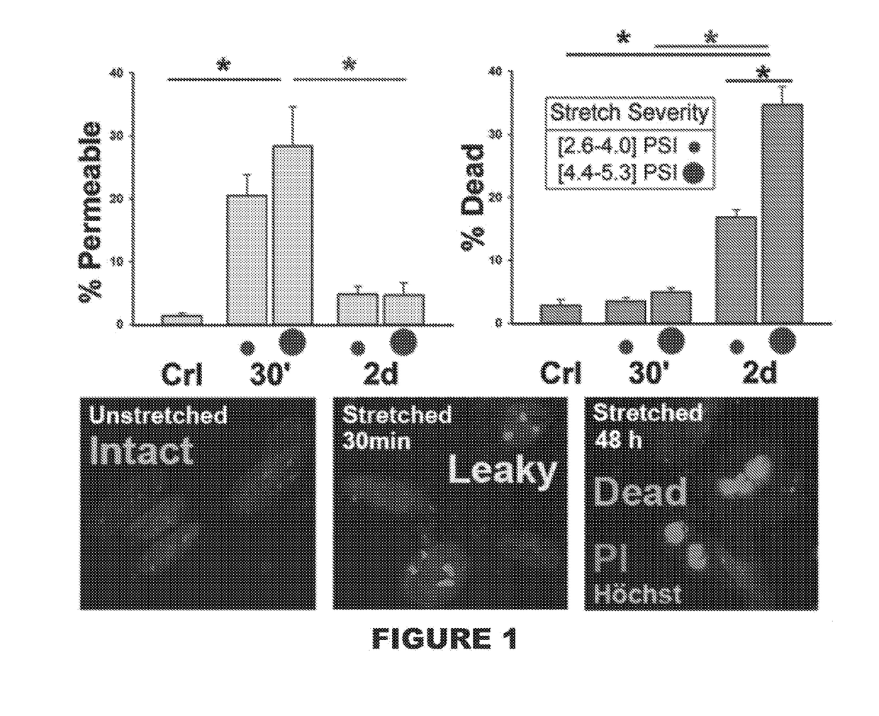
[0074]This Example shows population scores of human astrocytes 30 minutes and 48 hours after abrupt pressure-pulse traumatic stretching using different severities (FIG. 1; PSI ranges from 2.6-4.0 for milder injury and 4.4-5.3 for severe injury). Human astrocytes were isolated from 16-18 week donated fetal neocortical brain specimen, purified and then differentiated on deformable membranes (Wanner, 2012). Cell membrane wounding / compromise, mechanoporation (Barbee, 2005), was determined using Propidium iodide (PI) uptake in living cultures accompanied by nuclear shape assessment (middle picture, stained nuclei, Hoechst stained after fixation, with little pink dots, nucleoli that are PI-stained). Cell death was determined by PI uptake accompanied by condensed nuclei (pyknotic nuclei with compacted chromatin and bright Hoechst and PI stains). Significant percent of cell integrity compromise is shown 30 min post-injury (left graph) whi...
example 2
lly Traumatized Human Astrocytes Show Prolonged Endurance in a Compromised State
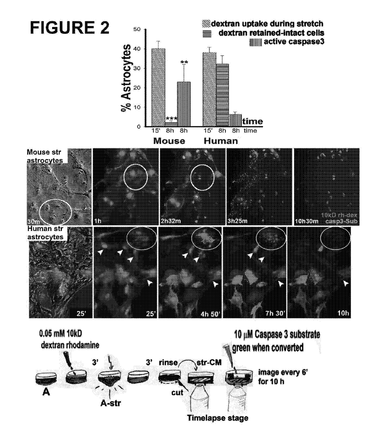
[0075]This Example demonstrates that mechanically traumatized human astrocytes show prolonged endurance in a compromised state after wounding versus mouse astrocytes (FIG. 2). Dye uptake (0.05 mM 10 kDa dextran rhodamine) during stretch as well as cell death by apoptosis (caspase activation using 10 μM caspase 3 substrate) was monitored using time-lapse imaging on a temperature and humidity controlled spinning disc confocal microscope (Levine et al., 2016). Human traumatized wounded and resealed human astroglia show prolonged endurance after integrity compromise.
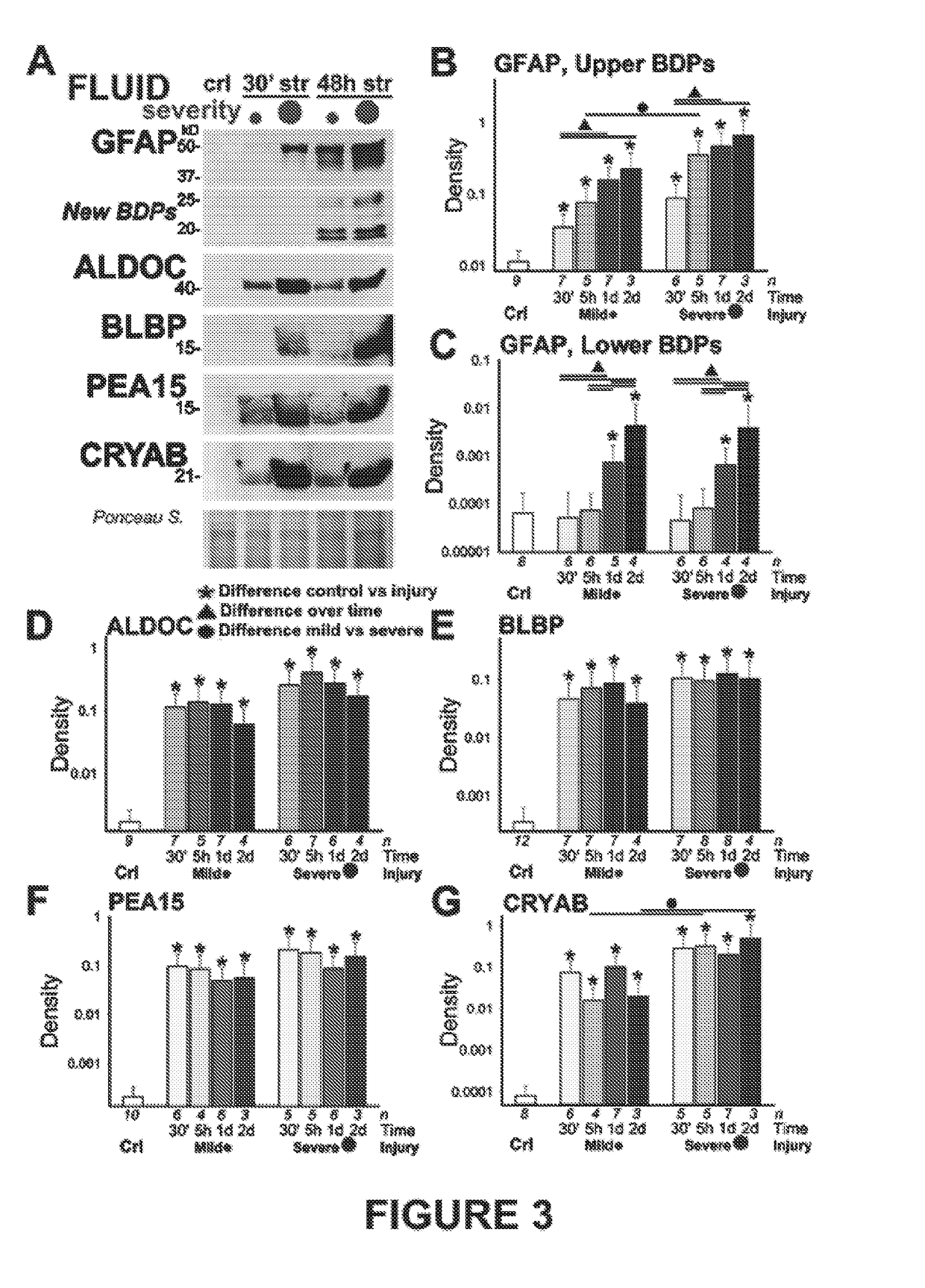
[0076]As shown in FIG. 3, mechanical trauma of human astrocytes causes significant release of astroglial marker into surrounding fluids. FIG. 3A shows immunoblots of concentrated, denatured conditioned media (Levine et al., 2016; Sondej et al., 2011) from traumatized astrocytes show different release profiles of glial fibrillary acidic proteins (...
example 3
ma Biomarkers Associated with Cell Fates of Human Traumatized Astrocytes
[0077]Shown in FIG. 4A are biplots of trauma-released astroglial marker levels (see FIG. 3) for ALDOC, BLBP, PEA15 and CRYAB over the percent membrane permeable cells (% wounded, red data, left) as well as correlated to percent of dead human traumatized astrocytes using PI-dye-update assay and nuclear morphology (see FIGS. 1+2). Regression lines (R2-value) and p-values indicate correlation significance. ALDOC, PEA15 and CRYAB show best correlation with human astroglial cell wounding. CRYAB, BLBP and ALDOC also correlated with extend of cell death after trauma.
[0078]FIG. 4B shows biplots of GFAP trauma-release, which show correlation of GFAP with cell death inflicted by mechanical trauma and weak / no correlation with cell wounding. Plots are separated by grouped GFAP fragment sizes (upper bands: 50-37 kD, lower bands: 25-19 kD).
PUM
 Abstract
Description
Claims
Application Information
 - R&D Engineer
- R&D Manager
- IP Professional
- Industry Leading Data Capabilities
- Powerful AI technology
- Patent DNA Extraction
Browse by: Latest US Patents, China's latest patents, Technical Efficacy Thesaurus, Application Domain, Technology Topic, Popular Technical Reports.
© 2024 PatSnap. All rights reserved.Legal|Privacy policy|Modern Slavery Act Transparency Statement|Sitemap|About US| Contact US: help@patsnap.com


