
Blood vessel model

Inactive Publication Date: 2018-12-13
FUJIFILM CORP +1
View PDF0 Cites 1 Cited by
  • Summary
  • Abstract
  • Description
  • Claims
  • Application Information

AI Technical Summary

Benefits of technology

The present patent provides a blood vessel model that enables the movement of red blood cells to be tested without being obstructed by a porous membrane. This is achieved by using a porous membrane with smaller thickness and opening diameters that allow red blood cells to pass through more easily. The membrane is also designed in a honeycomb pattern with communication holes and flattened shapes that facilitate the passage of red blood cells. Additionally, non-porous regions can be added to regulate the flow of red blood cells inside the microchannels and improve the accuracy of the extravasation test. Overall, this model provides a more accurate and reliable tool for testing the movement of red blood cells during extravasation.

Problems solved by technology

Namely, in the conventional internal organ models, there were cases in which the level of drug induced injury to a layer of cells provided at a surface of the porous membrane may not be accurately evaluated.

Method used


Image

  • Blood vessel model
  • Blood vessel model
  • Blood vessel model

Examples


Embodiment Construction

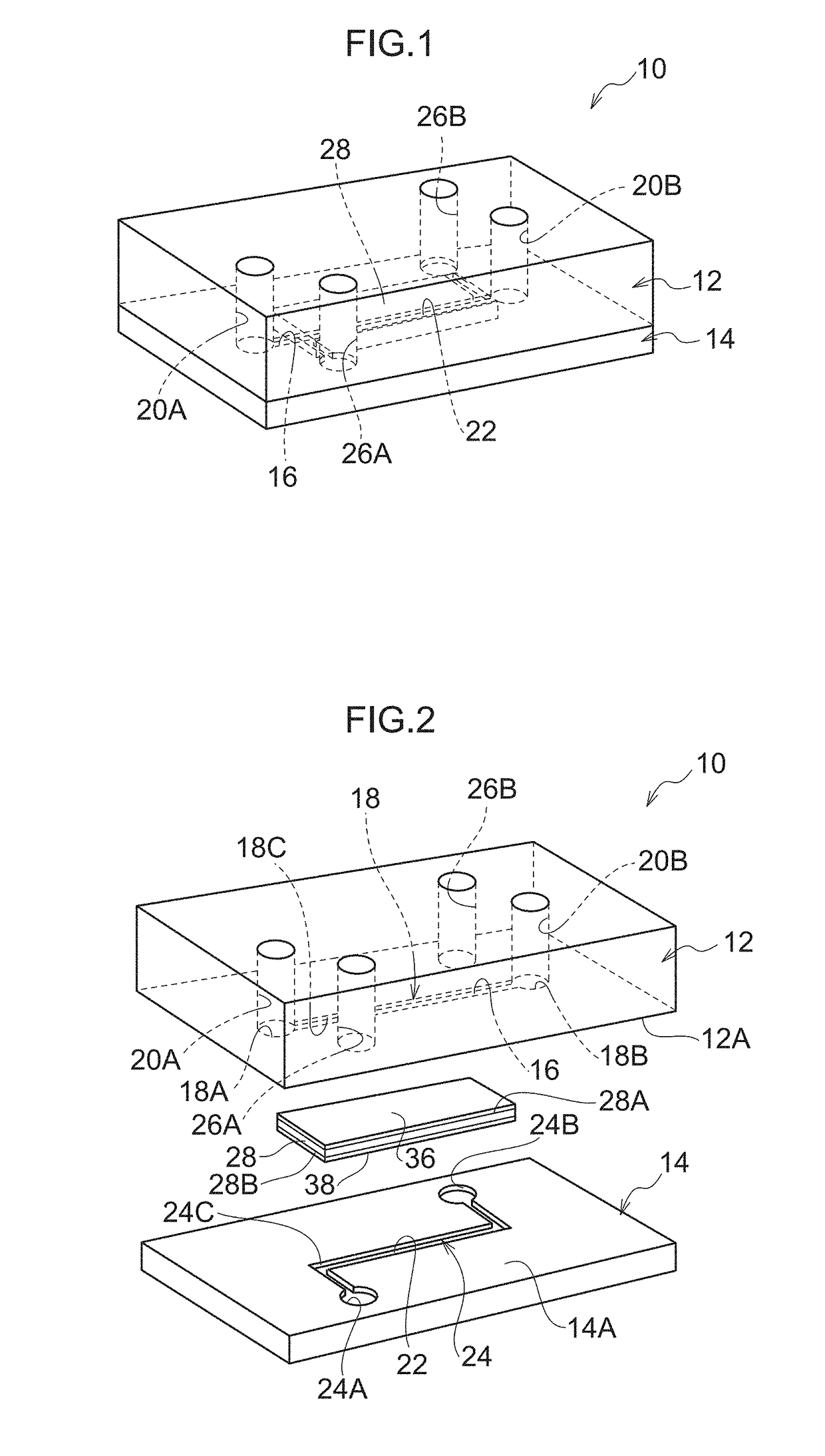
[0030]Explanation follows regarding an example and modified examples of an exemplary embodiment of the present disclosure, with reference to FIG. 1 to FIG. 6. Note that the following exemplary embodiment is merely an example of the present disclosure, and does not limit the scope of the present disclosure. Also note that the dimensions of various configuration in the drawings are modified as appropriate in order to facilitate explanation of the various configuration. Accordingly, the scale in the drawings may differ from the scale in actual practice.

[0031]As illustrated in FIG. 1 and FIG. 2, a blood vessel model 10 of an exemplary embodiment includes an upper channel member 12 and a lower channel member 14 stacked on one another. The upper channel member 12 and the lower channel member 14 are, for example, configured from an elastic material such as polydimethylsiloxane (PDMS), and have substantially rectangular plate shapes.

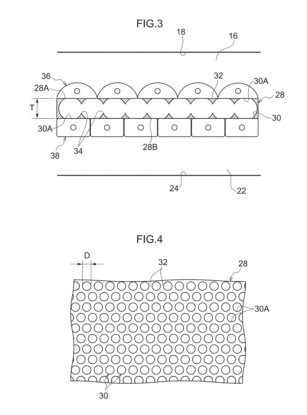
[0032]A concave portion 18, that defines an upper microcha...


PUM


Abstract

The present disclosure provides a blood vessel model including: a pair of channel members, mutually opposing each other, each of which includes an opposing face in which a respective microchannel is formed; and a porous membrane that includes plural through-holes penetrating in a thickness direction, that is disposed between the opposing faces of the pair of channel members, and that partitions between the microchannels, wherein the porous membrane is provided with a vascular endothelial cell layer so as to cover one face facing one of the microchannels, an average opening diameter of the through-holes is from 1 μm to 20 μm, and an opening coverage ratio of the through-holes is from 30% to 70%.

Description

BACKGROUNDTechnical Field[0001]The present disclosure relates to a blood vessel model.Related Art[0002]Recently, there have been attempts to model internal organs such as blood vessels, intestines, livers, and lungs, using devices including what are referred to as microchannels, which include channels with micrometer order widths. United States Patent Application Publication (US) No. 2011 / 0053207, Japanese Patent Application Publication (JP-B) No. 5415538, and JP-B No. 5815643, for example, each discloses an internal organ model including a porous membrane having a layer of cells provided on a surface thereof, and at least two microchannels partitioned by the porous membrane.[0003]Various experiments and tests can be performed using an internal organ model such as that disclosed in US 2011 / 0053207, JP-B No. 5415538, and JP-B No. 5815643. For example, what is called an extravasation test may be performed by running blood containing a drug through one of the microchannels, and then me...

Claims


Application Information

Patent Timeline
IPC IPC(8): G01N33/50
CPCG01N33/5088G01N33/5064G01N33/5029
Inventor ITO, KOJUKAKINUMA, CHIHAYAMIMA, SHINJIHIKIMOTO, DAICHINEVILLE, CRAIG MSUNDBACK, CATHRYN A
Owner FUJIFILM CORP
Learn More