Anti-dc-hil antibodies for cancer diagnosis, prognosis and therapy
a technology of anti-dc-hil and cancer, applied in the field of immunodiagnostics, oncology, medicine, oncology and cancer therapy, can solve the problems of insufficient understanding of exact signals responsible for suppressive function, endogenous expansion of mdsc,
- Summary
- Abstract
- Description
- Claims
- Application Information
AI Technical Summary
Benefits of technology
Problems solved by technology
Method used
Image
Examples
example 1
Materials And Methods
[0174]Reagents. Ab against CD3 (145-2C11), CD11b (M1 / 70), CD11c (N418), CD14 (61D3), CD19 (eBio1D3), CD28 (37,51), CD80 (16A-10A1), CD86 (GL1), Gr-1 (RB6-8C5), HLA-DR (LN3), IFN-γ (XMG1.2), IL-10 (JES5-16E3), PD-L1 (MIH5), TGF-β1 (9016), Thy1.1 (HIS51) and control Ab were purchased from eBioscience; anti-phosphotyrosine (4G10) from Upstate Biotechnology; and all recombinant cytokines from Pepro Tech. The inventors generated 1E4 rat anti-mouse DC-HIL and UTX103 rabbit anti-mouse DC-HIL as described previously (Chung et al., 2009). DC-HIL-Fc fusion protein was produced by COS-1 cells and purified (Chung et al., 2007). The chemical inhibitors, L-NG-monomethyl-arginine citrate, N6-(1-iminoethyl)-L-lysine, N-hydroxyl-nor-arginine, 1-methyl-tryptophan, and catalase and superoxide dismutase, were purchased from Sigma-Aldrich. hgp100 peptide (KVPRNQDWL), OVA257-264 H-2Kb-class I (SIINFEKL), and OVA323-339 H-2Kb-class II peptide (ISQAVHAAHAEINEAGR) were synthesized by th...
example 2
Results
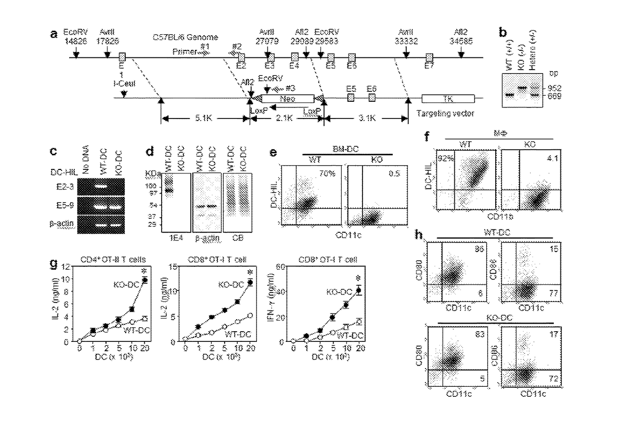
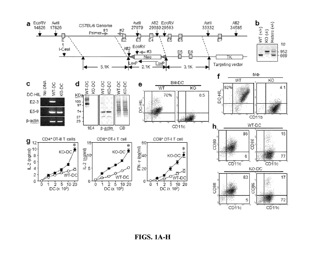
[0193]DC-HIL gene disruption enhances the T cell-stimulatory capacity of DC. To study the in vivo significance of the DC-HIL receptor on APC, the inventors created DC-HIL gene-knocked out (KO) mice by replacing a fragment spanning exons 2-4 with a targeting vector (FIG. 1A) to produce a frame-shift replacement in downstream exons caused by mismatched acceptor-donor sites for RNA splicing between exons 1 and 5. The targeted DC-HIL mutation was introduced into C57BL / 6 mice, and the KO allele confirmed by PCR analysis of genomic DNA (FIG. 1B).
[0194]DC-HIL mRNA expression in KO mice was examined by RT-PCR analysis of total RNA isolated from BM-derived DC (BM-DC) using 2 primer sets: the first to amplify exons 2-3 (within targeted region), and the second for exons 5-9 (downstream) (FIG. 1C). The first primer amplified RNA from DC of WT mice, but not of KO mice; and the second primer produced PCR product from both RNA samples, indicating that KO mRNA lacked targeted exons but bore ...
example 3
Discussion
[0223]While the important detrimental effect of an expanding population of circulating MDSC in patients with growing cancers is well established, the mechanism underlying the profound immunosuppression induced by MDSC remain unclear. Like tolerogenic APC, soluble inhibitory mediators and coinhibitory receptors were both reported to mediate MDSCs suppressor function, but no data has bridged these 2 mechanisms. The inventors now provide such linkage through the DC-HIL receptor that can trigger the IFN-γ / NOS-2 axis. Since DC-HIL expression confers the greatest suppression to CD11b+Gr-1+ MDSC generated by melanoma, DC-HIL may be considered an activation marker for MDSC, akin to CD80 / CD86 costimulatory receptors for immune-stimulatory APC, but with a completely polar effect.
[0224]Because CD80, CD86 (as ligands of CTLA-4), and PD-L1 are coinhibitory ligands that deliver negative signals to T cells through their corresponding receptors (Egen et al., 2002 and Keir et al., 2011), t...
PUM
| Property | Measurement | Unit |
|---|---|---|
| Fraction | aaaaa | aaaaa |
| Fraction | aaaaa | aaaaa |
| Fraction | aaaaa | aaaaa |
Abstract
Description
Claims
Application Information
 - R&D
- Intellectual Property
- Life Sciences
- Materials
- Tech Scout
- Unparalleled Data Quality
- Higher Quality Content
- 60% Fewer Hallucinations
Browse by: Latest US Patents, China's latest patents, Technical Efficacy Thesaurus, Application Domain, Technology Topic, Popular Technical Reports.
© 2025 PatSnap. All rights reserved.Legal|Privacy policy|Modern Slavery Act Transparency Statement|Sitemap|About US| Contact US: help@patsnap.com


