
Method and System for Analysis of Myocardial Wall Dynamics

a myocardial wall and dynamic analysis technology, applied in the field of image processing for understanding and diagnosing, can solve the problems of poor image quality, limited access to all cardiac structures, and difficulty in strain calculation, and achieve the effect of minimizing the cost function

Inactive Publication Date: 2017-03-09
CIRCLE CARDIOVASCULAR IMAGING INC
View PDF3 Cites 16 Cited by
  • Summary
  • Abstract
  • Description
  • Claims
  • Application Information

AI Technical Summary

Benefits of technology

The patent describes a method for identifying and tracking sample points in a series of frames, using a reference frame to help with the process. The method involves identifying epi-points, endo-points, and midpoints based on the contours of the reference frame. For each frame in the cine data set, points are identified that correspond to epi-points and endo-points of the previous frame. These points are then transferred to the current frame and spatially translated to improve the match with the previous frame's epi-points and endo-points. This method helps to accurately track the movement of the sample points through the frames and can be useful in various applications such as video analysis and sports motion detection.

Problems solved by technology

There are a number of difficulties associated with strain calculation using the above methods.
While echocardiography is known for superior temporal resolution (˜10 ms), it suffers from poor image quality and limited access to all cardiac structures as compared to MRI.
However, this technique suffers as the RF tags fade over a period of time.
Consequently, tracking and quantification of the tags over the entire cardiac cycle becomes difficult resulting in poor temporal resolution.
In addition, image acquisition and quantitative post-processing consumes a significant amount of time.
Therefore, this technique is not used in routine clinical diagnostics, but is mostly used for research purposes.
However, lack of features in the myocardium as well as through-plane motion of the myocardium makes the method less accurate.
Because any point in the myocardium moves through 3-dimensions in a normal cardiac cycle and different parts of the myocardium are visible in the image plane during the cardiac cycle, it is difficult to get a high level of accuracy in static short axis (SAX) or long axis (LAX) slices.

Method used


Image

  • Method and System for Analysis of Myocardial Wall Dynamics
  • Method and System for Analysis of Myocardial Wall Dynamics
  • Method and System for Analysis of Myocardial Wall Dynamics

Examples


Embodiment Construction

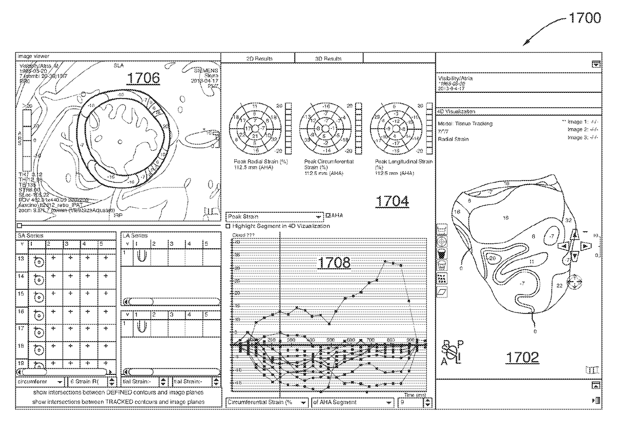
[0065]Generally, the present disclosure describes methods and systems for image processing for understanding, diagnosing, as well as improving existing and developing new treatments for diseases. More particularly, the present disclosure describes methods and systems for the qualitative and quantitative analysis of myocardial wall dynamics medical image datasets (e.g. 2D cine data sets), such as CT and MRI datasets, derived over a cardiac cycle.

[0066]The methods and systems of the present disclosure may be used in many diagnostic and therapeutic areas. For example, the methods and systems of the present disclosure may aid early detection of myocardial insufficiency in a sub-clinical state (or a pre-clinical state when a patient has not been diagnosed with a particular disease or disorder) as well as in both acute and chronic ischemia. Often patients are not treated because all of the functional parameters appear to be normal, such as normal ejection fraction (that is, a fraction of ...


PUM


Abstract

A method to determine myocardial wall dynamics and tissue characteristics using a method provided to determine myocardial wall dynamics and tissue characteristics using a 3D model of the myocardium. The method comprises generating an epicardial surface and an endocardial surface from a plurality of SAX and LAX slices; identifying a plurality of nodes on the epicardial and endocardial surfaces or in between these surfaces within the myocardium in a reference frame; defining a set of coefficients, each coefficient being associated with the respective location of the corresponding node in a phase; determining the coefficients and in this way determining the model; determining the myocardial wall dynamics in terms of strain values and displacements.

Description

CROSS REFERENCE TO RELATED APPLICATIONS[0001]This application claims the benefit of priority of U.S. Provisional Patent Application No. 61 / 989,214 filed May 6, 2014, which is hereby incorporated by reference in its entirety.FIELD OF THE INVENTION[0002]The present disclosure relates generally to image processing for understanding, diagnosing, as well as improving existing and developing new treatments for diseases. More particularly, the present disclosure relates to the qualitative and quantitative analysis of myocardial wall dynamics from medical image datasets, such as Computed Tomography (CT) and Magnetic Resonance Imaging (MRI) datasets, derived over a cardiac cycle.BACKGROUND OF THE INVENTION[0003]Medical imaging is used as a diagnostic tool as well as an experimental tool to study the anatomy and physiology of humans and other animals. It is also used to guide targeted treatment of some illnesses, for example cancer. Various medical imaging techniques include X-ray, ultrasound...

Claims


Application Information

Patent Timeline
Patent Type & Authority Applications(United States)
IPC IPC(8): A61B6/00A61B5/055G06T7/00A61B6/03
CPCA61B6/5217A61B6/032A61B5/055A61B6/503A61B2034/105A61B6/466G06T2207/30048G06T2200/04G06T7/0016A61B6/486A61B6/507A61B5/02028A61B5/1073A61B2576/023G06T2207/10076G06T2207/10081G06T2207/10088G06T7/12G06T7/149G16H50/30
Inventor CHIRVASA, MIHAELAGAO, XUEXINSKRINJAR, OSKARBARCKOW, KAI PHILIPPCHERNIWCHAN, KELLY
Owner CIRCLE CARDIOVASCULAR IMAGING INC
Learn More