Peptides derived from ncapg2 and their use
a technology of ncapg2 and peptides, which is applied in the field of peptides derived from ncapg2, can solve the problems of dissimilar regulation of chromosome segregation by each condensin component, defects in chromosome alignment or segregation, etc., and achieve the effect of inhibiting cell division
- Summary
- Abstract
- Description
- Claims
- Application Information
AI Technical Summary
Benefits of technology
Problems solved by technology
Method used
Image
Examples
example 1
C. elegans Strains and RNA Interference
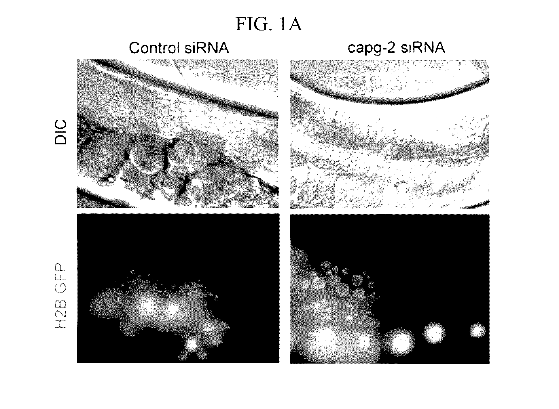
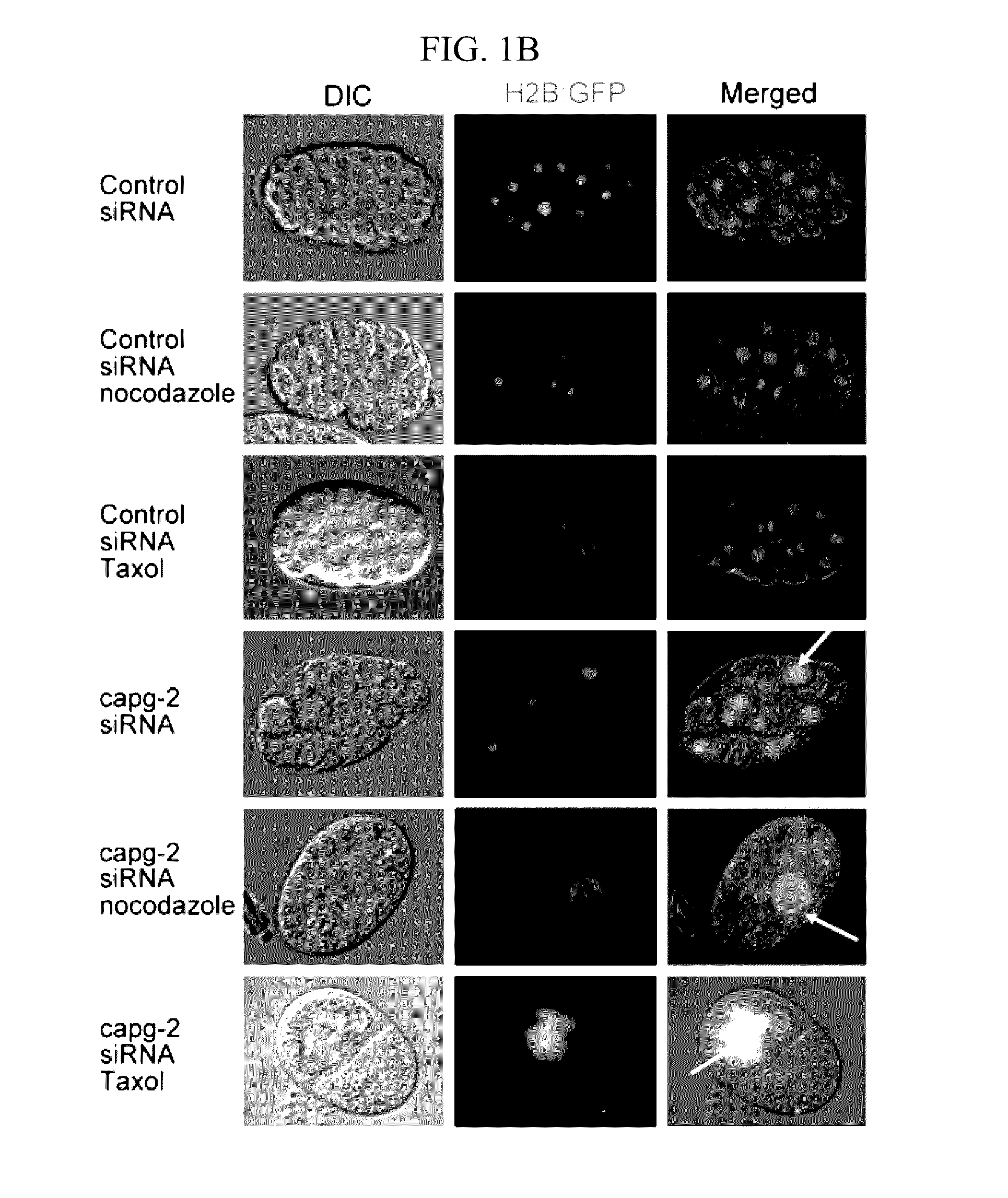
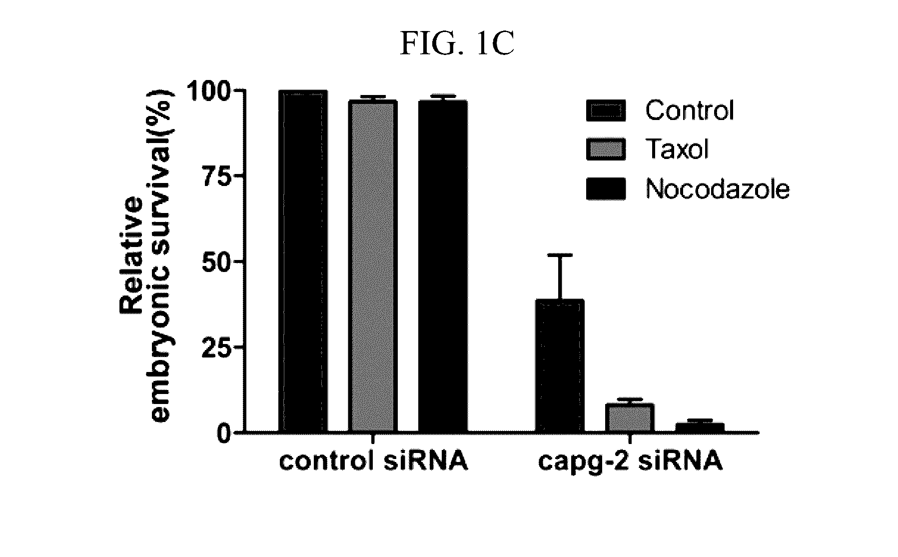
[0041]C. elegans were cultivated on NGM plates at 20° C. as a standard culture method (Brenner, S. Genetics 77, 71-94 (1974)). The capg-2 RNAi clone was obtained from Dr. J. Ahringer's bacteria-feeding RNAi library, which was a kind gift from Dr. J. Lee (Seoul National University). The wild-type N2 strain was used for measuring embryo lethality when capg-2 RNAi was treated with nocodazole. RW10006 (HIS-72::GFP), XA3501 (GFP::HIS-11 and GFP::TBB-2), JG479 (NPP-1::GFP, mCherry::HIS-58 and GFP::TBB-2) and SAl27(GFP::PLK-1) were used to determine the effects of capg-2 knockdown on mitosis. JG479 was generated by crossing XA3501 and OCF3 (NPP-1::GFP and mCherry::HIS-58). HIS-72 is histone 3, and TBB-2 is β-tubulin. Both HIS-11 and HIS-58 are histone 2B proteins (www.wormbase.org). Most strains were provided by the CGC, which is funded by the NIH Office of Research Infrastructure Programs (P40 OD010440). For capg-2 RNA interference, L4 worms were tra...
example 2
Experimental Conditions
[0042]MDA-MB-231 and HEK293 cells, which were purchased from the American Type Culture Collection (ATCC), were grown in Dulbecco's modified Eagle's media (DMEM; Invitrogen) supplemented with 10% FBS (Invitrogen) and 1% penicillin-streptomycin (P / S) in a 5% CO2 atmosphere at 37° C. Small interfering RNAs (siRNAs) for NCAPG2 were synthesised using 5′-GCU UCA UAG GGU CAU UUA UTT-3′ (SEQ ID NO: 1) and 5′-GAA GAA UGA UGC UGA AAC ATT-3′ (SEQ ID NO: 2) sequences (ST Pharm. Co. LTD). Expression vectors or siRNAs were transferred into the cells using the Amaxa Nucleofector system (Amaxa Biosystems) according to the manufacturer's instructions. For synchronising cells into a specific phase, 50 ng ml−1 nocodazole (Sigma-Aldrich) or 100 μM monastrol (Sigma-Aldrich) was used.
example 3
Immunofluorescence Imaging
[0043]Cell were grown on coverslips, rinsed twice with PHEM buffer (60 mM PIPES, 25 mM HEPES, 10 mM EGTA, and 2 mM MgCl2, pH 6.9 with KOH), permeabilised with 0.5% Triton X-100 in PHEM buffer at 4° C. for 1 min, and fixed with 4% paraformaldehyde in PHEM buffer. The fixed cells were incubated at 4° C. for 1 hr with each primary antibody, followed by incubation with a secondary antibody plus 100 ng ml−1 DAPI for 3 hr. The acquired images were analysed using a confocal microscope (Zeiss 510 Meta, Carl Zeiss). Live cell imaging was detected every 15 sec for 72 hr, 0.5 sec exposures were acquired using 2×NA0.75 objective on an LSM500 META Confocal Microscope (Carl Zeiss).
PUM
| Property | Measurement | Unit |
|---|---|---|
| volumes | aaaaa | aaaaa |
| pH | aaaaa | aaaaa |
| pH | aaaaa | aaaaa |
Abstract
Description
Claims
Application Information
 - R&D
- Intellectual Property
- Life Sciences
- Materials
- Tech Scout
- Unparalleled Data Quality
- Higher Quality Content
- 60% Fewer Hallucinations
Browse by: Latest US Patents, China's latest patents, Technical Efficacy Thesaurus, Application Domain, Technology Topic, Popular Technical Reports.
© 2025 PatSnap. All rights reserved.Legal|Privacy policy|Modern Slavery Act Transparency Statement|Sitemap|About US| Contact US: help@patsnap.com


