
Image processing device and method, eye fundus image processing device, image photographing method, and eye fundus image photographing device and method

a technology of image processing and eye fundus, applied in image enhancement, medical science, diagnostics, etc., can solve the problems of difficult diagnosis, blackness in the macular region and peripheral region, etc., and achieve the effect of high quality

Active Publication Date: 2016-06-16
SONY CORP
View PDF0 Cites 12 Cited by
  • Summary
  • Abstract
  • Description
  • Claims
  • Application Information

AI Technical Summary

Benefits of technology

The patent technology can help create better quality images.

Problems solved by technology

Moreover, even if photographing is performed so as not to cause the blown-out highlights in the optic disk region, the macular region and the peripheral region may be filled with black and become difficult to be diagnosed.

Method used


Image

  • Image processing device and method, eye fundus image processing device, image photographing method, and eye fundus image photographing device and method
  • Image processing device and method, eye fundus image processing device, image photographing method, and eye fundus image photographing device and method
  • Image processing device and method, eye fundus image processing device, image photographing method, and eye fundus image photographing device and method

Examples


first embodiment

Outline of the Present Technology

[0044]First, the outline of the present technology is described.

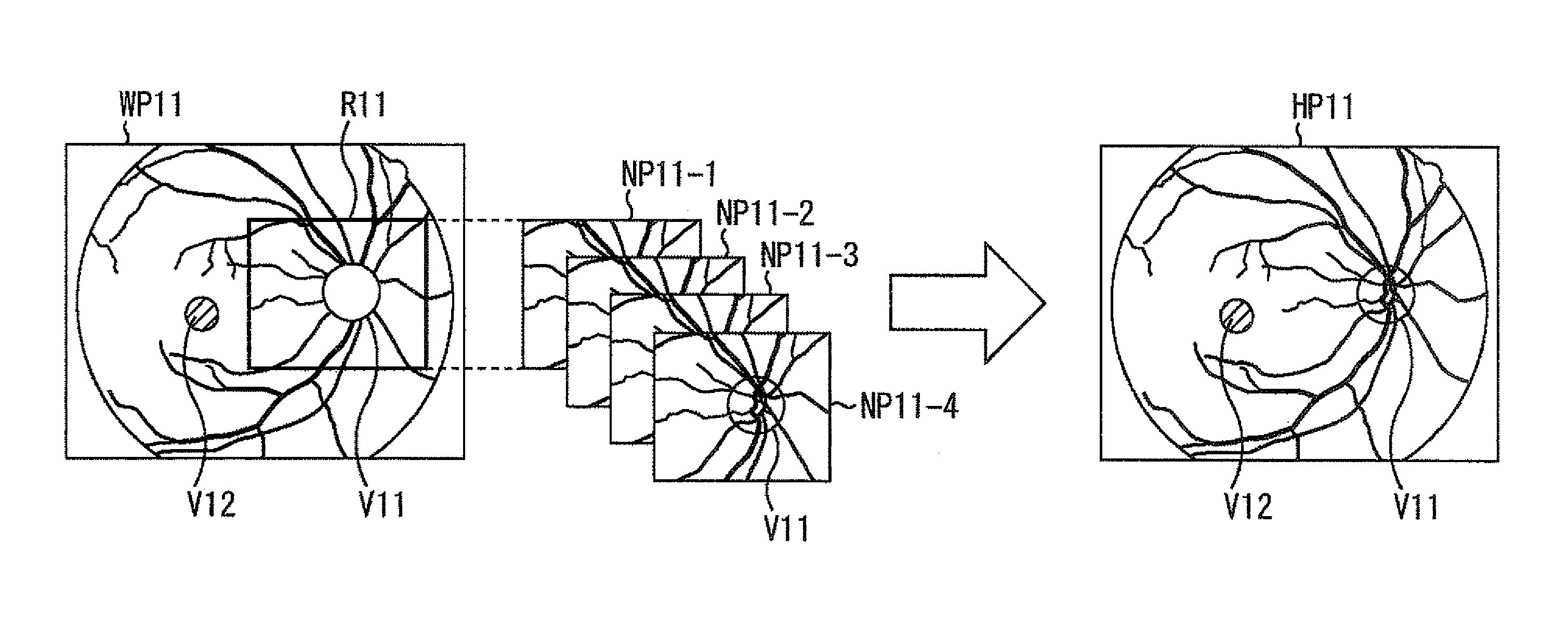
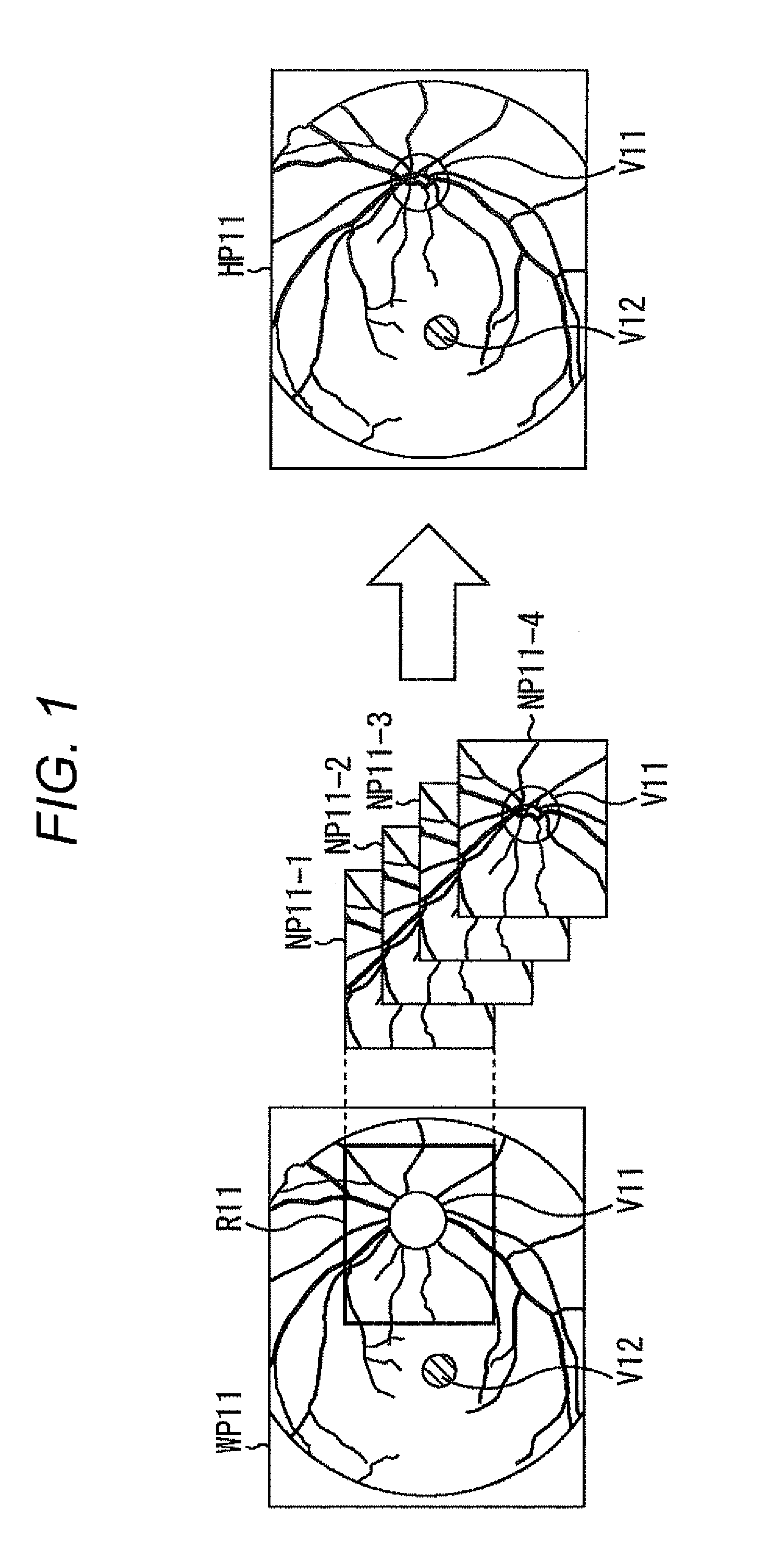
[0045]According to an embodiment of the present technology, by synthesizing an image of the entire eye fundus photographed under high illumination intensity and an image of the optic disk region of the eye fundus photographed under low illumination intensity, it is possible to acquire an eye fundus image of high quality with a wide dynamic range.

[0046]Specifically, in an eye fundus image photographing device to which the present technology applies, for example, as the first photographing processing as illustrated in FIG. 1, an eye fundus area that is an object is photographed under high illumination intensity and wide-angle eye fundus image WP11 is acquired as a result.

[0047]Wide-angle eye fundus image WP11 is an eye fundus image photographed with a wide angle of view. In this example, wide-angle eye fundus image WP11 includes optic disk region V11 and macular region V12. Generally, it i...


PUM


Abstract

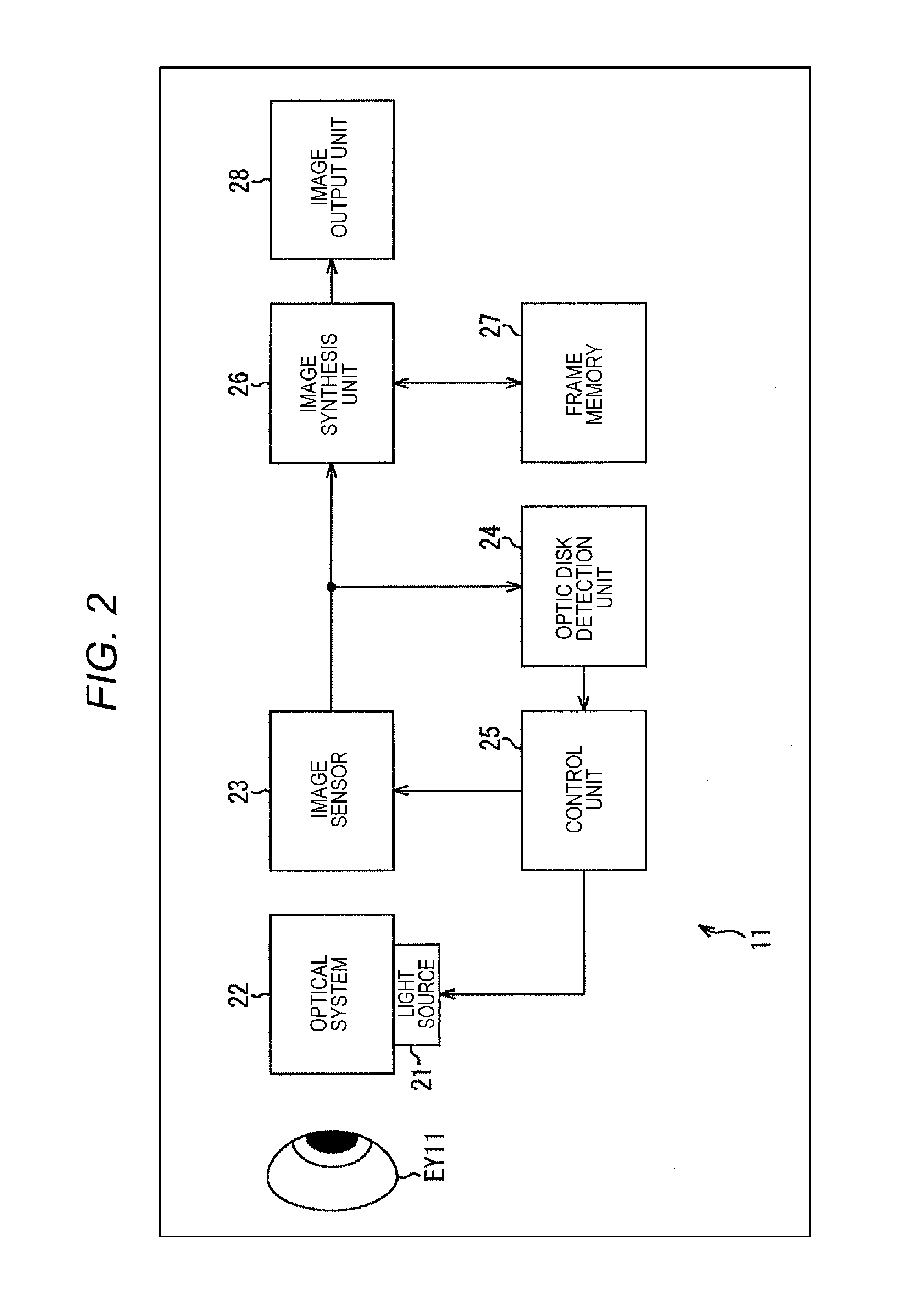
An eye fundus image photographing device and method that can acquire images of higher quality. The eye fundus image photographing device photographs a wide-angle eye fundus image on an appropriate exposure condition with respect to a peripheral area different from an optic disk region of an eye fundus area and detects the optic disk region from the wide-angle eye fundus image. Moreover, based on the detection result of the optic disk region, the eye fundus image photographing device photographs a narrow-angle eye fundus image which assumes an area of the optic disk region as an object, on an appropriate exposure condition with respect to the area of the optic disk region. Further, the eye fundus image photographing device generates a synthetic eye fundus image of a high dynamic range by synthesizing the wide-angle eye fundus image and the narrow-angle eye fundus image.

Description

TECHNICAL FIELD[0001]The present technology relates to an image processing device and method, an eye fundus image processing device, an image photographing method and an eye fundus image photographing device and method. Especially, the present technology relates to an image processing device and method, an eye fundus image processing device, an image photographing method and an eye fundus image photographing device and method that can acquire images of higher quality.CROSS REFERENCE TO RELATED APPLICATIONS[0002]This application claims priority to Japanese Priority Patent Application JP 2013-163781 filed on Aug. 7, 2013, the entire contents of which are incorporated herein by reference.BACKGROUND ART[0003]For example, in an eye fundus camera that photographs patient's eye fundus image in the checkup or the ophthalmologic examination, a photographing function in a high dynamic range is desired to stably acquire an eye fundus image of high quality.[0004]In an eye fundus image assuming ...

Claims


Application Information

Patent Timeline
IPC IPC(8): A61B3/00A61B3/14A61B3/12
CPCA61B3/0025A61B3/14A61B3/12G06T2207/10144G06T2207/30041G06T5/50G06T3/047
Inventor KOBAYASHI, SEIJI
Owner SONY CORP
Learn More