Determination of intracellular bacteria
a technology of intracellular bacteria and determination method, applied in the direction of microbiological testing/measurement, biochemistry apparatus and processes, etc., can solve the problems of inability to detect bacteria, inherently difficult to diagnose by traditional methods, and further complicated analysis, so as to reduce increase sensitivity. , the effect of reducing the risk of false positive results
- Summary
- Abstract
- Description
- Claims
- Application Information
AI Technical Summary
Benefits of technology
Problems solved by technology
Method used
Image

Examples
example 1
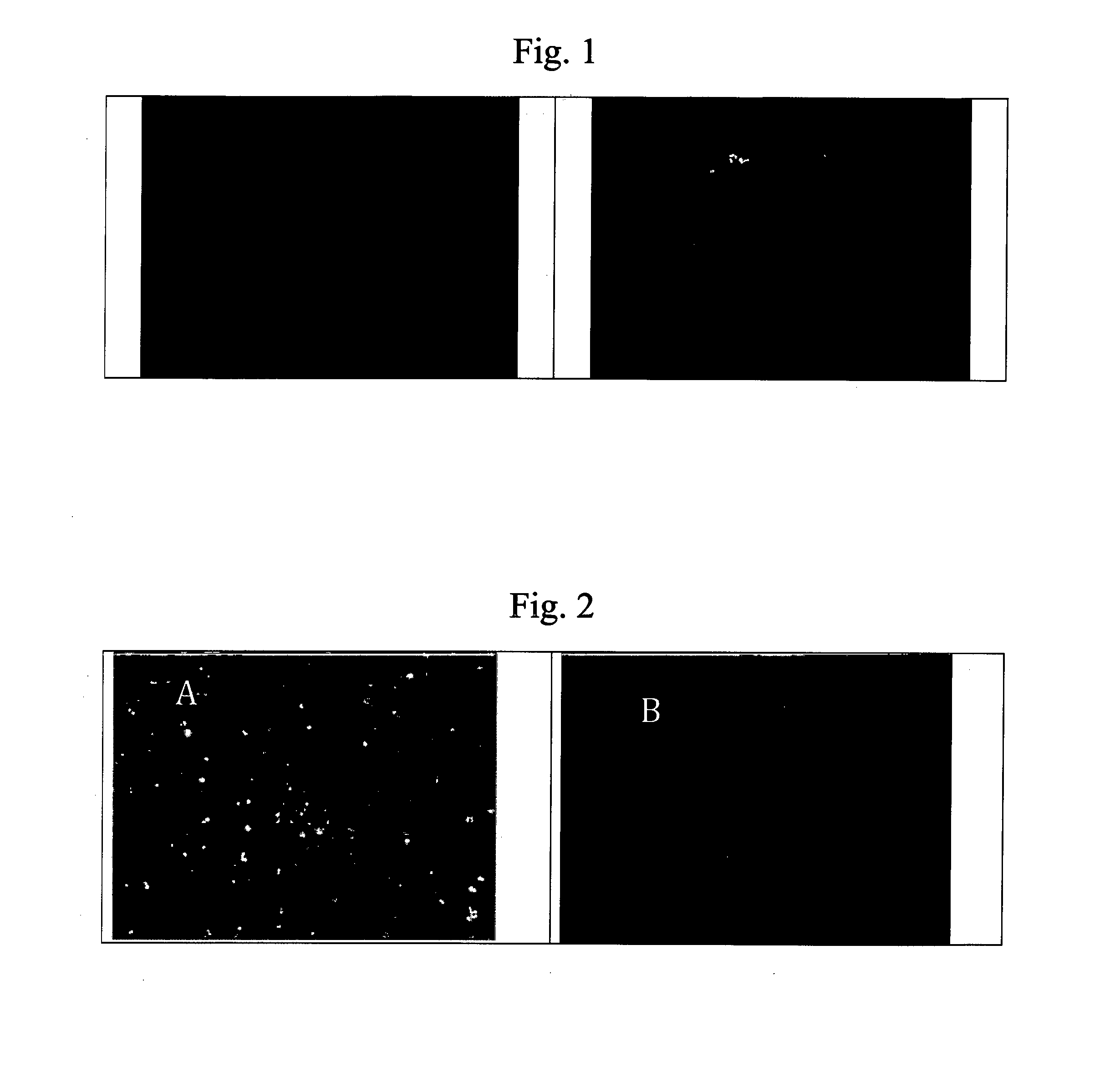
[0097]Staphylococcus aureus were experimentally spiked into donor blood and incubated 15-45 min for ingestion of S. aureus by phagocytic cells. The PMN fraction was purified and subsequent analyzed by S. aureus PNA FISH on microscope slides in accordance with Oliveira et al., J. Clin. Microbiol 40:247-251 (2002). Briefly, the samples was fixed onto microscope slides and hybridized with fluorescein-labeled PNA probes in hybridization buffer containing formamide for 30 minutes. Unbound PNA probes were removed by stringent wash for 30 minutes at 55° C. and counterstained with DAPI for visualization of both PMNs (blue) and S. aureus (blue). Examination by flourescence microscopy showed PMNs, including S. aureus (A) using DAPI (blue) filter and only S. aureus (B) using FITC / Texas Red filter. (blue cocci are observed within the PMN with the same morphology and position as the green cocci), See FIG. 1. In conclusion, intracellular bacteria in phagocytic cells can be determined by fluoresce...
example 2
[0098]S. aureus and Staphylococcus epidermidis were analyzed by fluorescence in situ hybridization in accordance with PLoS ONE 6(10): e25527 modified with PNA probe sequence and temperature (55° C.) from J. Clin. Microbiol 40:247-251 (2002). Briefly, the bacteria were fixed in solution using saline ethanol and washed twice (pre-hybridization) with hybridization buffer (0.75 M NaCl, 5 mM EDTA, 0.10 M Tris HCl, pH 7.8) followed by hybridization with PNA probe for 1 hour at 55° C. Unbound probe was removed by washing and the samples were mounted onto microscope slides. Examination by flourescence microscopy using FITC / Texas Red filter showed strong fluorescence of S. aureus (A) and low / none fluorescence of S. epidermidis (B), See FIG. 2. In conclusion, S. aureus can be determined by fluorescence in situ hybridization using PNA probes in buffered saline as hybridization buffer and without immobilizing the bacteria on microscope slides during the hybridization.
PUM
| Property | Measurement | Unit |
|---|---|---|
| temperature | aaaaa | aaaaa |
| pH | aaaaa | aaaaa |
| fluorescent | aaaaa | aaaaa |
Abstract
Description
Claims
Application Information
 - R&D
- Intellectual Property
- Life Sciences
- Materials
- Tech Scout
- Unparalleled Data Quality
- Higher Quality Content
- 60% Fewer Hallucinations
Browse by: Latest US Patents, China's latest patents, Technical Efficacy Thesaurus, Application Domain, Technology Topic, Popular Technical Reports.
© 2025 PatSnap. All rights reserved.Legal|Privacy policy|Modern Slavery Act Transparency Statement|Sitemap|About US| Contact US: help@patsnap.com