Wound healing via autologous stem cell mobilization
a technology of autologous stem cells and wound healing, applied in the field of wound healing, can solve the problems of imperfect treatment and high cost of these conditions, and achieve the effect of simple, rapid, and convenient treatmen
- Summary
- Abstract
- Description
- Claims
- Application Information
AI Technical Summary
Benefits of technology
Problems solved by technology
Method used
Image

Examples
example 1
AMD3100 Plus Low-Dose Tacrolimus Accelerated Wound Healing after Full-Thickness Skin Excision
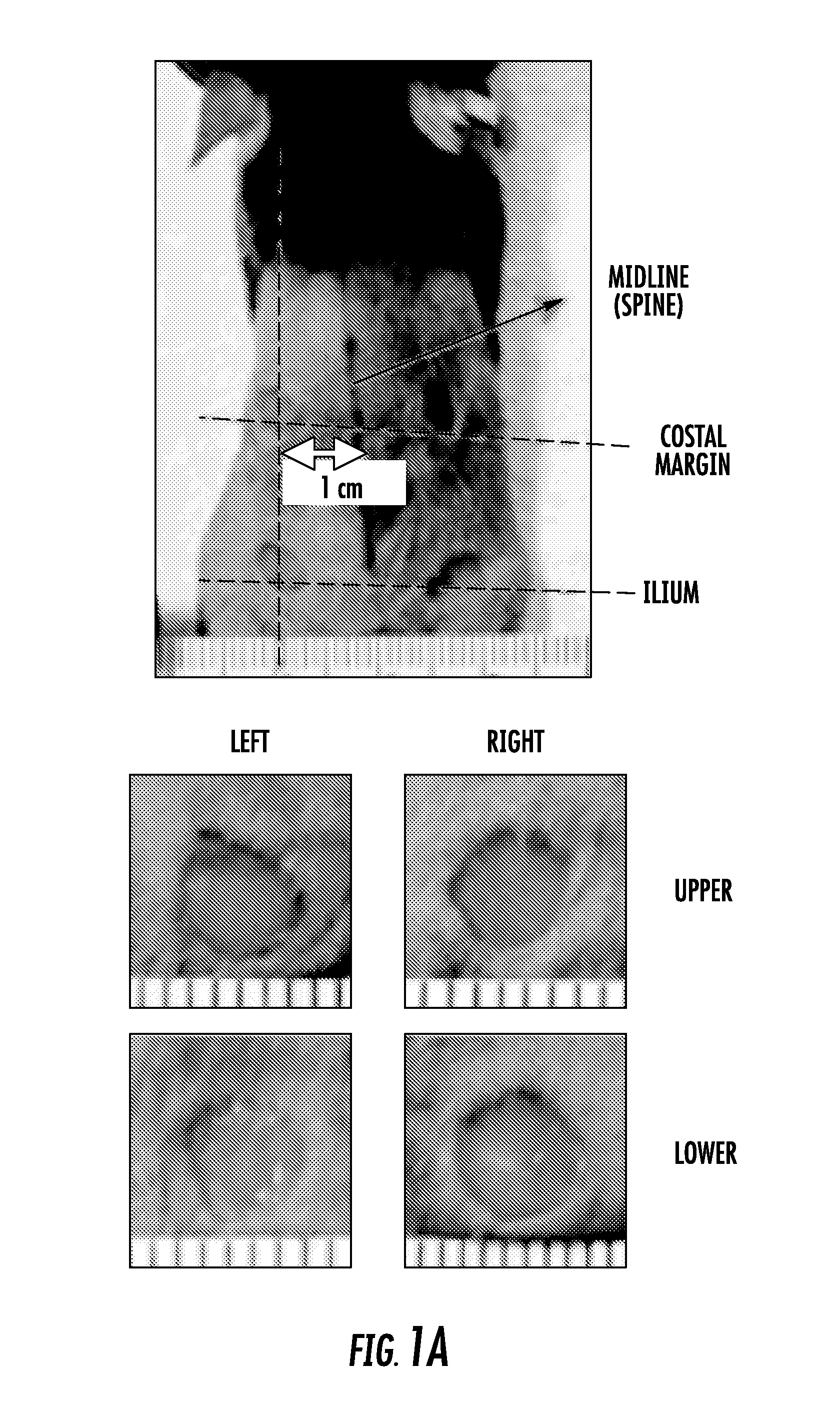
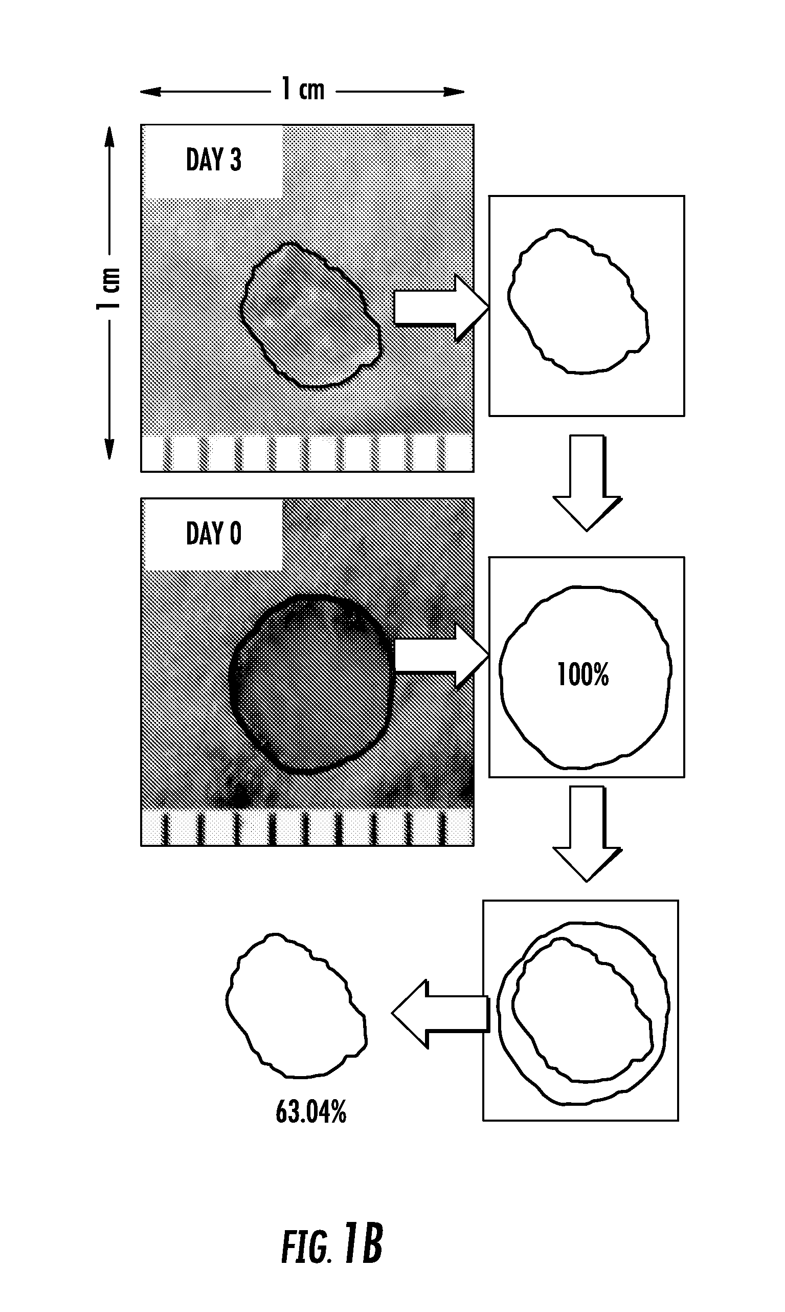
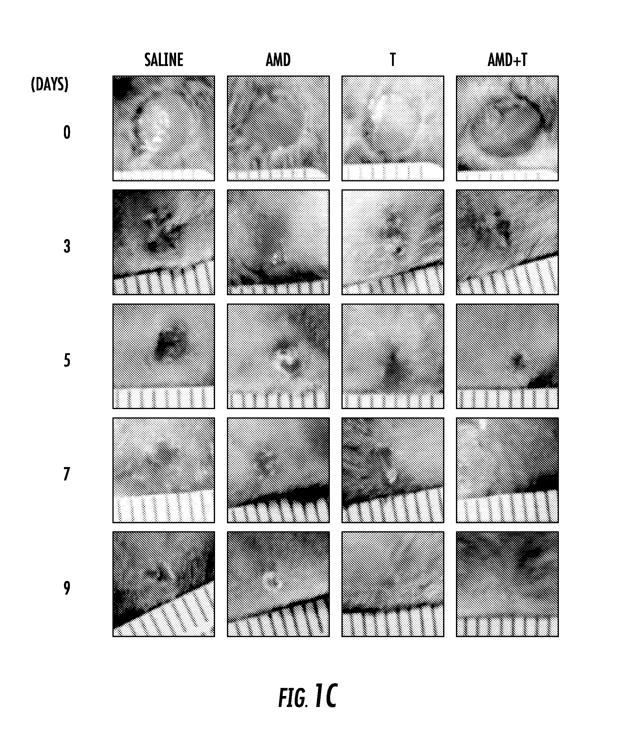
[0117]Four full-thickness wounds were generated by 5-mm diameter circular excisions on the shaved back of a wild-type C57 / B6 mouse (FIG. 1A). Each wound site was photographed digitally at the indicated time intervals, and wound areas were calculated using Adobe Photoshop software. Changes in wound areas over time were expressed as the percentage of the initial wound areas (FIG. 1b). Wounded mice were divided randomly into four experimental groups and received subcutaneous injections of saline or drugs immediately after wounding until complete healing: 1) Control group treated with saline, 2) Tacrolimus group treated daily with low-dose (0.1 mg kg-1), 3) AMD3100 group treated every other day (1.0 mg kg-1) and 4) Combination group given low dose Tacrolimus and AMD3100. All wound evaluations were double blinded.
[0118]Wounds reached complete closure on day 12 after surgery in group 1 (n=6) (FIG....
example 2
AMD3100 Plus Low-Dose Tacrolimus Ameliorated Scar Formation and Promoted Hair Follicle Regeneration
[0120]Dermal wound repair commences with the arrest of hemorrhage followed by an inflammatory response, formation of granulation tissue within the wound space, fibrosis, and re-epithelization of the wound, culminating in the production of a scar.
[0121]Histologically, the re-epithelialized wound in the control groups on day 15 showed a disorganized epidermis, with blurring of the boundary between the epidermis and the dermis (FIG. 2b). There were few hair follicles in the group 1, 2 and 3 mice and collagen was abundant and disorganized (FIG. 2c), which is in agreement with the results of published studies (Devine et al., 2004). By contrast, the dual drug treated animals had a thin and well organized epidermis with well-formed hair follicles and much better organized collagen (FIG. 2b and c; lower panels). The most striking finding was that hair appeared only in the reepithelialized woun...
example 3
AMD3100 and Low-Dose Tacrolimus Synergistically Mobilized Lineage Negative (Lin-) c-Kit+ CD34+ CD133+ Stem Cells Whereas Low-Dose Tacrolimus Increased the Number of SDF-1 Producing Macrophages
[0122]We performed flow cytometry on blood samples to determine the stem cell constituents mobilized by treatment with Plerixafor and Tacrolimus. The numbers of Lin-, c-Kit, CD34+, CD133+ cells and Lin-Triple positive (c-Kit+ CD34+ CD133+) cells were significantly greater in peripheral blood (FIG. 3a) in the animals receiving combination drug treatment at 5 days post-injury Immunofluorescence staining of wound tissue sections showed that the number of CD133+ was increased by combination treatment as were c-Kit+ and CD34+ cells (FIG. 3b). These cells were found particularly in newly formed granulation tissues of the wounds. Interestingly, at this 5 day time point, many CD133+ cells co-stained with CD31, a marker of endothelial cells (FIG. 3b; right panels).
[0123]Semi-quantitative PCR analysis of...
PUM
| Property | Measurement | Unit |
|---|---|---|
| Concentration | aaaaa | aaaaa |
| Concentration | aaaaa | aaaaa |
| Dimensionless property | aaaaa | aaaaa |
Abstract
Description
Claims
Application Information
 - R&D
- Intellectual Property
- Life Sciences
- Materials
- Tech Scout
- Unparalleled Data Quality
- Higher Quality Content
- 60% Fewer Hallucinations
Browse by: Latest US Patents, China's latest patents, Technical Efficacy Thesaurus, Application Domain, Technology Topic, Popular Technical Reports.
© 2025 PatSnap. All rights reserved.Legal|Privacy policy|Modern Slavery Act Transparency Statement|Sitemap|About US| Contact US: help@patsnap.com