
Method and system for characterizing tissue in three dimensions using multimode optical measurements

Inactive Publication Date: 2015-12-31
SPECTRAL MOLECULAR IMAGING
View PDF9 Cites 29 Cited by
  • Summary
  • Abstract
  • Description
  • Claims
  • Application Information

AI Technical Summary

Benefits of technology

This patent describes a method and system for characterizing biological tissue by illuminating it with multiple wavelengths of light and analyzing the resulting images. This approach can help overcome limitations of existing methods for early diagnosis of melanomas and other abnormalities. The model used for analyzing the tissue can be generated based on measurements of normal tissue or a tissue phantom. The system can also separate the concentration of melanin in order to accurately measure the distribution of oxy-and-deoxy hemoglobin. Overall, this technology can provide biologically plausible measurements and help improve the diagnosis and treatment of tissue abnormalities.

Problems solved by technology

Melanoma is a serious and challenging disease.
It is an increasingly lethal form of skin cancer, especially when detected in later stages.
Despite great effort worldwide, no significant advancements in treatment have occurred.
; Verisante Aura is claimed as a trademark by The BC Cancer Agency and the University of British Columbia) Although all of these optical systems provide high sensitivity, they have not achieved the desired level of specificity in diagnosis.
Unfortunately many systems employing these methods have shown reductions in performance as the studies move from smaller to larger populations.
The MelaFind® device data shows unavoidable rates of false-positives and false-negatives.
The MelaFind® device data was not validated and the device cannot be used for lesions with foreign material present such as dirt, ink or splinters, or with skin erosion, ulcers or bleeding and others defects.
Some private practice dermatologists find that they cannot justify its use.
As reflected in such data the statistical classification approach is encountering fundamental barriers to success as promising clinical devices fail when they are evaluated in larger studies.
A key problem is that in order to adequately validate these statistical models large numbers of patients must have biopsy confirmed measurements to develop these models or else resulting diagnostic algorithms will have poor performance.
This means large and thus expensive clinical trials are required.
Another more fundamental limitation is that the “black box approach” is only indirectly linked to tissue physiology.
The limited biological plausibility has kept clinicians and dermatologists from embracing this method.
When considered with the modest improvement of specificity from the current dermatologist examination specificity of 3% to the 10% to 13% range of specificity of such devices, it is difficult to justify their adoption.
This is especially true when both the change of procedure and the equipment expense are considered.
It is clear that such attempts to achieve early detection have shown disappointing reductions in specificity when clinical trials proceed from smaller to larger study populations.
These devices are very expensive, and the interpretation of the information requires the skills of a pathologist.
These statistical classifiers are used to decide whether a tissue has a particular pathology, but there is little information that can be directly related to the tissue biology providing a model that does not distinguish between correlation and causation.
This makes it difficult to evaluate the algorithm for the biological plausibility that usually engenders clinical confidence in a medical device Bergstrom, K. G. MelaFind was approved by FDA; where does it fit in dermatology?, J Drug Dermatol, 11, 420-422 (2012).
In skin studies, using SIAscopy, the limited multi-wavelength measurements appear to be inadequate for the light-tissue model being applied, Moncrieff, M., Cotton, S., Claridge, E., & Hall, P. Spectrophotometric intracutaneous analysis: a new technique for imaging pigmented skin lesions., Br J Dermatol 146, 448-57 (2002), because the results do not adequately correlate with pathology, Terstappen, K., Suurkiila, M., Hallberg, H., Ericson M. B., & Wennberg, A. M., Poor correlation between spectrophotometric intracutaneous analysis and histopathology in melanoma and nonmelanoma lesions., J Biomed Opt, 18, 061223 (2013).
However, it has become evident that, even with complex algorithms, misestimation of chromophore concentrations has been reported.
High skin melanin content usually leads to over-estimation of deoxy-hemoglobin and total hemoglobin and consequent under-estimation of hemoglobin oxygenation.
The problem persists in complex models where dark-skinned subjects always seem to have much lower oxygenation compared to Caucasian subjects, as presented by Yudovsky et al.
Poor correlation between spectrophotometric intracutaneous analysis and histopathology in melanoma and nonmelanoma lesions, J Biomed Opt, 18, 061223 (2013) showed a poor correlation between the SIA scans and histopathological findings in pigmented skin lesions, and attributed this error to misrepresentation of melanin and blood content due to high concentrations of melanin disturbing the quantification algorithm determining blood and collagen distributions.
However, they showed in other work that this method was only partially effective in a benign pigmented nevus with a high melanin concentration.

Method used


Image

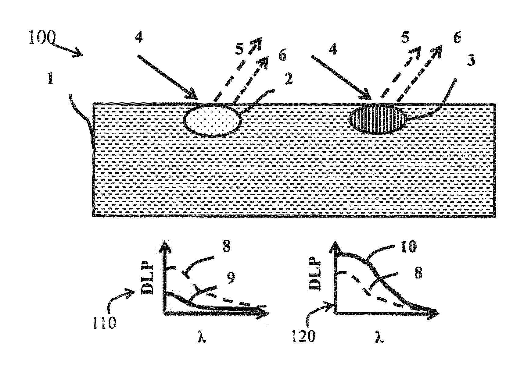
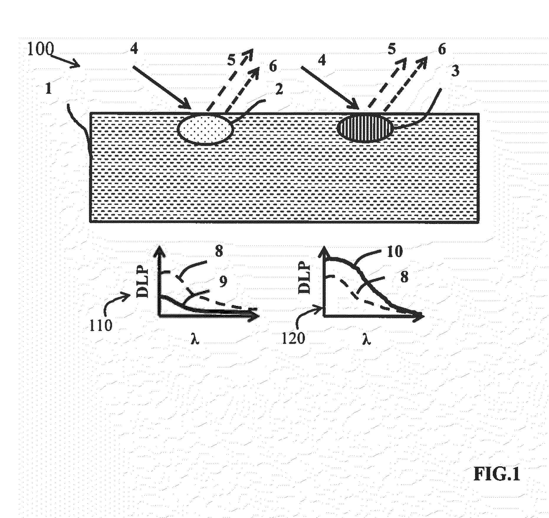
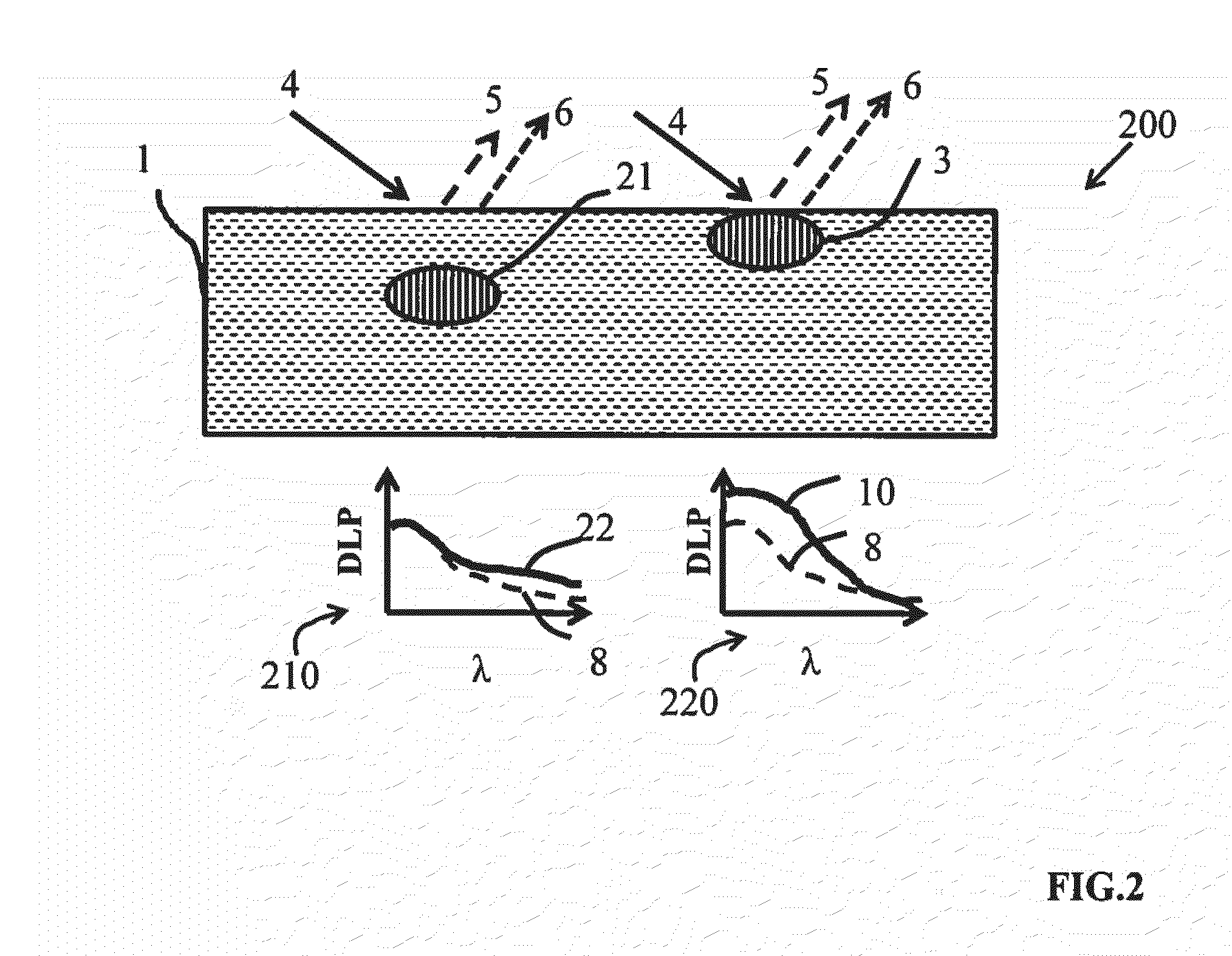
  • Method and system for characterizing tissue in three dimensions using multimode optical measurements
  • Method and system for characterizing tissue in three dimensions using multimode optical measurements
  • Method and system for characterizing tissue in three dimensions using multimode optical measurements

Examples


Embodiment Construction

Introduction

[0071]This disclosure presents preferred embodiments of a system and method that enable the production of a three dimensional map that provides volumetric information about tissue biology from analysis of multimode hyperspectral data cubes. Data cubes are sets of images taken under multiple modalities which can be analyzed. The images for the datasets are captured by a multimode imaging system such as the SkinSpect™ multimode imaging system developed by Spectral Molecular Imaging, Beverly Hills, Calif., which combines hyperspectral, polarization, reflection, scattering, fluorescence and bio-fluorescence imaging modalities. The three-dimensional optical map created from this data provides information to the physicians that helps to diagnose tissue abnormalities with higher precision than with imaging data sets having fewer modalities and combinations thereof.

[0072]Hyperspectral imaging is the capture of a sequence of images of a target such as tissue at multiple wavelengt...


PUM


Abstract

A method and system are provided for characterizing a portion of biological tissue. A surface of the tissue is illuminated with light having a known wavelength spectrum capable of materially penetrating the tissue. The intensity of the illumination light remitted from the tissue in response to the illumination over a known measurement window is measured over a hyperspectral range of wavelengths for at least two distinguishable polarization components. Based on a model of the response of the tissue and the preceding measurements, data representative of the location and one or more characteristics of an abnormal portion of the tissue are produced. A method is provided to eliminate the masking effect of melanin to obtain accurate estimations of an anomaly.

Description

RELATED APPLICATIONS[0001]This application claims the benefit under 35 U.S.C. §120 of international application Serial No. PCT / US2014 / 014330, filed Jan. 31, 2014 and designating the U.S., which claims the benefit under 35 U.S.C. §119(e) of U.S. patent application Ser. No. 61 / 759,910, filed Feb. 1, 2013, both of which are incorporated by reference herein in their entireties.BACKGROUND OF THE INVENTION[0002]Melanoma is a serious and challenging disease. It is an increasingly lethal form of skin cancer, especially when detected in later stages. Melanoma risk during a lifetime increased from 1:1500 in 1935 to 1:58 in 2009, and is still the fastest growing cancer both in the U.S. and worldwide. The National Cancer Center has estimated that 76,250 patients will be diagnosed with melanoma of the skin in 2012 and that 9,180, or more than one patient per hour, will die.[0003]Survival rates strongly favor early diagnosis, ranging from 98.2% for early, primary site detection to at best 15.1% f...

Claims


Application Information

Patent Timeline
IPC IPC(8): A61B5/00A61B5/145
CPCG01N21/21G01N21/31A61B5/0071A61B5/0075A61B5/443A61B5/444A61B5/14546A61B5/7278A61B5/0035A61B5/14558A61B5/0059
Inventor FARKAS, DANIEL L.VASEFI, FARTASHMACKINNON, NICHOLAS
Owner SPECTRAL MOLECULAR IMAGING
Learn More