
Non-implantable medical device coated with nano-carriers for delivering one or more drugs to a body site

a medical device and nano-carrier technology, applied in the field of medical devices, can solve the problems of ineffective penetration of micro-sized particles of drugs into tissues inflammation at the target site, and affecting the effect of drug delivery,

Inactive Publication Date: 2015-03-19
CONCEPT MEDICAL
View PDF7 Cites 3 Cited by
  • Summary
  • Abstract
  • Description
  • Claims
  • Application Information

AI Technical Summary

Benefits of technology

The invention is about a medical device that can deliver drugs to a specific area in the body, such as a blood vessel, organ cavity, or sac. The device is a drug-delivering insertable medical device, specifically a balloon catheter, which is designed to efficiently deliver drugs to the target site for a short period of time. The device uses nano-carriers of drugs to avoid inflammation and provide enhanced bioavailability. The device also ensures that the drugs are released from the surface of the device in a controlled manner, reducing the amount of drugs wasted during the procedure. The invention addresses the need for a device that can efficiently deliver drugs to the maximum area of a lesion and provide enhanced bioavailability with an optimum amount of drugs loaded on the device.

Problems solved by technology

The polymers used may cause inflammation at the target site.
The remaining amount of the drug present on the DEB may get washed away in the blood stream while the DEB is being withdrawn through the blood vessel thereby producing unwanted side effects.
The micro-sized particles of the drug may not penetrate tissues at the target site efficiently.
Thus, the current DEBs may not exhibit an efficient in-tissue diffusion of the drug.

Method used


Image

  • Non-implantable medical device coated with nano-carriers for delivering one or more drugs to a body site
  • Non-implantable medical device coated with nano-carriers for delivering one or more drugs to a body site
  • Non-implantable medical device coated with nano-carriers for delivering one or more drugs to a body site

Examples


example 1

[0070]Soya phospholipid was obtained from Lipoid GMBH, Batch No.: 776114-1 / 906. Sirolimus was obtained from Fujan Chemicals, China with purity greater than 99.5%. The water, other solvents and reagents used were of HPLC grade. A polyamide catheter system with COPAN Co-Polyamide angioplasty balloon (herein after “the balloon system”) coated with Hydrophilic coating (hereinafter “the hydrophilic surface”) was obtained from Minvasys, Paris, France.

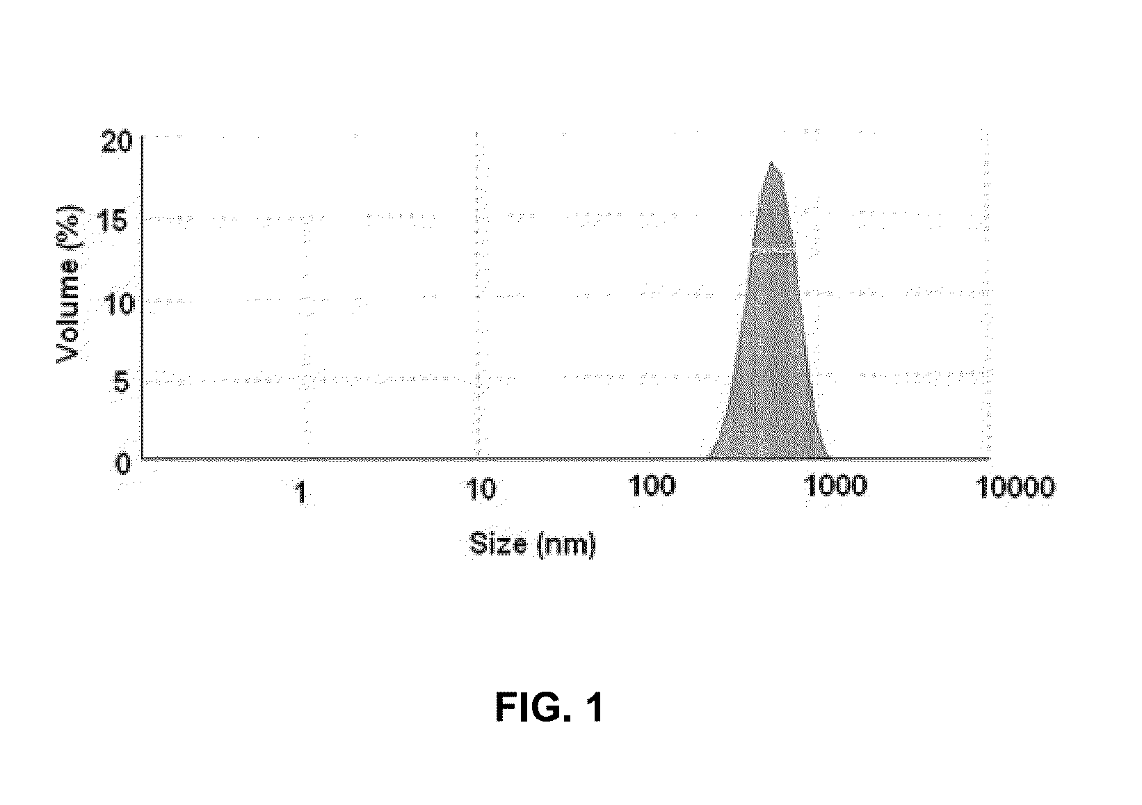
[0071]Soya phospholipid (20 mg w / w) was added to HPLC grade water (20 ml) followed by Tween 80 (5 mg) to obtain aqueous solution of soya phospholipid. The aqueous solution of soya phospholipid (20 ml) was subjected to a high speed homogenization at 15000-20000 rpm for 20 to 25 minutes in ice-cold water bath to obtain Solution A1. The Solution A1 thus obtained contained nano-particles of soya phospholipid. The solution A1 was subsequently analyzed for particle size detection using Malvern ZS90 (Malvern, UK) size detector. FIG. 1 illustrates th...

example 2

[0076]The coated balloon was further evaluated in-vivo in 17 male New Zealand rabbits, 5 to 6 months old and weighing between 3 and 4 Kg (hereinafter “animals”) for PharmacoKinetic (PK) study and histological evaluation or Light Microscopy (LM). FIG. 3 is a table illustrating numbers assigned to 17 animals, stent type and location of stent in each animal and the type of study (PK / LM) conducted on the animals. Out of the 17 animals 9 (Animals 66 to 74) were used for PK study and 8 (Animals 75 to 82) were used for histological evaluation.

[0077]The coated balloons were inserted into both the iliofemoral arteries of the animals used for the PK study. The coated balloons were inflated twice in the iliofemoral arteries. The coated balloons were first inflated for 70 seconds at 7 ATM and then deflated. Again, the coated balloons were inflated for the second time for 60 seconds at 7 ATM and then deflated and withdrawn.

[0078]A pre-mounted stent (3.0 mm× 12-14 mm) i.e. a stent mounted on the ...

example 3

[0085]6 Brazilian pigs (Hereinafter “the animals”) weighing about 25 to 30 kg were selected for the study. Bare metal stent (Cronus®, obtained from, Scitech, Brazil), with sizes of stent ranging from 2.5*13 mm to 3.0*13 mm and the coated balloons with sizes of about 3.0*15 mm were used. The stents were deployed in three vessels i.e. LAD (Left Interior Descending), LCX (Left Circumflex) and RCA (Right Coronary Artery of each animal by using: a) the coated balloon (Sirolimus), b) the coated balloon (Paclitaxel) and c) a bare balloon. The Paclitaxel coated balloon was prepared by replacing sirolimus in example 1 with Paclitaxel and rest of the process was kept same. The balloon to artery ratio for each coated balloon was approximately 1.1:1.0 to 1.2:1.0. Each of the coated balloon and the bare balloon were inflated for 60 seconds. The animals with sirolimus coated balloon were labeled “Sirolimus”, the animals with Paclitaxel coated balloon were labeled “Paclitaxel” and the animals with...


PUM

PropertyMeasurementUnit
average diameteraaaaaaaaaa
diameteraaaaaaaaaa

Abstract

A drug-delivering medical device for delivering a drug to a target site in a human body is disclosed. The drug-delivering medical device may have a hydrophilic surface, with one or more portions of the hydrophilic surface coated with one or more nano-carriers bearing one or more drugs. Each nano-carrier may include a drug surrounded by an encapsulating medium. As the drug is surrounded by the encapsulating medium, the surface of each nano-carrier can be devoid of the respective drug. A non-implantable medical device coated with nano-carriers can deliver one or more drugs to a blood vessel, organ cavity, sac, capsule, lining, layer, coating, membrane, connective tissue, fluid surrounding an organ, and so forth.

Description

RELATED APPLICATIONS[0001]This continuation-in-part application claims the benefit of priority to U.S. patent application Ser. No. 12 / 920,812, filed Sep. 2, 2010, now U.S. Pat. No. 8,801,662, which is a National Phase Application of International PCT Application No. PCT / IN2010 / 000349, filed May 21, 2010, which claims priority to India Patent Application No. 1337 / MUM / 2009, filed Jun. 2, 2009.FIELD OF THE INVENTION[0002]The invention generally relates to a medical device for administrating a drug to a target site in the body, such as a blood vessel, organ cavity, sac, capsule, lining, layer, coating, membrane, connective tissue, fluid surrounding an organ, and so forth. More specifically, the invention relates to a drug-delivering instrument coated with nano-carriers of one or more drugs for efficient delivery of the one or more drugs to the target site in the body.BACKGROUND OF THE INVENTION[0003]Drug-delivering insertable medical devices are used for localized delivery of a drug to ...

Claims


Application Information

Patent Timeline
Patent Type & Authority Applications(United States)
IPC IPC(8): A61L29/08A61K31/436A61K31/337
CPCA61L29/08A61K31/436A61K31/337A61L29/085A61L29/16A61M25/0045A61M25/10A61K9/5123A61M25/1027A61M25/104A61L2300/416A61L2300/42A61L2300/624A61L2300/626A61L2400/12A61M2025/105A61M2025/1088A61K38/00
Inventor DOSHI, MANISHSOJITRA, PRAKASHNETO, PEDRO A LEMOS
Owner CONCEPT MEDICAL
Learn More