
Dermoscopy illumination device with selective polarization and orange light for enhanced viewing of pigmented tissue

a dermoscopy and illumination device technology, applied in semiconductor devices for light sources, diagnostic recording/measuring, lighting and heating apparatus, etc., can solve the problems of difficult to see the deep pigmentation structures in the deep pigmentation area, different transillumination technology, etc., to maximize the viewing of the skin pigmentation area, the effect of less glare and high magnification and clarity

Active Publication Date: 2015-02-05
DERMLITE LLC
View PDF12 Cites 39 Cited by
  • Summary
  • Abstract
  • Description
  • Claims
  • Application Information

AI Technical Summary

Benefits of technology

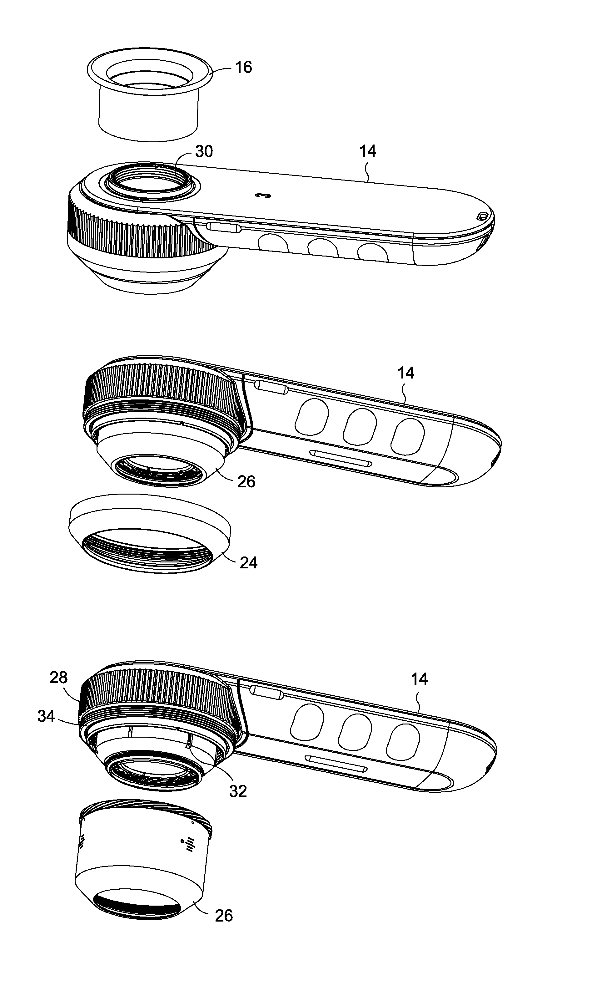
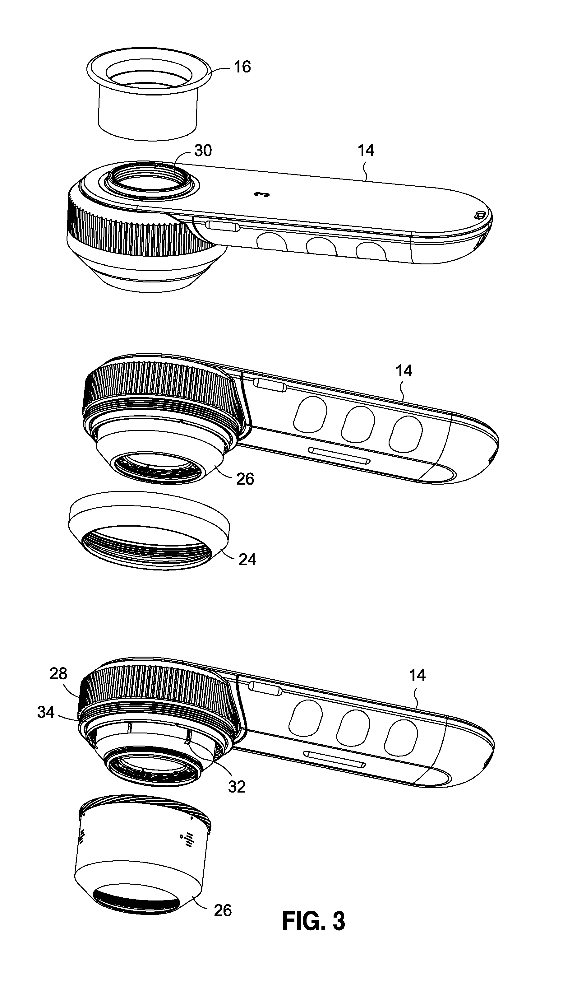
The present invention is an epiluminescence microscopy device that uses multiple sets of LEDs to view skin lesions with high clarity. The device includes a high-quality lens for color correction and reduced image distortion. The use of orange light in a specific range enhances the ability of white light LED based dermatoscopes for imaging deeper pigmentation or blood vessels. The device also allows for the independent use of cross-polarized white light and cross-polarized orange light, and provides a enhanced perspective of the skin for a doctor, researcher or healthcare professional. The device also provides the ability to toggle between several modes of operation and enhance the imaging of deeper pigmentation when using a glass-liquid interface and white light.

Problems solved by technology

However, the technology of transillumination is different than imaging skin with direct surface light.
When using the blue tinted white light in dermatoscopes, it is difficult to see the pigmentation structures in the deeply pigmented areas that are deep.

Method used


Image

  • Dermoscopy illumination device with selective polarization and orange light for enhanced viewing of pigmented tissue
  • Dermoscopy illumination device with selective polarization and orange light for enhanced viewing of pigmented tissue
  • Dermoscopy illumination device with selective polarization and orange light for enhanced viewing of pigmented tissue

Examples


Embodiment Construction

[0027]It is generally known that light is highly absorbed by the pigmentation in skin lesions for the central part of the visible spectrum from 500 to 570 nm. This high absorption is a problem with visualizing deep pigmented areas because the light gets blocked or absorbed superficially. The absorption of light in pigmentation for the frequencies above 570 nm decreases and this light can be used to see into deeply pigmented areas better.

[0028]Surface light falling on human skin in the visible spectrum can include a range of colors with Blue light (450-500 nm) penetrating the shortest distance in skin tissue. Blue light is known to be highly absorbed in pigmentation and blood and as such its depth is limited to less than 1 mm. Green light (500-550 nm) penetrates slightly deeper than blue light and is highly absorbed by hemoglobin in the blood. Yellow-orange light (560-610 nm) light penetrates between 1 and 2 mm depth in skin tissue, while red color light (620-670 nm) penetrates deepe...


PUM


Abstract

The present invention relates generally to an illumination device used in dermoscopy. More particularly, the invention comprises an improved apparatus for enhanced imaging and illumination of the skin for medical examination and treatment. An array of LEDs encircle a magnifying lens assembly. One set of surrounding LEDs provides cross-polarized white light to aid in canceling the reflected light from the skin and creating less glare. Another set of surrounding LEDs provides non-polarized white light for traditional immersion fluid dermoscopy or for non-polarized viewing of the skin. A third set of surrounding LEDs provides polarized colored light, wherein the wavelength color is selected at a color wavelength that maximizes the viewing of skin pigment regions which is important for early detection of skin cancers and other skin diseases. In an embodiment of the invention the colored LEDs comprise orange light at a wavelength of 588 nm. The present invention also discloses that the range of colored LEDs in the orange portion of the visible spectrum, namely at 581 nm to 600 nm wavelengths.

Description

CROSS-REFERENCE TO RELATED APPLICATIONS[0001]Not ApplicableSTATEMENT RE: FEDERALLY SPONSORED RESEARCH / DEVELOPMENT[0002]Not ApplicableBACKGROUND[0003]1. Field of the Invention[0004]The present invention relates generally to an illumination device used in dermoscopy. More particularly, the invention comprises an improved apparatus for enhanced imaging and illumination of the skin for medical examination and treatment.[0005]2. Background[0006]When using surface illumination to view human skin in medical examination, a great deal of surface light is reflected from the top layer of skin. Hand held dermoscopy devices that use light along with magnification can utilize polarizers or liquid-glass interface to reduce surface reflection and see deeper in to the skin. Dermoscopy apparatuses that employ light polarization to aid in viewing human skin surfaces and deeper tissue and structures of the skin are known and described U.S. Pat. Nos. 7,006,223 and 7,167,243 both issued to Mullani, the s...

Claims


Application Information

Patent Timeline
Patent Type & Authority Applications(United States)
IPC IPC(8): F21V9/14F21K99/00
CPCF21V9/14F21W2131/20F21K9/00A61B5/0059A61B5/441A61B5/0077
Inventor MULLANI, NIZAR
Owner DERMLITE LLC
Learn More