
Method of detecting residual genomic DNA and a kit thereof

a genomic dna and kit technology, applied in the field of methods of detecting residual genomic dna, can solve the problems of lack of sensitivity and specificity, limited traditional methods of quantitating residual host cell dna, and slow tim

Active Publication Date: 2015-01-29
BIOCON LTD
View PDF0 Cites 4 Cited by
  • Summary
  • Abstract
  • Description
  • Claims
  • Application Information

AI Technical Summary

Benefits of technology

The present disclosure describes a method and a kit for detecting and measuring the amount of residual genomic DNA from a rodent family in a protein product. This is accomplished using specific primers and probes to amplify specific sequences, making the process sensitive and accurate. The technical effect is the ability to reliably detect small amounts of genomic DNA in protein products, which can be useful in various applications.

Problems solved by technology

Traditional methods of quantitating residual host cell DNA have been limited by laborious sample preparation protocols, lack of sensitivity and specificity, and slow time to results.
The purified protein from the bulk harvest is likely to carry traces of CHO host cell genomic DNA into the final formulation thereby posing additional risk.
Traditional methods of quantifying levels of residual host cell DNA have been limited by lack of sensitivity and specificity, poor assay reliability, low sample capacity, slow time-to-results and high cost per sample tested.
However, the methods described here are not sensitive enough to detect contamination in picogram levels.
This reduces the overall ease of operation as well as the sensitivity.

Method used


Image

  • Method of detecting residual genomic DNA and a kit thereof
  • Method of detecting residual genomic DNA and a kit thereof
  • Method of detecting residual genomic DNA and a kit thereof

Examples


examples

Example: 1

Biological Sample

[0151]Any monoclonal antibodies or therapeutic proteins expressed in CHO—S cell line (obtained from Invitrogen cat#11619012).

Preparation of Standard DNA:

[0152]Genomic DNA from CHO cells is extracted from monoclonal antibody production strain by following phenol chloroform extraction method. DNA is resuspended in 10 mM Tris-1 mM EDTA, pH 8.0, the concentration determined, and diluted to a storage concentration of 100 ng / μL and a working concentration of 10 ng / μL. This DNA is used as Standard in qPCR.

Extraction of DNA from Biological Sample:

[0153]DNA from biological sample is extracted by using silica based DNA extraction columns where DNA binds to the silica membrane due to change in pH to acidic and DNA is eluted from the membrane with the addition of water which changes pH to neutral.

Example: 2

Materials and Method

[0154]The primers are obtained from Eurofins (Germany) and probe from Eurogentec (Belgium). Real time PCR machines Mx3000P from Stratagene and R...

example 10

Sequence Comparison of the Target Sequence with Other Alu Targets

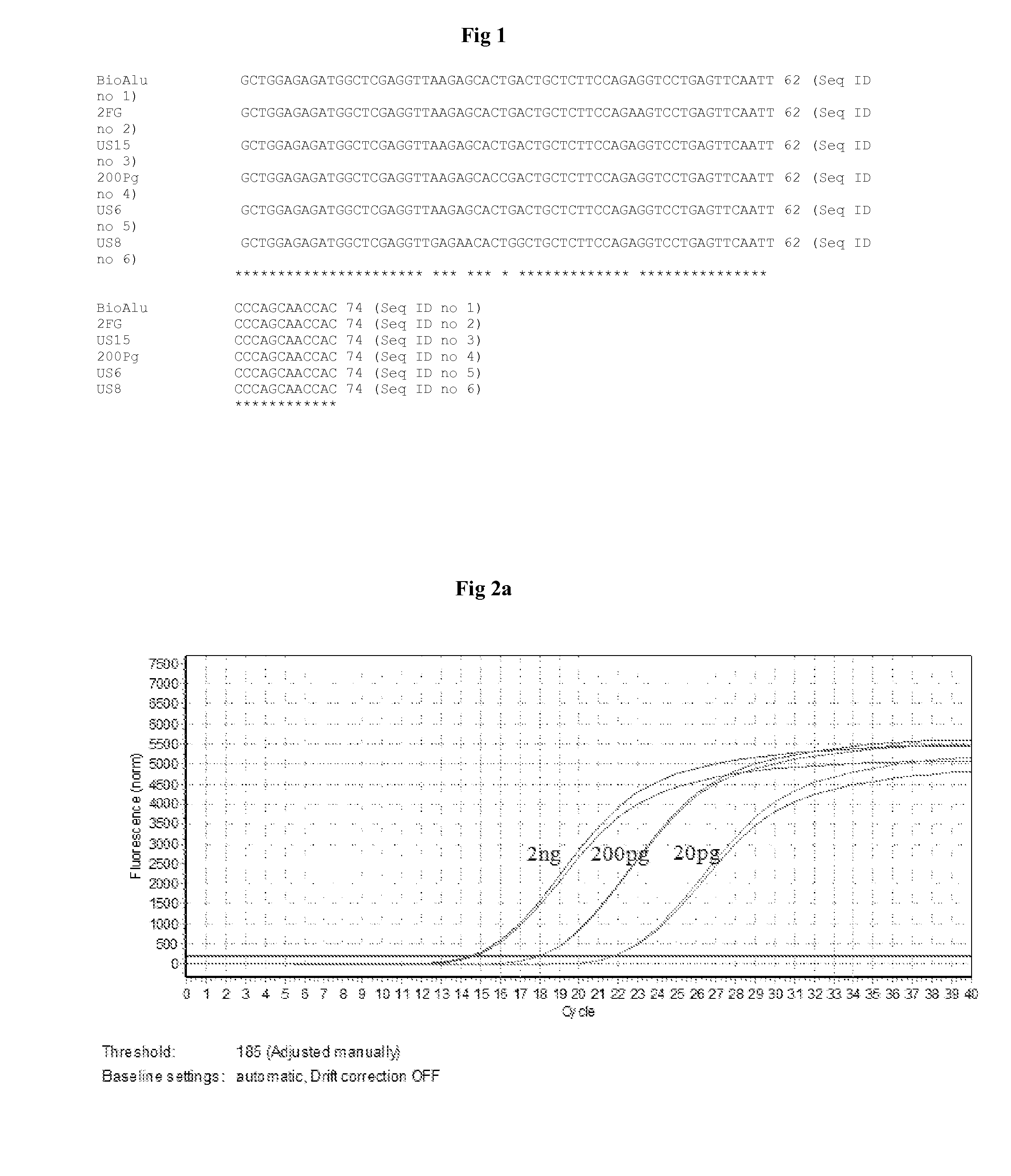
[0204]The target sequence designated as SEQ 1 is compared with the Alu-equivalent sequences available in public domain. Since the rational of seq ID 27 & Seq ID 28 is being used as target Alu sequence for residual DNA quantification from biological proteins as described in patents US2009325175 and U.S. Pat. No. 5,393,657 respectively. Therefore the following sequence comparison of Seq ID 1 with Seq ID 27 & 28 was performed to identify the sequence variations between the target identified in present disclosure and those reported in public domain.

[0205]The published sequences designated as SEQ ID 27 and SEQ ID 28 are compared with the seq ID 1 using the CLUSTAL W online software. The percentage homology observed with the sequence ID 27 is 50% and hence the percentage variance is 50% (FIG. 21a). The comparison of sequence ID 1 with the sequence 28 revealed percent homology as 71.6% and hence the percent variance is 28.4% ...

example 11

Presence of Other Alu Targets in the Genomic DNA

[0206]The two PCR products amplified from CHO genomic DNA by using the primers SEQ ID 25 and SEQ 26 and SEQ ID 29 and SEQ ID 30 are sequenced. Sequencing results revealed the presence of similar sequences with 5% variation with that of the SEQ ID 27 (FIG. 22a) and 21% variance with that of the SEQ ID 28 (FIG. 22b).


PUM

PropertyMeasurementUnit
temperatureaaaaaaaaaa
concentrationaaaaaaaaaa

Abstract

The present disclosure relates to a highly specific and sensitive method of detecting host cell impurities in a biological sample by using quantitative real time polymerase chain reaction (q PCR). The present disclosure also provides novel designed primer and probe to amplify only the specific Alu family of dispersed repetitive sequences from Chinese hamster ovary cells used for expression of therapeutic proteins.

Description

TECHNICAL FIELD[0001]The present disclosure describes a sensitive method of determining and detecting residual DNA in a biological sample wherein specific region of Alu equivalent sequences in Chinese Hamster Ovary (CHO) genomic DNA is detected with the help of designed primers and probe. The designed probe helps in increasing the sensitivity as well as the accuracy of the method. Hence the identification of specific regions within the CHO genomic DNA increases the overall sensitivity and efficiency of the detection process.BACKGROUND AND PRIOR ART OF THE DISCLOSURE[0002]Biopharmaceuticals are medical drugs produced using biotechnology. They include proteins (including antibodies), nucleic acids (DNA, RNA or antisense oligonucleotides) and living microorganisms like virus and bacteria where the virulence of viruses and bacteria is reduced by the process of attenuation. They can be used for therapeutic or in vivo diagnostic purposes, and are produced by means other than direct extrac...

Claims


Application Information

Patent Timeline
Patent Type & Authority Applications(United States)
IPC IPC(8): C12Q1/68
CPCC12Q1/686C12Q1/6888C12Q2600/166
Inventor FADNIS, RAHUL SHARADRAGHUNANDAN, REENA NICHINMETIA
Owner BIOCON LTD
Learn More