
Method of intra-operative coating therapeutic agents onto sutures composite sutures and methods of use

a technology of composite sutures and therapeutic agents, which is applied in the field of intraoperative coating therapeutic agents onto sutures composite sutures and methods of use, can solve the problems of inability to implantation of these implants into humans using these techniques, lack of long-term follow-up data, and inability to disclose the general literature on coating growth factors onto sutures. to achieve the effect of enhancing anterior stability

Inactive Publication Date: 2006-12-21
DEPUY SYNTHES PROD INC +1
View PDF33 Cites 57 Cited by
  • Summary
  • Abstract
  • Description
  • Claims
  • Application Information

AI Technical Summary

Benefits of technology

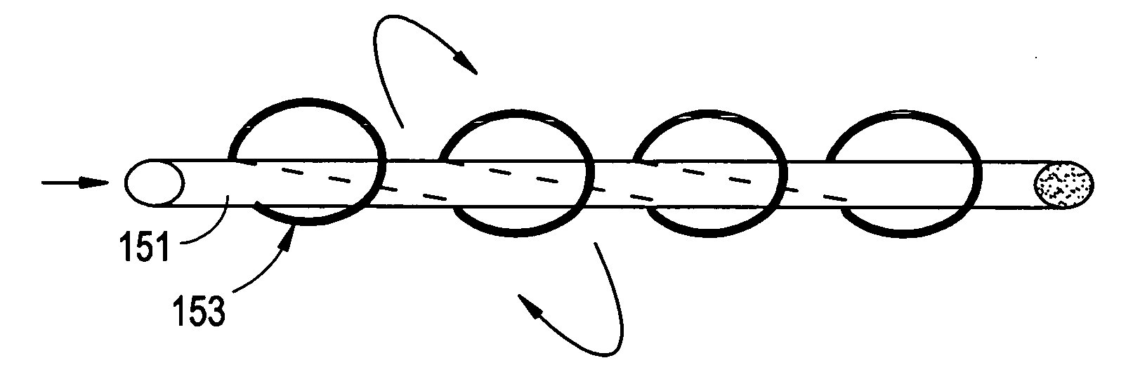
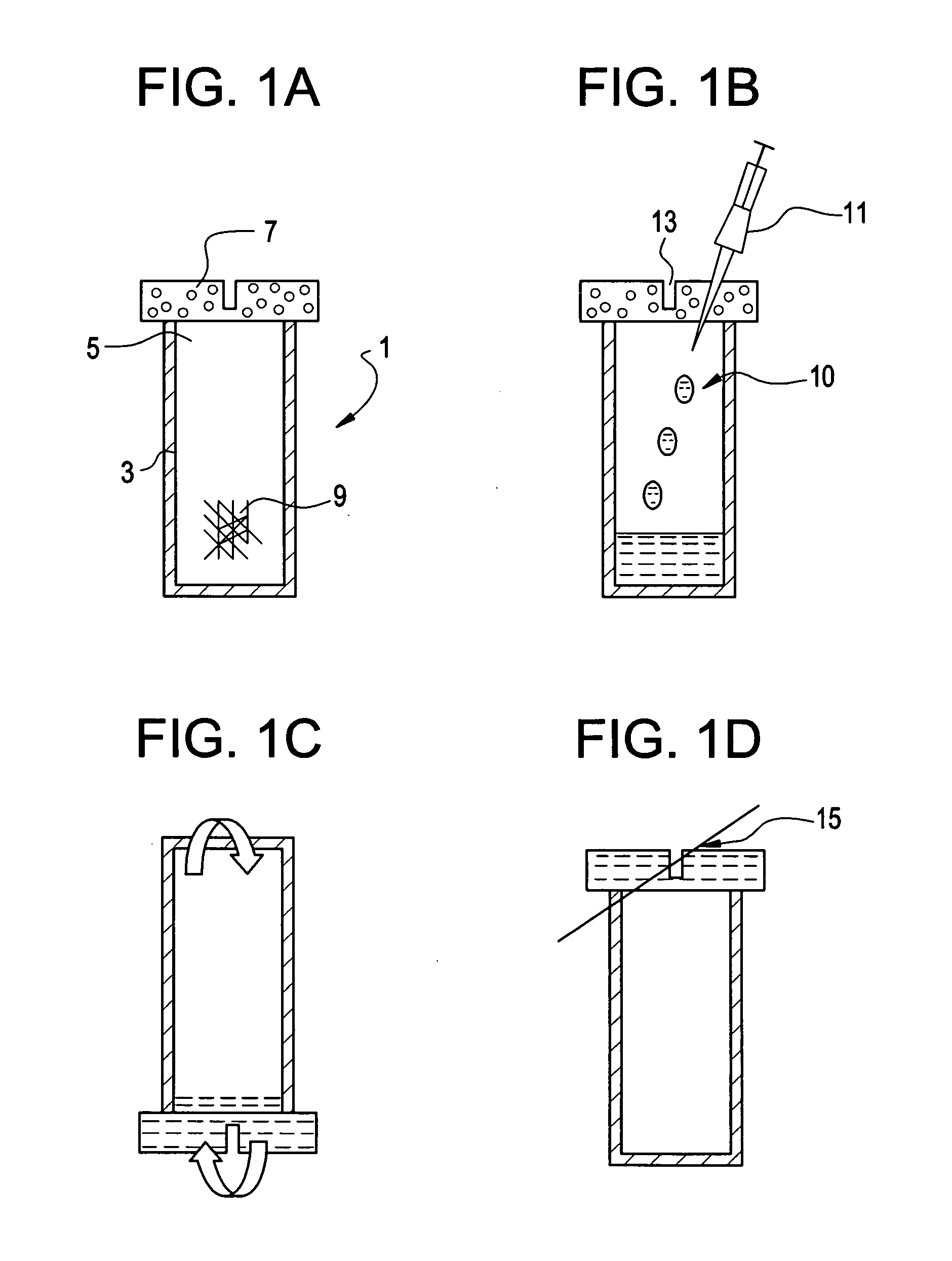
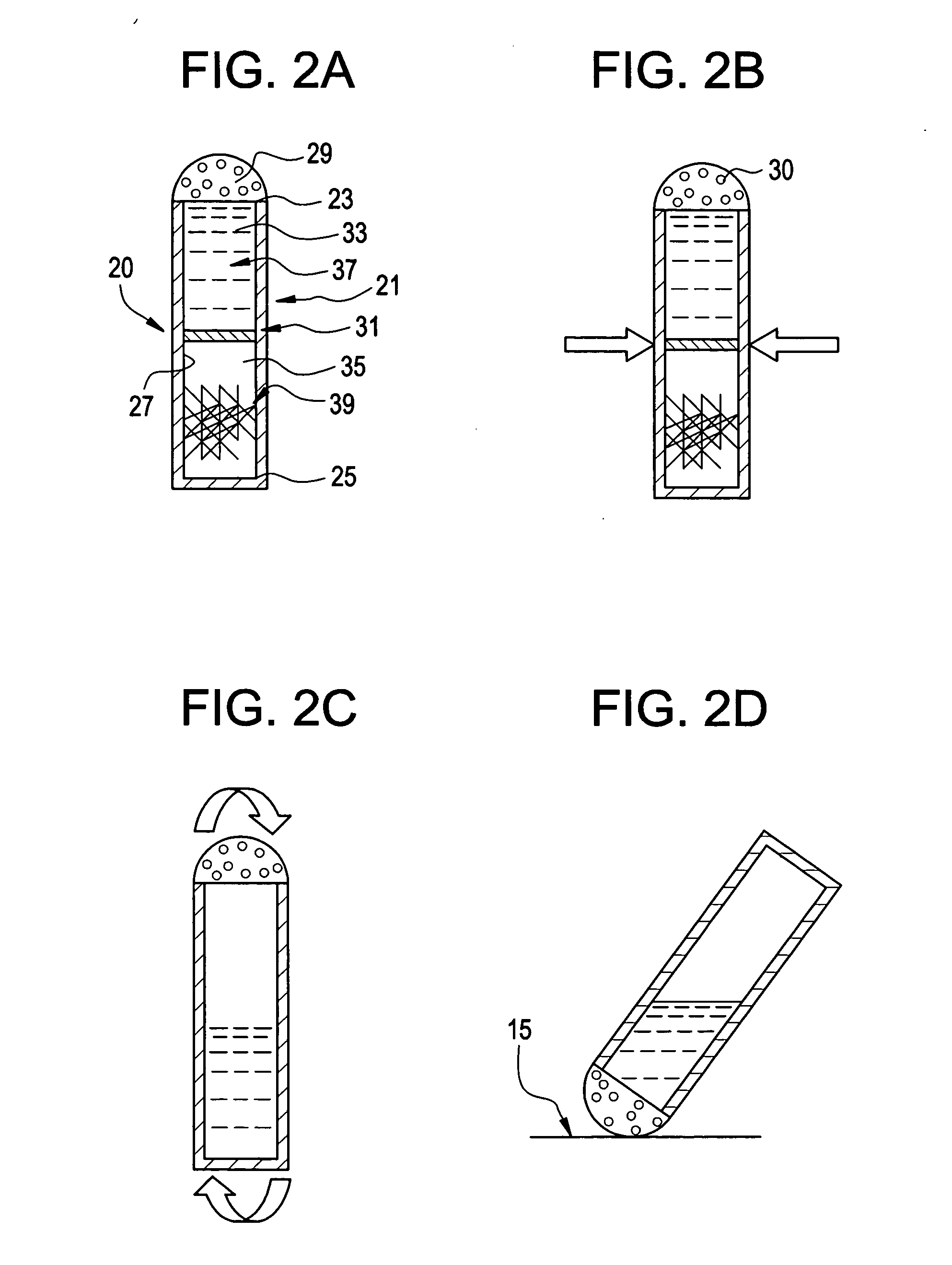
[0056] The present invention offers a novel method for creating coated sutures intra-operatively. In addition, it provides surgeons with the flexibility to (i) determine the desired amount of therapeutic agent to administer, and (ii) to select the length of suture that requires coating. Moreover, the present invention will provide many advantages over the conventional methods of coating sutures:
[0058] Second, the present invention will provide the surgeon with an ability to vary the dose of the therapeutic agent present on the suture.
[0059] Third, the present invention will provide the surgeon with the ability to coat sutures of different sizes and needles types. This allows the surgeon to coat portions of a single suture with the therapeutic agent (such as a growth factor), more specifically in areas where its contact with the host tissue is intended to have a therapeutic effect and leave other portions of the same suture uncoated.
[0060] Fourth, the present invention will provide the surgeon with the potential for providing sterile long-term storage of vials of therapeutic agents (such as growth factors like rhGDF-5) at either frozen, refrigerated or at room temperature.
[0061] Fifth, the present invention will provide the surgeon with the potential for providing growth factor-coated sutures for soft tissue repair, such as but not limited to repair of anterior cruciate ligament (ACL), medial collateral ligament of the knee or elbow, reconstruction of the anterior shoulder capsule to enhance anterior stability, reattachment of tendons especially near the bony insertion, reconstruction of the lateral joint capsule of the ankle, meniscus, Achilles tendon and rotator cuff tears.
[0062] Sixth, the present invention will provide the surgeon with the potential for providing new ways of using therapeutic agents such as growth factors independent of a specific implant.

Problems solved by technology

In fact, the literature in general does not disclose methods of coating growth factors onto sutures.
However, implantation of these implants into humans using these techniques are not currently possible, as in vitro models require further development and additional data are required to better characterize the in vivo models.
However, as the study reported on by Wright lasted only 6 weeks, long-term follow-up data are lacking.

Method used


Image

  • Method of intra-operative coating therapeutic agents onto sutures composite sutures and methods of use
  • Method of intra-operative coating therapeutic agents onto sutures composite sutures and methods of use
  • Method of intra-operative coating therapeutic agents onto sutures composite sutures and methods of use

Examples


example i

[0148] A 4-0 VICRYL (Polyglactin 910) Suture (Ethicon, Somerville, N.J.) was coated with rhGDF-5 and gelatin. The coating solution comprised of 4 ml gelatin solution and 2 ml of rhGDF-5 growth factor solution. The gelatin component was prepared by heating a 10 wt % solution of medical grade soluble bovine collagen (Semed-S, Kensey-Nash, Exton, Pa.) to 80° C. for 10 minutes followed by incubation at 37° C. rhGDF-5 (Biopharm GmbH, Heidelberg, Germany) was reconstituted with 10 mM HCl at concentrations of 3, 0.6, and 0 mg / ml. The resulting concentrations in the coating solutions were 1000, 200, and 0 μg / ml, respectively. The coating solutions were kept at 37° C. until use.

[0149] Prior to coating, the sutures were pretreated with a bath of 70% ethanol solution for 10 minutes, followed by a wash with saline. The suture was then placed in the coating solution and incubated at 37° C. for 30 minutes with gentle agitation. The suture was then removed from the solution and was then air-dried...

example ii

[0151] A 0 ETHIBOND EXCEL Polyester Suture (Ethicon, Somerville, N.J.) was coated with rhGDF-5 and gelatin in a similar manner as described in Example I. A rhGDF-5 solution was concentrated to 30 mg / ml with a centrifugal filter device (Centriplus YM-10, Regenerated Cellulose 10,000 MWCO, Amicon Bioseparations). The coating solution comprised of 0.5 ml concentrated rhGDF-5 solution and 1 ml 10 wt % gelatin solution. The concentration of rhGDF-5 on the coated suture, as quantified by ELISA, was 6.5 μg / cm.

[0152] Sutures were pulled through goat ACL tissue to evaluate if any of the growth factor coating is sheared off during its use. The concentration of rhGDF-5 post-surgery was 5.9 μg / cm, indicating that gelatin is effective in maintaining the growth factor on the suture even while passing through tissue.

example iii

[0153] A 0 Plain Surgical Gut Suture (Ethicon, Somerville, N.J.) was coated with rhGDF-5. The coating solution comprised of 1 ml rhGDF-5 solution concentrated to 13.9 mg / ml with a centrifugal filter device (Centriplus YM-10, Regenerated Cellulose 10,000 MWCO, Amicon Bioseparations). The gut suture was pretreated in a bath of 200 mM NaH2PO4 (pH 11.2) for 10 minutes followed by a wash in PBS prior to coating. The concentration of rhGDF-5 on the coated gut suture, as quantified by ELISA, was 26.3 μg / cm.


PUM

PropertyMeasurementUnit
concentrationsaaaaaaaaaa

Abstract

Intra-operative coating of sutures with therapeutic proteins, particularly growth factors such as rhGDF-5. including contacting a suture to a device containing a therapeutic agent, methods of repairing soft tissue defects with coated sutures and composite sutures coated with therapeutic agents.

Description

CONTINUING DATA [0001] This application claims the benefit of co-pending U.S. patent application Ser. No. 11 / 153,285, filed Jun. 15, 2005, entitled Method of Intraoperative Coating Therapeutic Agents onto Sutures” (Prajapati et al.) (DEP5505USNP), the specification of which is incorporated by reference in its entirety.BACKGROUND OF THE INVENTION [0002] The journal and patent literature describe the use of rhGDF-5 for its ability to form tendon, cartilage, bone and ligament-like structures. For example, Rickert et al., Growth Factors, 19, 2001, 115-126, discloses the use of recombinant human Growth and Differentiation Factor-5 (rhGDF-5) upon sutures to stimulate tendon healing in an Achilles tendon model in rats. However, the method of coating the rhGDF-5 to the sutures is described as simple pipetting of a solution containing rhGDF-5 onto the suture. In fact, the literature in general does not disclose methods of coating growth factors onto sutures. See for example, U.S. Pat. No. 5,...

Claims


Application Information

Patent Timeline
Patent Type & Authority Applications(United States)
IPC IPC(8): A61L17/00
CPCA61L17/145
Inventor PRAJAPATI, RITA T.BOWMAN, STEVENTIMMER, MARKDE DYNE, PATRICKHAMMER, JOSEPHYANG, CHUNLIN
Owner DEPUY SYNTHES PROD INC
Learn More