
Method to prepare an interventional and/or diagnostic imaging procedure with at least two different medical imaging modalitites

Active Publication Date: 2014-08-07
SIEMENS HEALTHCARE GMBH
View PDF4 Cites 3 Cited by
  • Summary
  • Abstract
  • Description
  • Claims
  • Application Information

AI Technical Summary

Benefits of technology

The present invention provides a method for simplifying preparation for interventional and diagnostic imaging by using information about the patient's volume elements and their properties. This information helps to quickly determine the compatibility of the patient with various medical imaging modalities. The method also allows for the selection of compatible additional procedures and the prevention of time-consuming preparations for examinations that may not be compatible with the patient. The compatibility value is based on a collision probability between the patient and the housing of the additional medical imaging modality. This helps to protect the patient from injury by preventing collisions and reducing the risk of injury during medical imaging examinations.

Problems solved by technology

In the positioning of the patient, the medical technician does not always succeed in taking into account all medical imaging modalities provided for the interventional and / or diagnostic imaging.
However, this can lead to a repositioning of the patient if an additional imaging measurement (data acquisition) is to take place with an additional medical imaging modality.
However, a repositioning of the patient hinders a fusing and / or superposition of image information and / or image data of the different medical imaging modalities.
In addition to this, the clinical workflow can be significantly disrupted and / or delayed, for example during a neurosurgical procedure on the head of a patient, wherein the head is clamped within a head mounting and is covered with sterile cloths.

Method used


Image

  • Method to prepare an interventional and/or diagnostic imaging procedure with at least two different medical imaging modalitites
  • Method to prepare an interventional and/or diagnostic imaging procedure with at least two different medical imaging modalitites

Examples


Embodiment Construction

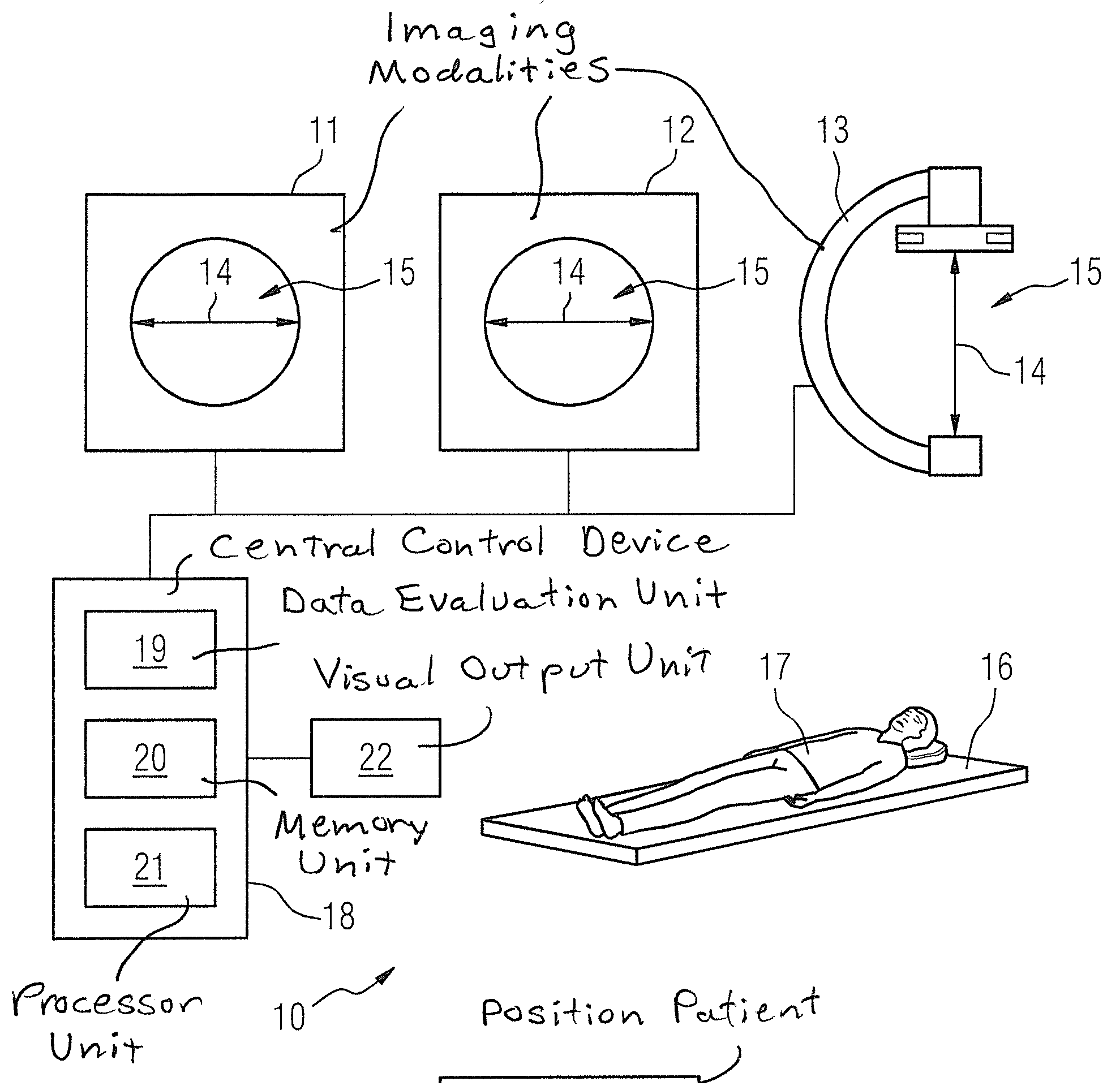
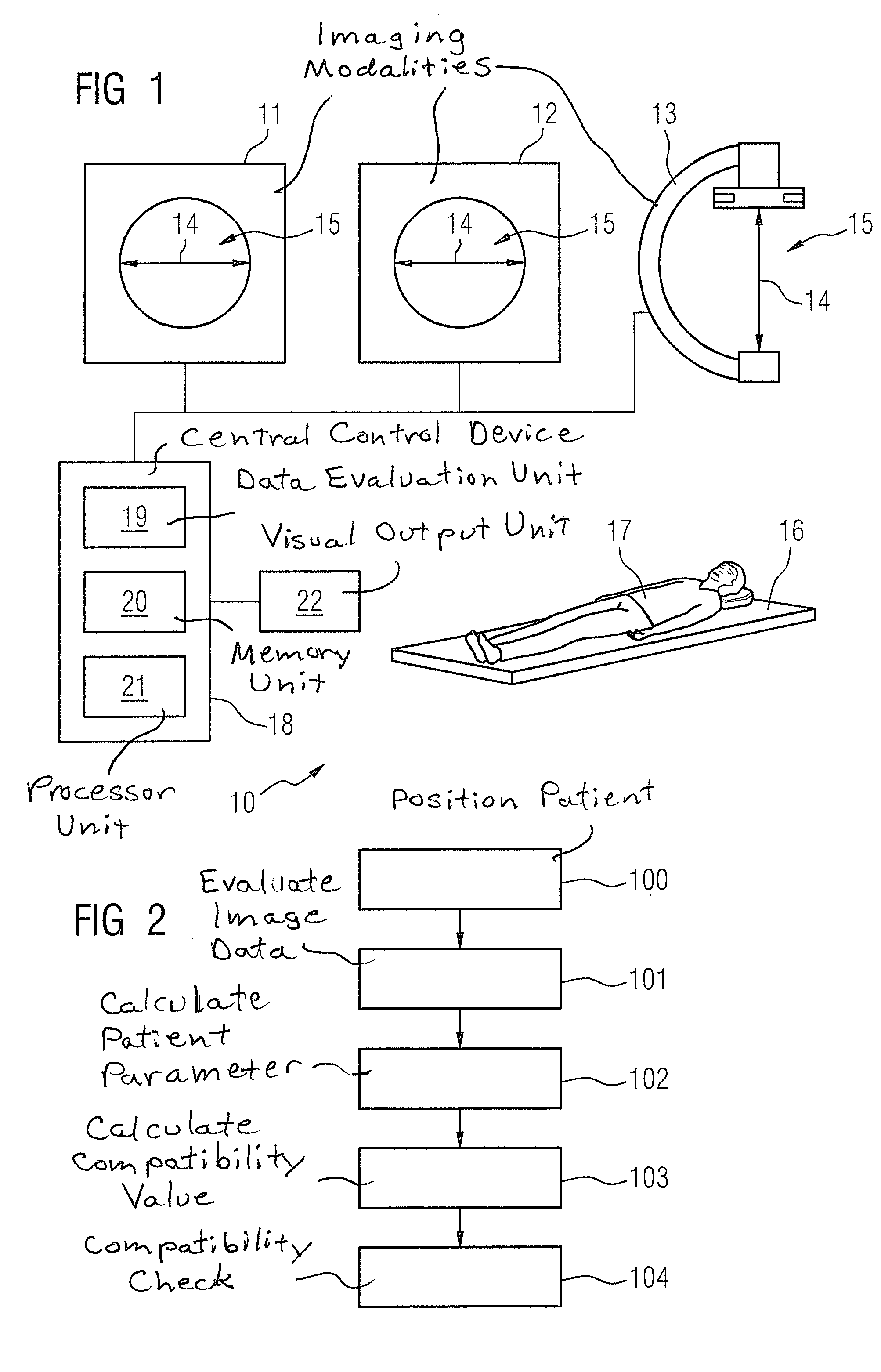
[0027]A medical imaging system 10 according to the invention is shown in FIG. 1. The medical imaging system 10 has multiple medical imaging modalities 11, 12, 13 that can be differentiated with regard to a detection type to acquire medical image data sets and / or with regard to a dimensioning given the same detection type. For example, the individual medical imaging modalities 11, 12, 13 can be formed by a magnetic resonance device, a computed tomography device, a PET device, an angiography device, etc. In addition, the individual medical imaging modalities 11, 12, 13 can be formed only by magnetic resonance devices, for example, wherein the individual magnetic resonance devices can be differentiated with regard to a magnetic field strength and / or an opening diameter 14 of a patient acquisition region 15 and / or additional parameters that are meaningful to those skilled in the art.

[0028]The medical imaging system 10 furthermore has at least one patient support device 16 on which a pat...


PUM


Abstract

In a method and medical imaging system to prepare an interventional and / or diagnostic imaging procedure to be conducted with at least two different medical imaging modalities of the system, a patient is positioned on a patient support device and the patient support device, together with the patient, are moved into a patient acquisition region of a first medical imaging modality of the system. A first image data set of the patient to be examined is acquired with the first medical imaging modality. The first image data set is automatically evaluated in a data evaluation unit. At least patient parameter is automatically calculated from the evaluated first image data set. At least one compatibility value is automatically calculated depending on the at least one patient parameter and depending on at least one apparatus parameter of at least one additional medical imaging modality in the system. The compatibility value indicates whether the additional modality is compatible with the patient in order to conduct another of the procedures using that additional modality.

Description

BACKGROUND OF THE INVENTION[0001]1. Field of the Invention[0002]The present invention concerns a method for preparing an interventional and / or diagnostic imaging procedure performed with a medical imaging system that has at least two different medical imaging modalities.[0003]2. Description of the Prior Art[0004]For interventional and / or diagnostic imaging, procedures performed on the patient and / or for treatment of the patient, the procedures are prepared and / or planned at least in part using medical image information that has been acquired by one or more medical imaging modalities, for example a magnetic resonance apparatus, a computed tomography apparatus, a PET (positron emission tomography) apparatus, etc. For this purpose, a reproducible, exact position of the patient is required in order to be able to fuse and / or combine image information of different medical imaging modalities to prepare and / or plan the intervention. However, these different medical imaging modalities freque...

Claims


Application Information

Patent Timeline
IPC IPC(8): G06T7/00
CPCG06T7/0012A61B6/5217A61B6/5247G16H30/20G16H50/30G06T2207/30004
Inventor EDER, HANNSGROSS, PATRICKHAUSOTTE, ANNEMARIERINGHOLZ, MARTINROTHGANG, EVA
Owner SIEMENS HEALTHCARE GMBH
Learn More