Electro-microneedle integrated body for in-situ cutaneous gene transfer and method of manufacturing same

Inactive Publication Date: 2014-05-22
RAPHAS +1
View PDF2 Cites 19 Cited by
  • Summary
  • Abstract
  • Description
  • Claims
  • Application Information

AI Technical Summary

Benefits of technology

This patent is about a device that can be used to release genes into the skin. The device combines a dissolving needle and an electrode that can be used to enhance the gene delivery. This allows for more efficient gene release in a specific location and increases the effectiveness of treatment. The patent also describes a method for preparing this device. Overall, the invention helps to deliver genes deeper into the skin and improve the efficiency of gene therapy.

Problems solved by technology

However, since the electroporation intended for intracellular transfection of a gene needed a sufficient gene concentration in an intracellular electric field pulse site, for this purpose, an additional transdermal delivery means which conventionally uses a disposal injection was required, and as such, an efficient intracutaneous gene delivery to a in-situ treatment site was not available with the said non-integrated system.
However, the transdermal delivery using a disposal injection caused an intracutaneous stimulus and pain, and further, accurate release of a gene in fine skin membranes was limited.
Johns Hopkins University introduced an electroporation gene gun in which a gene gun and electroporation were integrated (US patent publication No. 20090030364 entitled “Electroporation gene therapy gun system”), yet the accurate transdermal delivery in the treatment site was also limited with the gene gun.
Talon Biotech suggested a system in which a gene-coated microneedle and an electrode for electroporation were integrated (“Smallpox DNA vaccine delivered by novel skin electroporation device protects mice against intranasal poxvirus challenge,”Vaccine 25, 1814-1823, 2007), though the coated microneedle had a limit on an amount of intracutaneous gene release because of a limited amount of gene load, and therefore, efficiency of percutaneous gene delivery was ultimately low.
However, it was difficult to prepare a monolithic integrated body in which the dissolving microneedle and the electrode for electroporation are integrated into one structure with a conventional molding method of preparing the dissolving microneedle, that is a micro-casting method.

Method used


Image

  • Electro-microneedle integrated body for in-situ cutaneous gene transfer and method of manufacturing same
  • Electro-microneedle integrated body for in-situ cutaneous gene transfer and method of manufacturing same
  • Electro-microneedle integrated body for in-situ cutaneous gene transfer and method of manufacturing same

Examples


Embodiment Construction

[0018]An electro-microneedle integrated body and a method of preparing the same according to the exemplary embodiments of the present invention will be described in detail below with reference to the accompanying drawings.

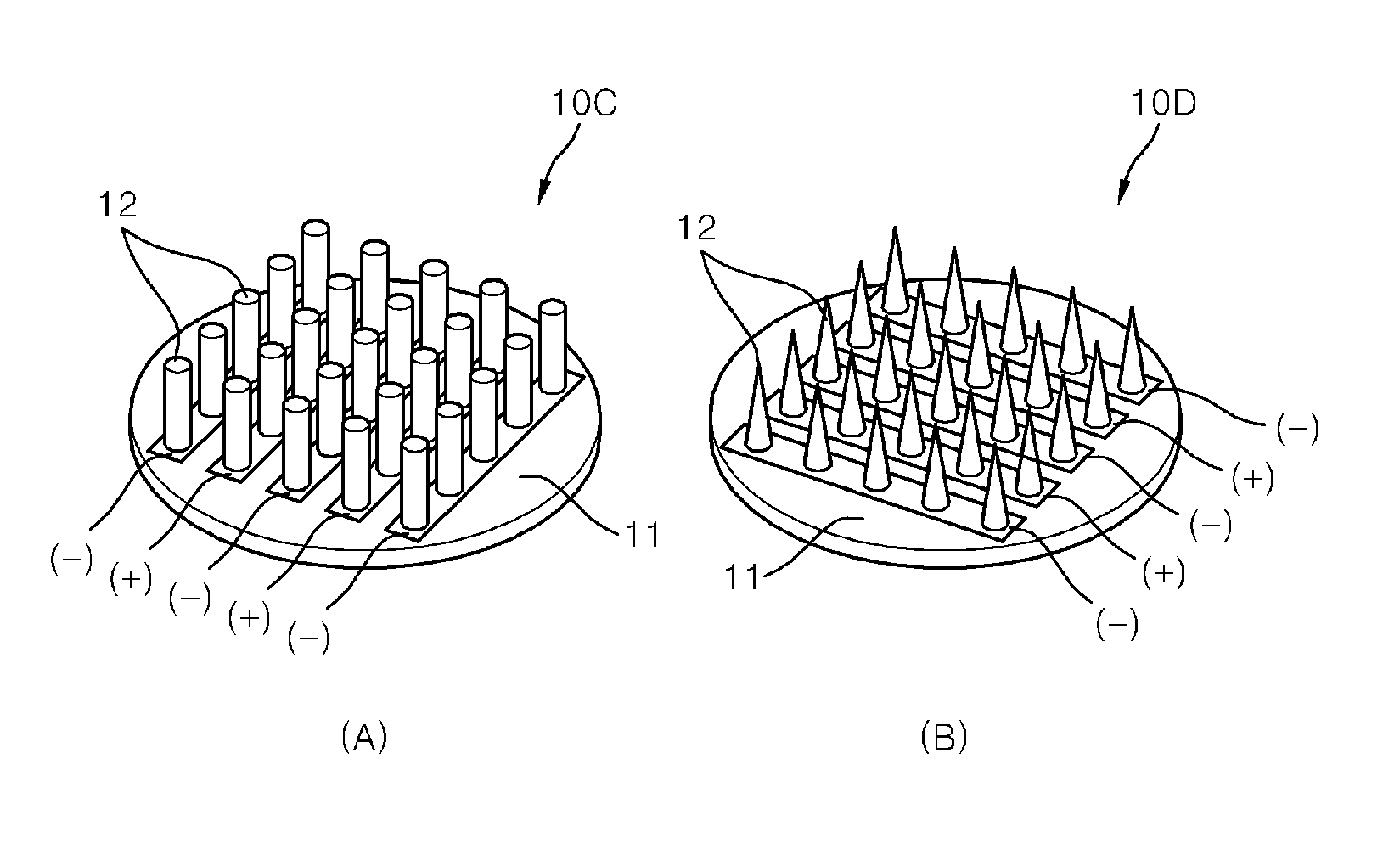
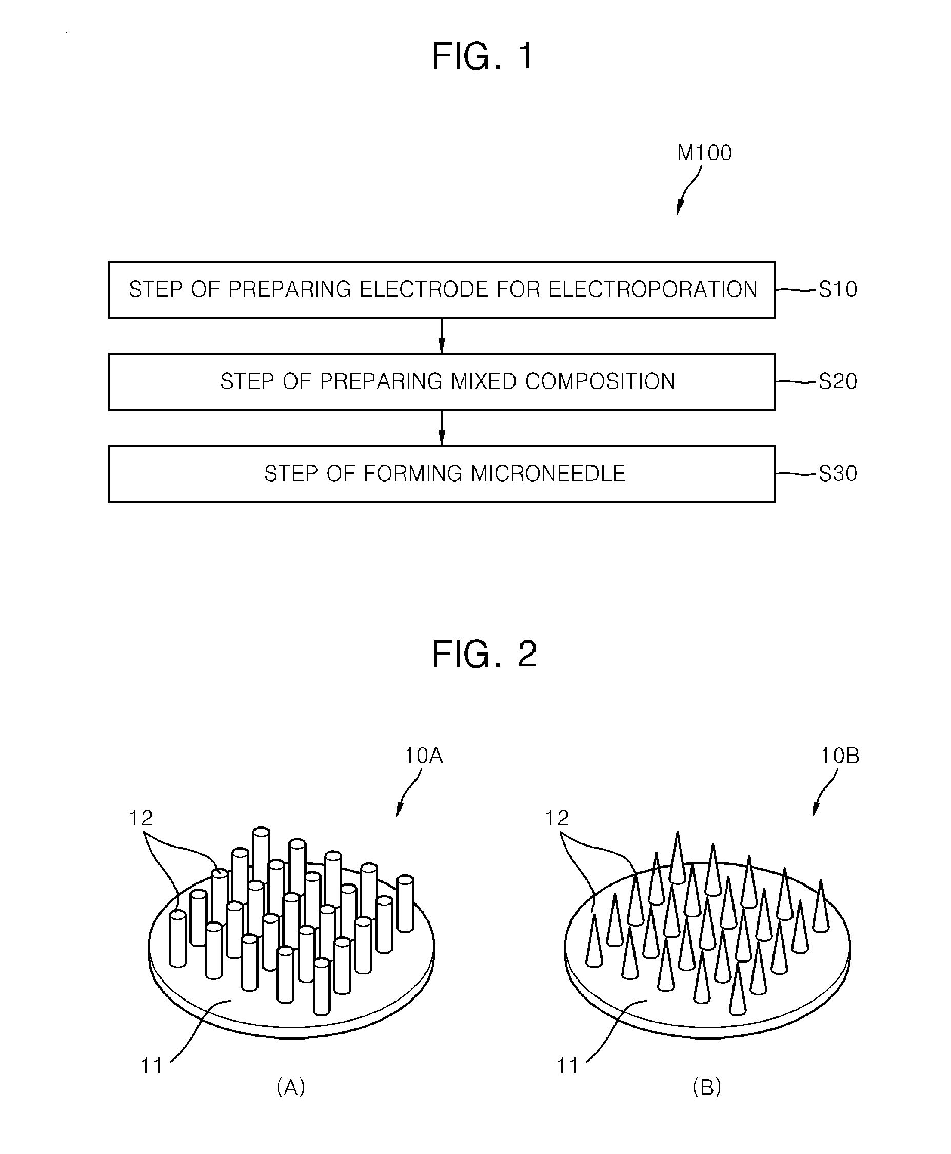
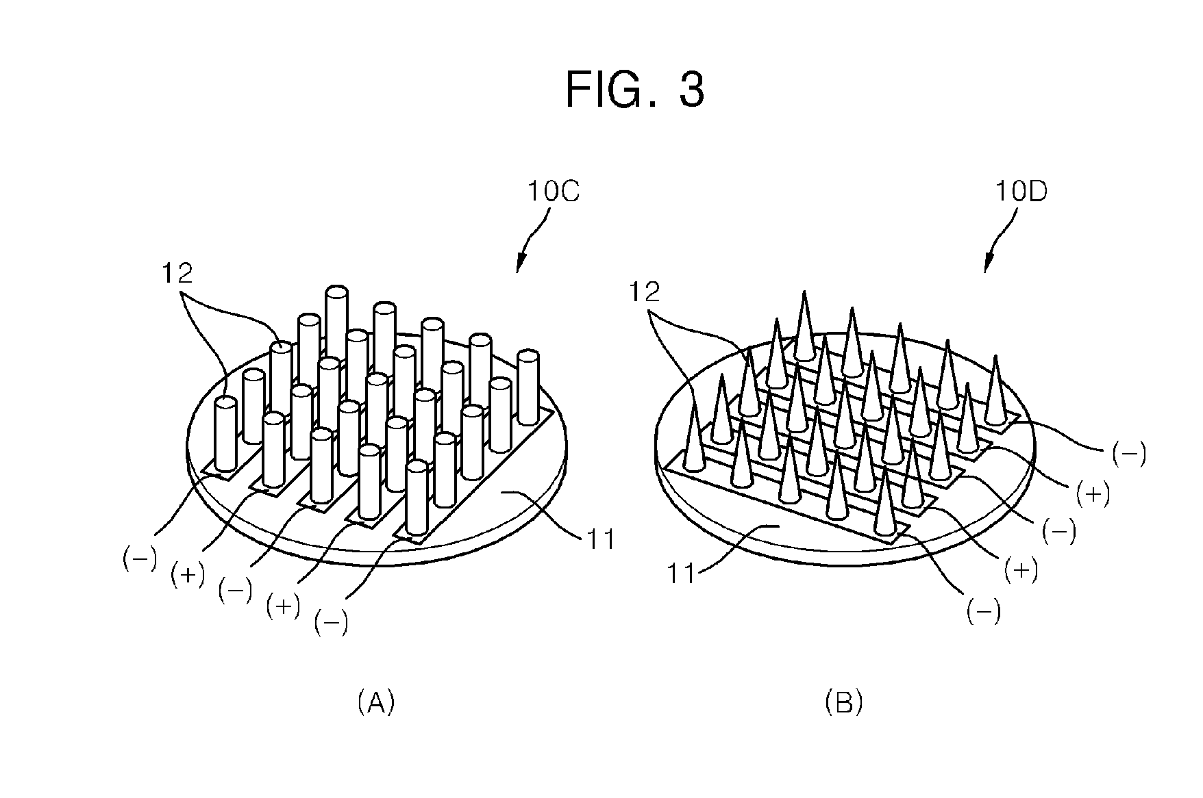
[0019]FIG. 1 illustrates a flowchart showing a method of preparing an electro-microneedle integrated body according to one embodiment of the present invention, FIG. 2 and FIG. 3 illustrate rough perspective views of an electrode for electroporation, and FIG. 4 and FIG. 5 illustrate rough flowcharts showing a method of forming a microneedle.

[0020]Referring to FIG. 1 to FIG. 5, the method of preparing an electro-microneedle integrated body according to the present embodiment (M100) includes preparing an electrode for electroporation (S 10), preparing a mixed composition (S20), and forming a microneedle (S30).

[0021]The preparing of the electrode for electroporation (S10) is a step to prepare an electrode for electroporation. The electrode for electroporation (10A to 1...


PUM

PropertyMeasurementUnit
Compositionaaaaaaaaaa
Biocompatibilityaaaaaaaaaa

Abstract

Provided is an electro-microneedle integrated body in which a dissolving microneedle and an electrode for electroporation are integrated into one, which enables a focused and efficient intracutaneous gene release for percutaneous gene delivery and intracellular gene delivery occurring in one in-situ treatment site The electro-microneedle integrated body according to the present invention includes an electrode for electroporation which is contacted with skin of a human body to apply an electric field pulse, including a base part and a plurality of electrode parts protruding from the base part, and a microneedle adhered to each electrode part and inserted into the skin of a human body, including a biocompatible and biodegradable viscous material and a genetic material, wherein the microneedle degrades within the skin, and the electric field pulse is applied through the electrode for electroporation in a site in which the microneedle is inserted.

Description

CROSS REFERENCE TO RELATED APPLICATIONS[0001]This applications is a continuation of International Application No. PCT / KR2012 / 005777 filed on Jul. 19, 2012, which claims priority to Korean Application No. 10-2011-0074250 filed Jul. 26, 2011, which applications are incorporated herein by reference.TECHNICAL FIELD[0002]The present invention relates to an electro-microneedle integrated body in which a dissolving microstructure and an electrode for electroporation are integrated into one structure for a percutaneous gene delivery in a treatment site, and to a method of preparing the same.BACKGROUND ART[0003]Purposes of gene delivery (gene transfer, delivery) and gene therapy are to treat diseases or to develop treatment models by introducing foreign genes to inside of a body to replace a gene having defective cells or by introducing a required gene to provide a particular function. In particular, attentions were made to cutaneous gene transfer and gene therapy thanks to its easy accessib...

Claims


Application Information

Patent Timeline
IPC IPC(8): A61N1/32B05D5/00
CPCB05D5/00A61N1/327A61K9/0009A61K9/0021A61K48/0075A61M37/0015A61M2037/0046A61M2037/0053A61N1/042A61N1/0424A61N1/0476A61N1/0502A61K48/00A61M5/158A61M37/00
Inventor JUNG, HYUNGILLEE, KWANGJEONG, DO HYEONKIM, JUNG DONG
Owner RAPHAS
Why Eureka
Try Eureka