Biomarker for breast cancer

a breast cancer and biomarker technology, applied in the field of breast cancer biomarkers, can solve problems such as difficult discrimination, and achieve the effects of poor prognosis, easy diagnosis, and effective breast cancer treatmen

Active Publication Date: 2014-04-17
J PHARMA
View PDF1 Cites 2 Cited by
  • Summary
  • Abstract
  • Description
  • Claims
  • Application Information

AI Technical Summary

Benefits of technology

[0039]According to the clauses (1) and (5) in the present invention, diagnosis can be easily conducted to show guidelines for the treatment of mammary gland tumors, which was difficult to do until now. More specifically, the examination of LAT1 expression in breast cancer by immunostaining using the anti-LAT1 antibody of mammary gland tumors enables conducting an effective cancer therapy for breast cancers of which the target is LAT1.
[0040]According to the clauses (2) and (6) in the present invention, the discrimination between benignancy (intraductal breast papilloma) and malignancy of intraductal breast tumors (ductal carcinoma in situ) can be easily conducted, of which diagnosis was difficult.
[0041]According to the clauses (3) and (7) in the present invention, most triple negative breast cancers that are negative for all of ER, PrG, and HER2 have a poor prognosis, but, guidelines for the treatment for triple negative breast cancer can be provided.
[0042]According to the clauses (4) and (8) in the present invention, highly accurate guidelines of the treatment can be provided in diagnosing breast cancer by the combined usage of an anti-LAT1 monoclonal antibody with ER, PgR and HER2 that have been conventionally known as markers.
[0043]In the present invention, mammary gland tumors are not particularly limited as long as mammary gland tissues contain tumors. Typical examples include, for example, benign tumors such as intraductal breast papilloma, adenoma of the nipple, and the like; malignant tumors such as noninfiltrating cancers (ductal carcinoma in situ, and the like), infiltrating carcinomas [ductal carcinoma in situ (papillotubular carcinoma, solid-tubular carcinoma, scirrhous carcinoma), specific type], Paget's disease, and the like. Moreover, mammary gland tumors are divided into breast cancer, which is malignant tumor, and benign tumors according to another classification.
[0044]The intraductal breast tumors are not particularly limited as long as tumors are contained inside of breast ducts Typical examples include, for example, intraductal breast papilloma (benign tumor) and ductal carcinoma in situ (malignant tumor).

Problems solved by technology

However, well-defined discrimination elements are not clear, so there are many lesions that are difficult to discriminate between benignancy and malignancy.

Method used


Image

  • Biomarker for breast cancer
  • Biomarker for breast cancer
  • Biomarker for breast cancer

Examples


reference example 1

Extraction of Experimental Samples

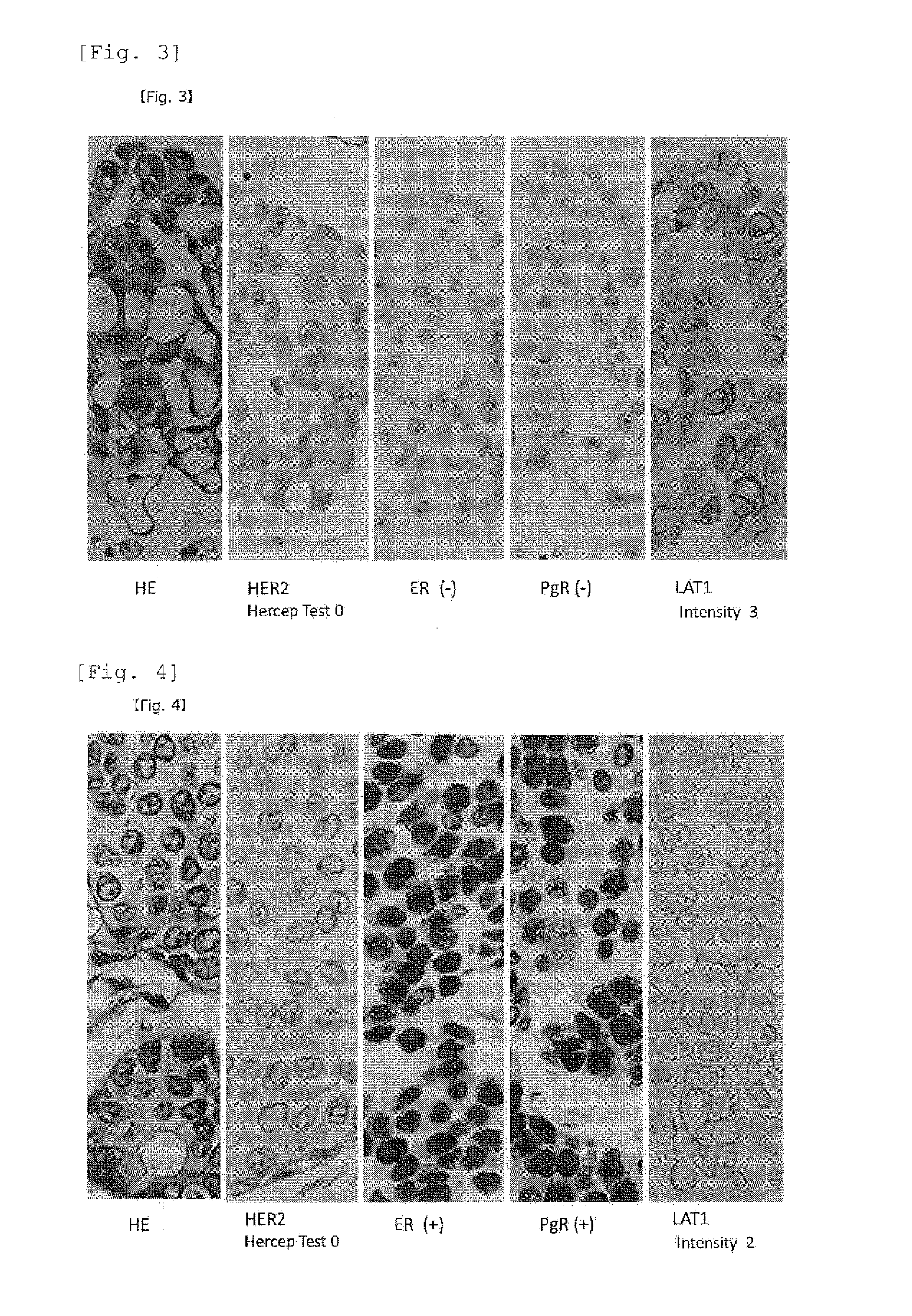
[0095]Neoplastic lesions of mammary glands, which were surgically resected between 2005 and 2009 at a facility where the present inventors belonged, were randomly extracted. Pathological samples of surgical materials in a total of 87 cases including 10 cases of intraductal breast papilloma, 10 cases of ductal carcinoma in situ (DCIS), and 67 cases of infiltrating cancer (invasive carcinoma) were used. Among them, 17 cases were triple negative infiltrating carcinomas that were negative for all of ER, PgR, and HER2.

example 1

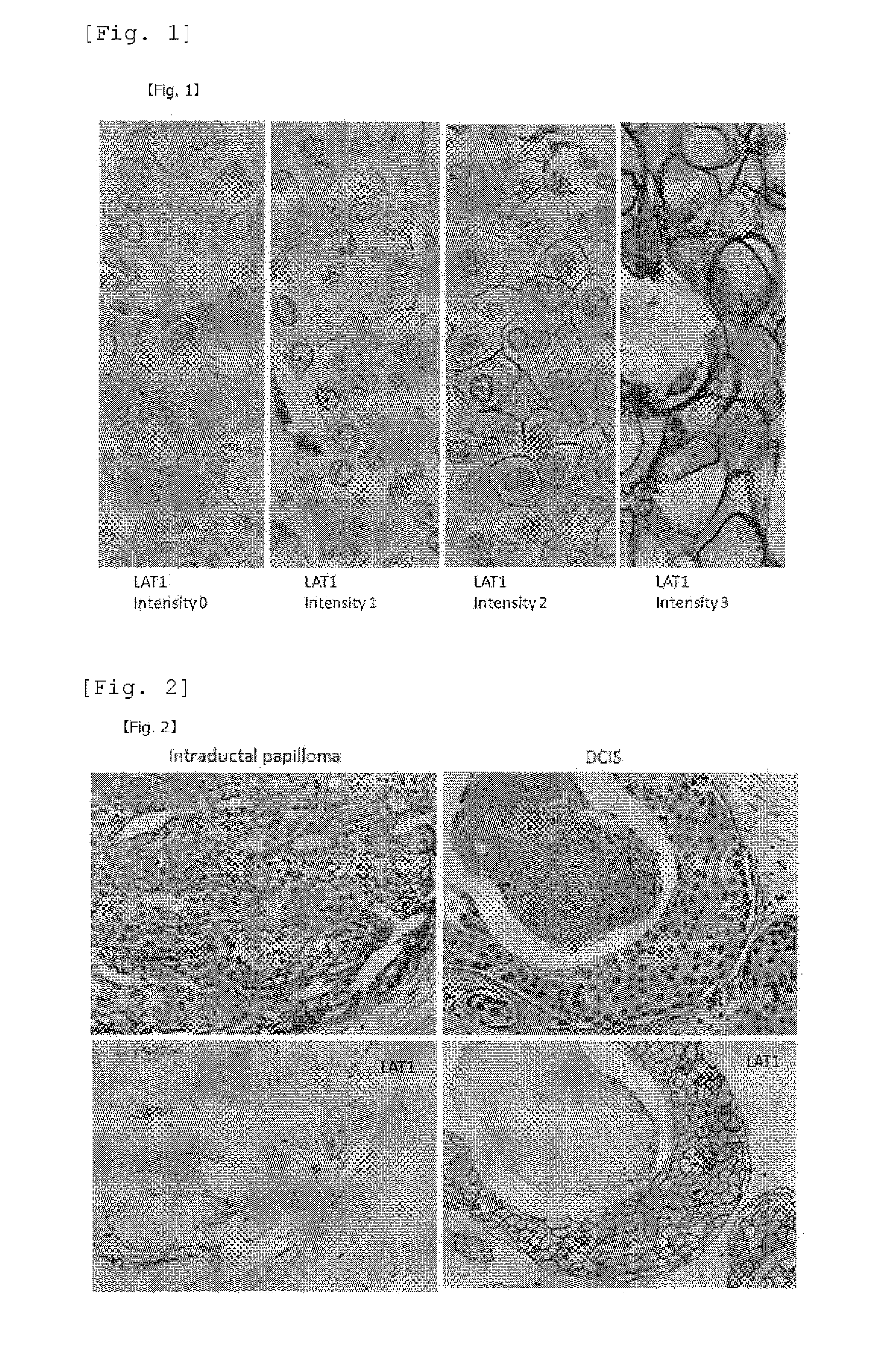
Staining Using an Anti-LAT Antibody

[0096]All the extracted neoplastic lesions of mammary glands in Reference Example 1 were subjected to 10% formalin fixation and a paraffin-embedded block was prepared, further prepared a thinly sliced section with a 3 μm in a thickness from a typical block having maximum divided face of neoplastic lesions. Furthermore, after activating with a microwave for 5 minutes, the sliced section was incubated with the anti-LAT1 mouse monoclonal antibody (refer to Non-Patent Literature 1 pp. 23, in FIG. 3A pp. 631) prepared by the present inventors at a temperature of 4° C. or lower overnight. Further, the section was added a universal antibody contained in a DAKO ChemMate ENVISION System HRP kit, and reacted at room temperature for 30 minutes, and then color was developed by reacting with a DAB (3,3′-diaminobenzidine tetrahydrochloride) substrate solution. For nucleus staining, methyl green or Mayer's Hematoxylin staining was carried out. For the immunostain...


PUM

PropertyMeasurementUnit
thicknessaaaaaaaaaa
temperatureaaaaaaaaaa
concentrationaaaaaaaaaa

Abstract

A method for the diagnosis of breast cancer using an anti-LAT1 monoclonal antibody. Among intraductal breast neoplastic lesions, duct carcinoma in situ, which is malignant, expresses LAT1 at a significantly high level, as compared to intraductal breast papilloma, which is benign. An anti-LAT1 monoclonal antibody is useful for the discrimination between these two lesions. Further, LAT1 is expressed at a high level in most of triple negative infiltrating carcinomas, which are negative for all of ER, PgR, and HER2 that are conventional molecular targeting markers for breast cancer. Therefore, LAT1 can be used as a novel molecular targeting marker for the triple negative breast cancer.

Description

TECHNICAL FIELD[0001]The present invention relates to a biomarker for breast cancers, specifically to a kit and a method for the diagnosis of mammary gland tumors.BACKGROUND ART[0002]A neutral amino acid transporter [L-type amino acid transporter (LAT)] is a transmembrane-type protein which engages in cellular uptake of neutral amino acids such as glycine, alanine, leucine, isoleucine, valine, serine, threonine, cysteine, asparagine, glutamine, methionine, phenylalanine, tyrosine, tryptophan, histidine, L-DOPA, and the like. The present inventors have been searching various LATs and have found LAT1 and isoforms thereof, namely, LAT2, LAT3, and LAT4.[0003]Among them, LAT1 is a 12-pass transmembrane spanning domain protein that has the ability to transport large neutral amino acids such as leucine, isoleucine, valine, methionine, phenylalanine, tyrosine, tryptophan, histidine, and the like; and L-DOPA, Na+ coexisting with an amino-acid transport activating factor, 4F2hc independently....

Claims


Application Information

Patent Timeline
Patent Type & Authority Applications(United States)
IPC IPC(8): G01N33/574
CPCG01N33/57415C07K16/28G01N33/48G01N33/53G01N33/574
Inventor ENDOU, HITOSHIOKAYASU, ISAO
Owner J PHARMA
Why Eureka
Try Eureka