Composition for improving inflammatory disease using abh antigens
a technology of inflammatory diseases and antigens, applied in the direction of dna/rna fragmentation, plant/algae/fungi/lichens ingredients, dispersed delivery, etc., can solve the problems of no success in these efforts so far, and achieve the effects of improving or relieving symptoms of inflammatory diseases carrying inflammation, enhancing skin barrier function, and accelerating antimicrobial peptide expression
- Summary
- Abstract
- Description
- Claims
- Application Information
AI Technical Summary
Benefits of technology
Problems solved by technology
Method used
Image
Examples
experimental example 1
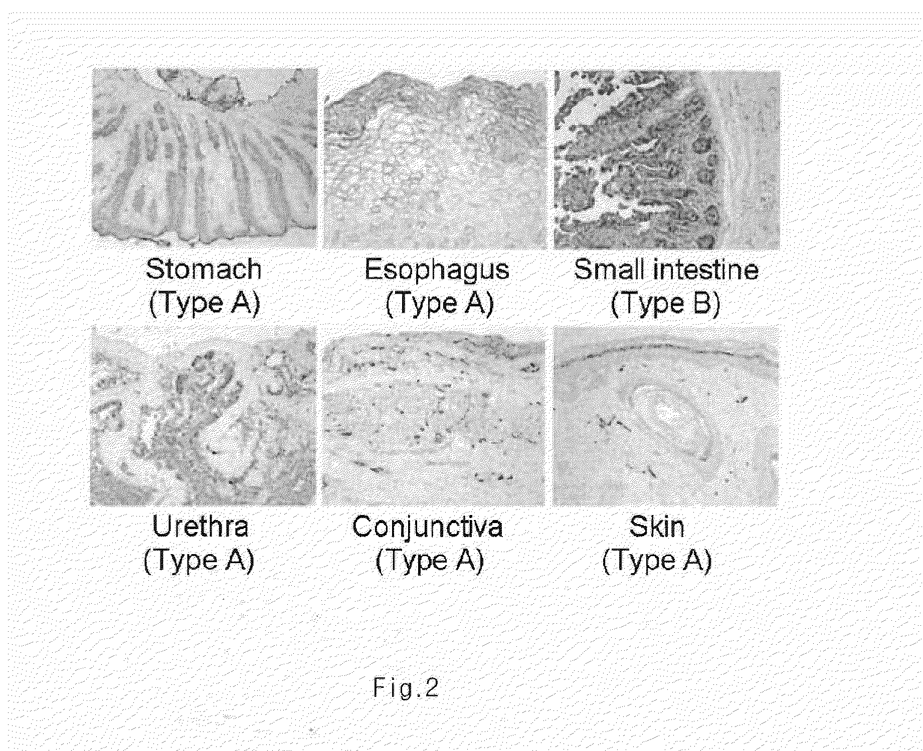
Investigation of the Expression of ABH Antigen in Different Human Tissues
[0044]Biopsy was performed to obtain different human tissues from various parts of human body. Immunohistochemical staining was performed by using ABH antigen specific antibody in order to investigate the expression of ABH antigen in each tissue. Particularly, the tissues obtained from biopsy were fixed in 10% formalin for overnight. Then, formalin was removed and the tissues were dehydrated, so as to make paraffin block. The tissue block was sliced into 0.4 μm thick sections, which were placed on slide. The slide was loaded in a 58° C. dry oven for one hour to melt paraffin. To eliminate paraffin completely, the slide was treated with xylem 4 times, 5 mines for each time, and then the slide was soaked in 100% ethanol twice, 1minute for each, in 95% ethanol for 1 minute, in 80% ethanol for 1 minute, and in 70% ethanol for 1 minute, leading to stepwise rehydration. The slide was washed with running tap water for...
experimental example 2
Down-regulation of the Expression of ABH Antigen by UV Irradiation
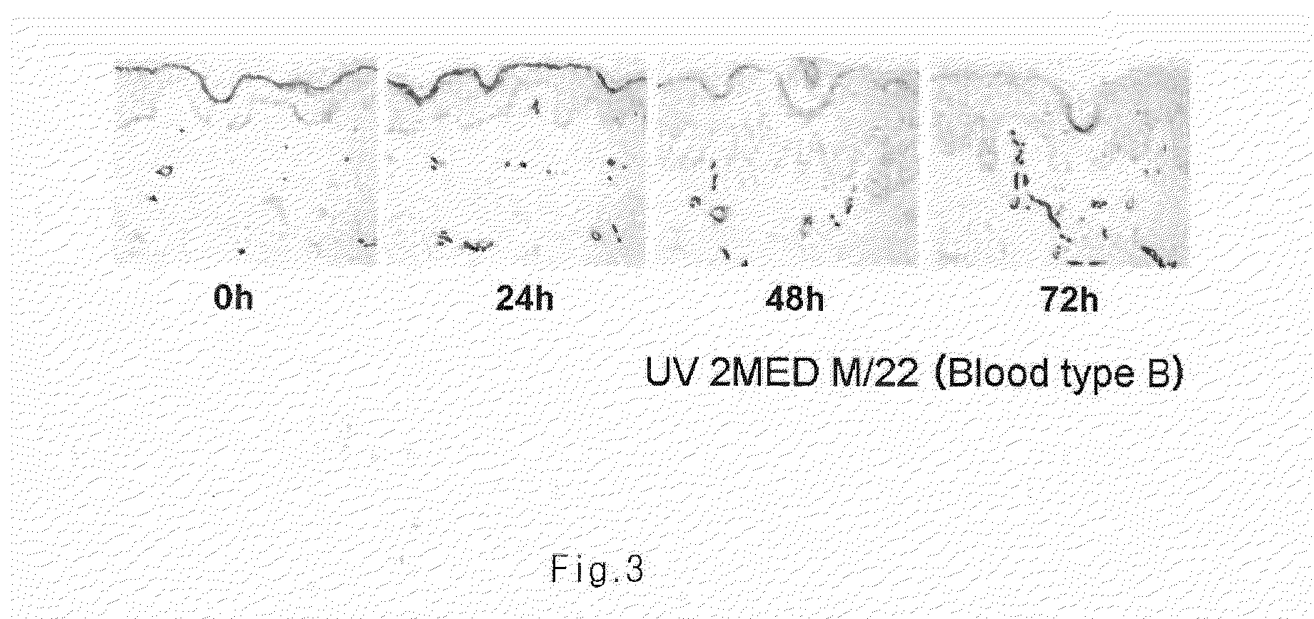
[0046]In normal tissues, the tissues of a person with blood type A are stained only by A antigen specific antibody, which usually takes place in granular layer in epidermis (Dabelsteen et al., J Invest dermatol 82:13-17(1984)). Minimal erythema dose (MED), indicating the strength with which the first erythema is observed after UV irradiation, was measured individually. Each skin was irradiated by 2MED, and then skin biopsy was performed 0, 28, 48, and 72 hours after the UV irradiation. Immunohistochemical staining was performed by the same manner as described in Experimental Example 1 to investigate the expression of ABH antigen.
[0047]As a result, ABH antigen was normally expressed until 24 hours after the UV irradiation, However, after 48-72 hours, the expression of ABH antigen was gradually decreased (FIG. 3).
experimental example 3
Down-regulation of the Expression of ABH Antigen in Various Skin Diseases
[0048]Based on the fact that the expression of ABH antigen is decreased by UV irradiation at the strength of inducing inflammation, the expression of ABH antigen was investigated in diverse skin diseases presumably carrying symptoms caused by irregulation of inflammation reaction. For the investigation, tissues were obtained by biopsy from patients having psoriasis (type A), atpoic dermatitis (type A), ichthyosis (type A), cellulitis (type A), discoid lupus (type A), acne (type B), etc. Immunohistochemical staining was performed with the tissues by the same manner as described in Experimental Example 1 to investigate the expression of ABH antigen.
[0049]As a result, the expression of ABH antigen in granular layer was deficient in most pathological tissues. In spite of individual difference, the expression of abnormal ABH antigen was commonly in spinous layer or basal layer (FIG. 4 and FIG. 5). This result indica...
PUM
 Abstract
Description
Claims
Application Information
 - Generate Ideas
- Intellectual Property
- Life Sciences
- Materials
- Tech Scout
- Unparalleled Data Quality
- Higher Quality Content
- 60% Fewer Hallucinations
Browse by: Latest US Patents, China's latest patents, Technical Efficacy Thesaurus, Application Domain, Technology Topic, Popular Technical Reports.
© 2025 PatSnap. All rights reserved.Legal|Privacy policy|Modern Slavery Act Transparency Statement|Sitemap|About US| Contact US: help@patsnap.com


