
Method for identifying or detecting genomic rearrangements in a biological sample

a biological sample and genomic technology, applied in the field of genomic rearrangement identification or detecting in biological samples, can solve the problems of insufficient reliability of diagnostic use, mainly limited by resolution, and the current use of probe designs that fail to allow cost- and time-efficient high resolution analysis of rearrangements, and achieve high precision/high-resolution detection. , the effect of increasing the risk of occurren

Active Publication Date: 2014-01-09
GENOMIC VISION
View PDF2 Cites 0 Cited by
  • Summary
  • Abstract
  • Description
  • Claims
  • Application Information

AI Technical Summary

Benefits of technology

The patent describes a method for designing short probes to detect and visualize DNA molecules. These probes can be used for diagnosis and assessment of various diseases, including colorectal cancer, and can also predict the outcome of a disease or the response to therapy. The method involves molecular combing, which allows direct visualization of single DNA molecules attached to a glass surface. The invention also includes designing a set of short and long probes and a technique to predict the course and outcome of a disease or condition using high resolution barcodes. Overall, this technology improves the analysis of DNA across the genome and has wide applications in disease diagnosis and treatment.

Problems solved by technology

Presently, the detection of genetic rearrangements using current technologies is often insufficiently reliable for diagnostics use.
Their use is mainly limited by their resolution.
Molecular Combing, on the other hand, may reach sufficient resolution, but probe designs currently used fail to allow cost- and time-efficient high resolution analysis of rearrangements.

Method used


Image

  • Method for identifying or detecting genomic rearrangements in a biological sample
  • Method for identifying or detecting genomic rearrangements in a biological sample
  • Method for identifying or detecting genomic rearrangements in a biological sample

Examples


examples

[0175]Application to HNPCC—Materials and Method

[0176]Probe Design v1

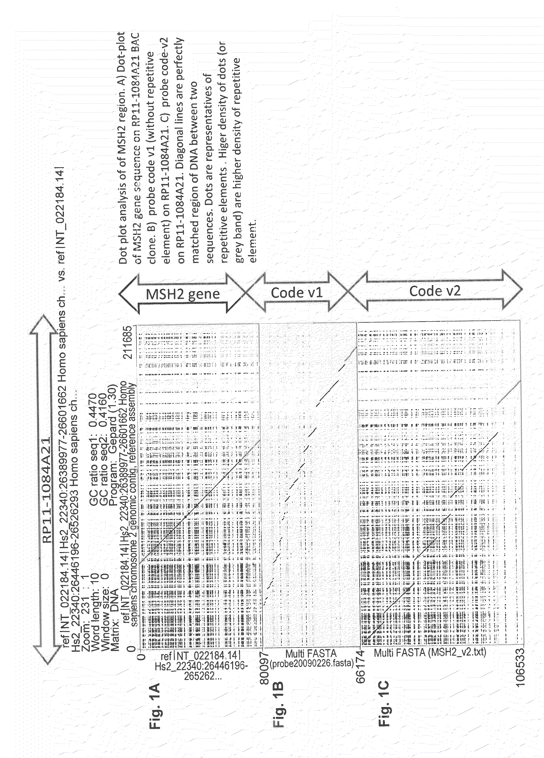
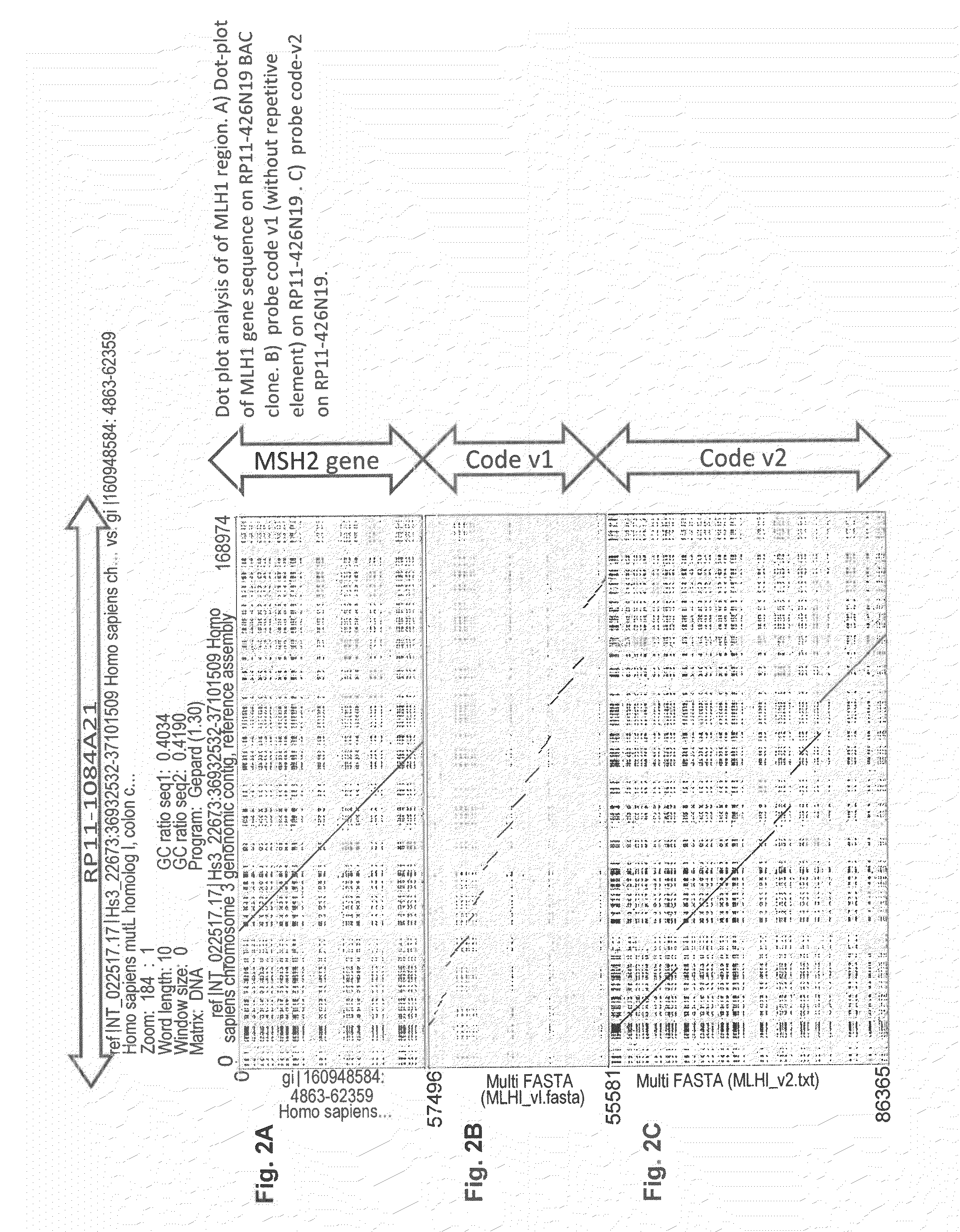
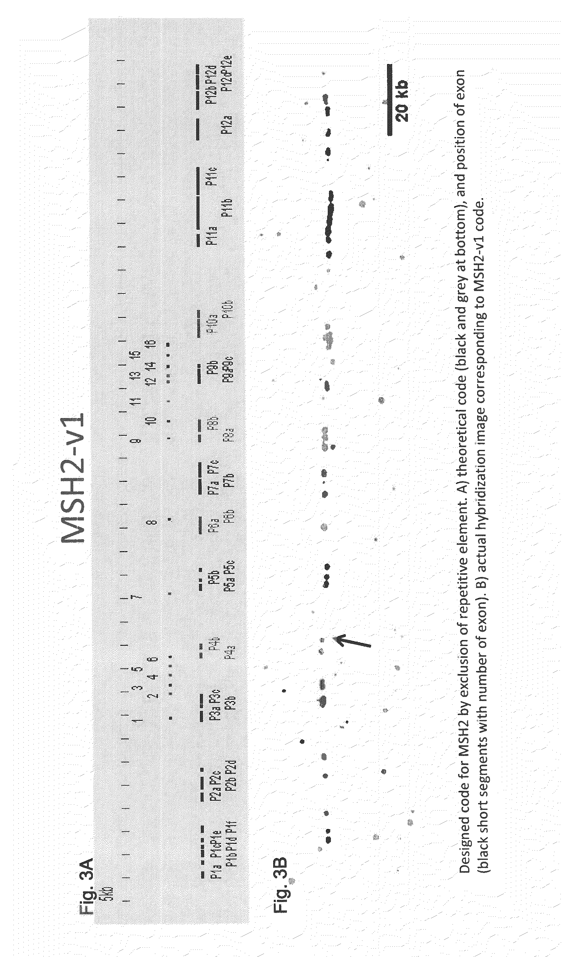
[0177]Each probe (probe means continuous hybridization signal, can consist of multiple cloned DNA fragments, e.g., probe 1 of MSH2-v2 covers a 15 kb stretch and consists of five cloned DNA fragments of 3 kb. Since gap or overlap of each junction of these five fragments are smaller than resolution (<50 bp), they are considered and indeed look like continuous single probe of 15 kb) on a region of gene sequence itself has a length between 3-6 kb. In case of larger rearrangement than probe or gap size, obvious change of color pattern of designed probe will be observed. As well as large rearrangement in probe region, such rearrangement is also detectable in gap region, meaning any rearrangement larger than 1 kb at any position in the target genes are detectable. This is a uniqueness of cartography method with high resolution probe hybridization. Other techniques (MLPA, aCGH) can detect only such rearrangement involving p...


PUM

PropertyMeasurementUnit
Fractionaaaaaaaaaa
Digital informationaaaaaaaaaa

Abstract

A method for detection, visualization and / or comparison of polynucleotide sequences of interest using specially designed sets of long and short probes that enhance resolution and simplify visualization and detection. Probe compositions useful for practicing this method and procedures for identifying useful probes and probe combinations. These methods are useful for the detection of genomic rearrangements, especially those associated with various diseases, disorders and conditions including cancer or for assessment of genomic rearrangements associated with therapy. The probe compositions may be used in kits for detection of genetic rearrangements or in companion diagnostic products or kits, such as kits for the diagnosis or assessment of predisposition to cancer such as colorectal cancer.

Description

CROSS-REFERENCE TO RELATED APPLICATIONS[0001]The present application claims priority to U.S. Provisional Application No. 61 / 553,889, filed Oct. 31, 2011, the entire contents of which are incorporated herein by reference. On Oct. 30, 2012, an International Application (PCT / IB / ______; submission number 1000168921) was also filed with the same title, the entire contents of which are incorporated herein by reference.BACKGROUND OF THE INVENTION[0002]1. Field of the Invention[0003]The invention relates to high-resolution, precise method for detecting genomic rearrangements in vitro using specially designed combinations of polynucleotide probes. The invention concerns accurate methods of detection and diagnosis of conditions, disorders and diseases associated with rearrangement of genomic DNA.[0004]2. Description of the Related Art[0005]The Multigenic Paradigm of Human Diseases[0006]Advances in genetic analysis of human diseases have provided better insights into the molecular mechanisms c...

Claims


Application Information

Patent Timeline
IPC IPC(8): C12Q1/68
CPCC12Q1/6881C12Q1/6827C12Q1/6841C12Q2600/156C12Q1/6886C12Q2523/303C12Q2537/143C12Q2565/102C12Q1/6883
Inventor KOMATSU, JUNWALRAFEN, PIERRECEPPI, MAURIZIOCONSEILLER, EMMANUEL
Owner GENOMIC VISION
Learn More