
Method for creating attenuation correction maps for pet image reconstruction

Active Publication Date: 2013-10-10
SIEMENS HEATHCARE GMBH
View PDF23 Cites 40 Cited by
  • Summary
  • Abstract
  • Description
  • Claims
  • Application Information

AI Technical Summary

Benefits of technology

The present invention provides a method for correcting attenuation of positron emission tomography (PET) image data of an area under examination by segmenting magnetic resonance (MR) images of the area into bone, soft tissue, and air using a patch-based segmentation technique. The MR images may comprise MR ultra-fast TE sequence images. The method also includes creating pseudo-CT images from the labeled MR images and performing attenuation correction of the PET image data from the pseudo-CT images. The technical effect of this invention is to improve the accuracy of PET imaging and provide more reliable data for diagnosis and treatment of various medical conditions.

Problems solved by technology

This can adversely affect the image interpretation and any medical diagnoses by the health professional.
However, MR imaging does not normally produce images with clearly defined bone segmentation, which is essential to performing accurate attenuation correction of PET imaging data.
Since manual segmentation by an expert is time-consuming and presents issues of intra and inter-reliability, different methods of automatic labeling have been developed during the last decade.
On MR images, intensity is similar in bones and air (i.e., air-filled body cavities), therefore a segmentation relying solely on the image is not feasible.
However, this assumption may not hold.

Method used


Image

  • Method for creating attenuation correction maps for pet image reconstruction
  • Method for creating attenuation correction maps for pet image reconstruction
  • Method for creating attenuation correction maps for pet image reconstruction

Examples


Embodiment Construction

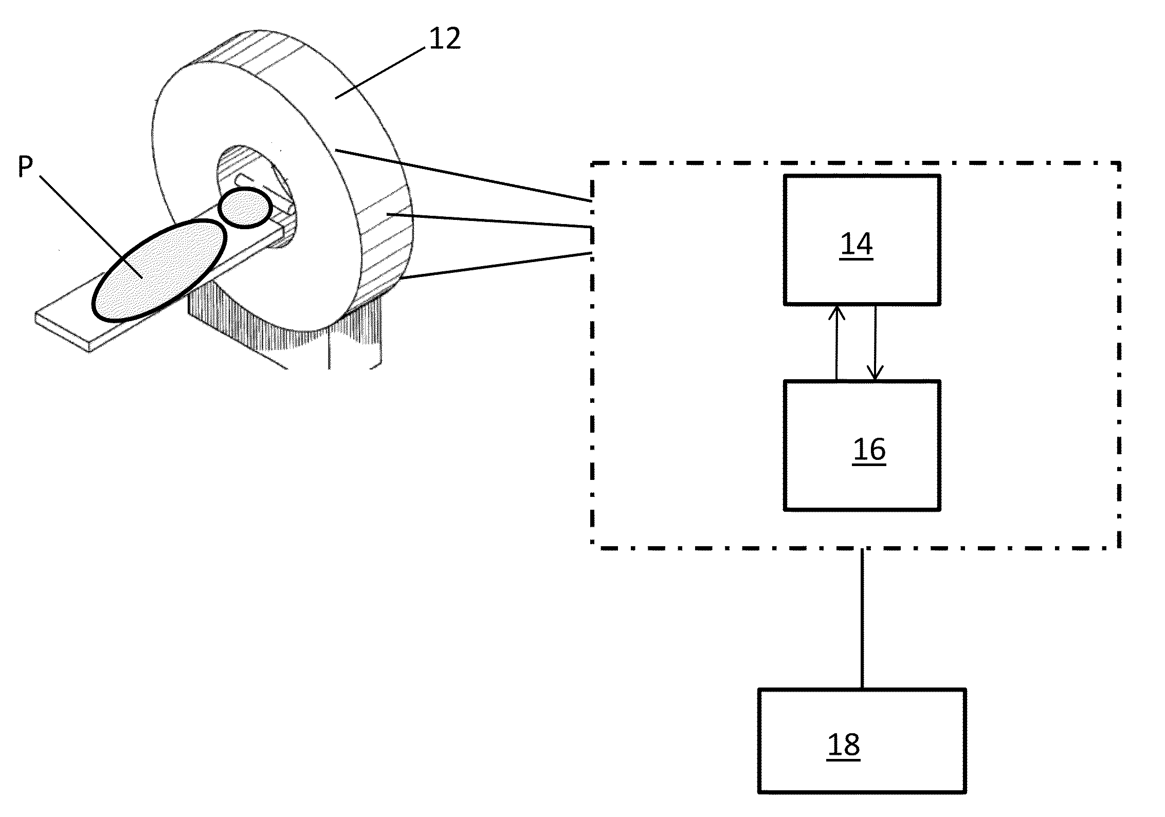
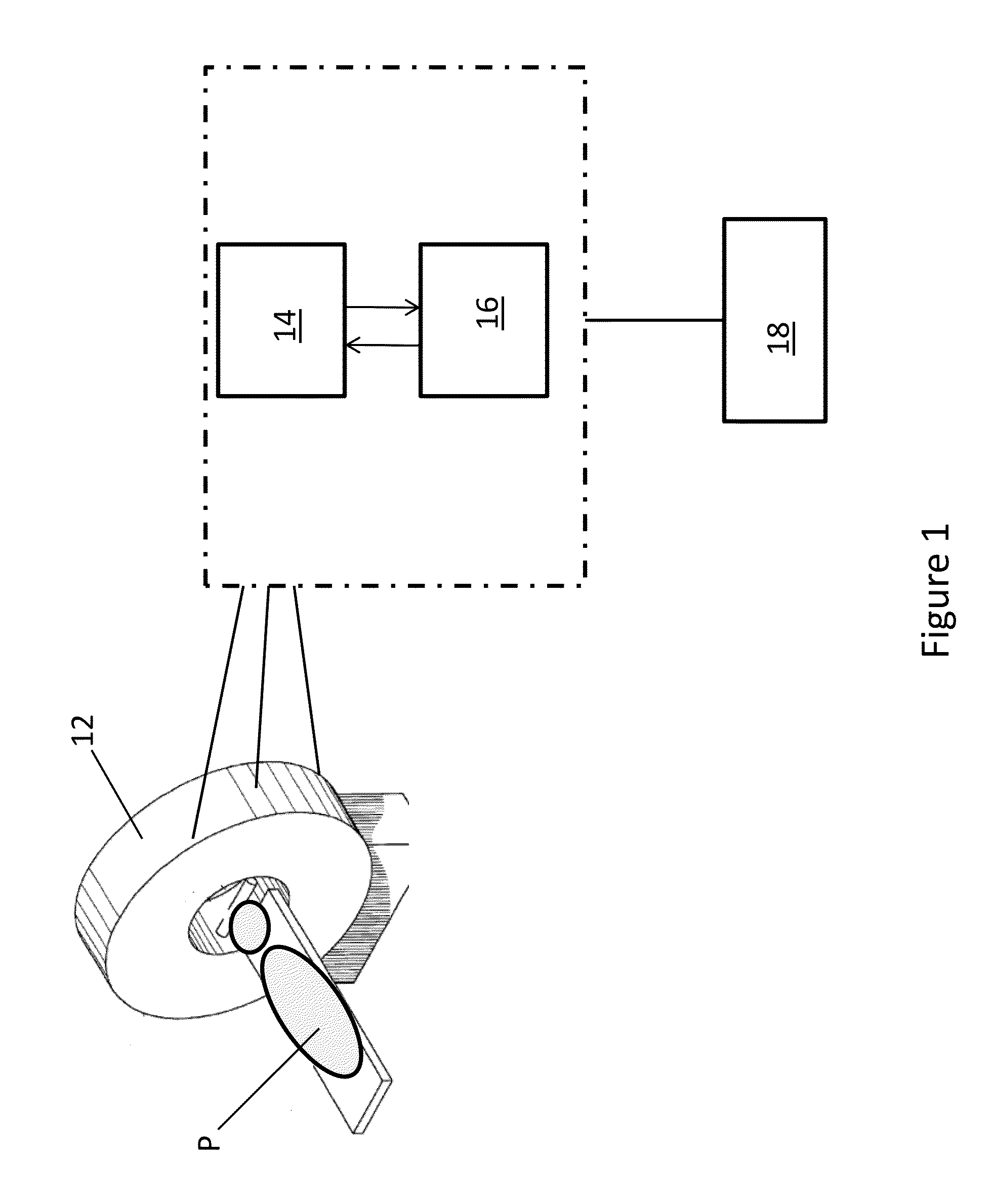
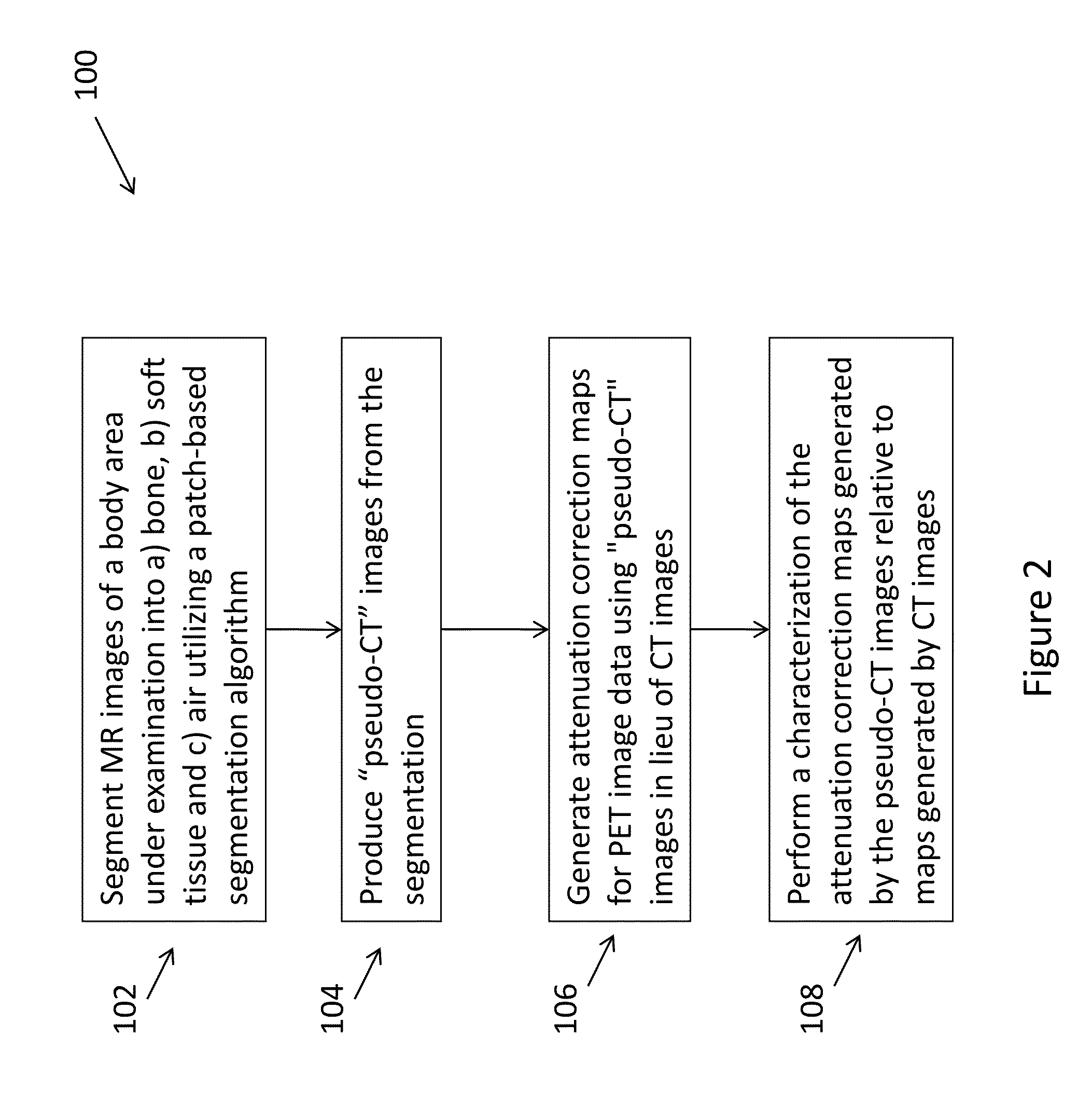
[0026]FIG. 2 shows an embodiment of a method 100 of the present invention to perform attenuation correction, using MR imaging, of PET imaging data. The PET imaging system 10 and, in particular, either processor 14, 16, is adapted to operate according to the method 100. The method 100 is described herein with reference to a patient's head / brain as the body P area under examination although it is understood that the method 100 is not so limited and may be applied to any body P area of interest.

[0027]Generally, the method 100 comprises utilizing a patch-based segmentation technique or algorithm to segment MR images of a body area under examination into bone, soft tissue, and air (Step 102). The method 100 then produces “pseudo-CT” images from the segmentation (Step 104). As further described below, the method 100 generates a pseudo-CT image by replacing the predictions of bone, soft tissue and air (from the patch mappings) with typical, fixed Hounsfield unit values for each of those in...


PUM


Abstract

A method (100) that generates attenuation correction maps for the reconstruction of PET data using MR images, such as, MR ultra-fast TE (UTE) images, Dixon MR images, as well as MR images obtained using other MR imaging methods.

Description

CROSS-REFERENCE TO RELATED APPLICATIONS[0001]This application claims the benefit of Provisional U.S. Patent Application Ser. No. 61 / 620,073 entitled, “Patch-based Segmentation of UTE Brain Images to Create Attenuation Correction Maps for PET Reconstruction”, filed in the name of Helene Chopinet, Francisco Pereira, James G. Reisman, and Christophe Chefd'hotel on Apr. 4, 2012 and Provisional U.S. Patent Application Ser. No. 61 / 644,029 entitled, “Accelerated. Multi-scale Patch-based Segmentation of MR Images to Identify the Bone Compartment and Create Attenuation Correction Maps for PET Image Reconstruction”, filed in the name of Francisco Pereira, James G. Reisman, and Christophe Chefd'hotel on May 8, 2012, the disclosures of which are also hereby incorporated herein by reference.FIELD OF INVENTION[0002]The present invention generally relates to simultaneous MRI / PET image data acquisition. More particularly, the present invention relates to generating attenuation correction maps for P...

Claims


Application Information

Patent Timeline
IPC IPC(8): G06T7/00
CPCG06T7/0012G06T5/008G06T2207/30016G06T2207/10088G06T2207/10104G06T5/50G06T5/94
Inventor PEREIRA, FRANCISCOCHOPINET, HELENEREISMAN, JAMES G.CHEFD'HOTEL, CHRISTOPHE
Owner SIEMENS HEATHCARE GMBH
Learn More