
Systems and methods for segmentation and processing of tissue images and feature extraction from same for treating, diagnosing, or predicting medical conditions

a tissue image and feature extraction technology, applied in image analysis, image enhancement, instruments, etc., can solve the problems of difficult segmentation of lumens, exacerbated problems, and under-segmentation of clustered nuclei

Active Publication Date: 2013-09-05
AUREON LAB INC +1
View PDF6 Cites 81 Cited by
  • Summary
  • Abstract
  • Description
  • Claims
  • Application Information

AI Technical Summary

Benefits of technology

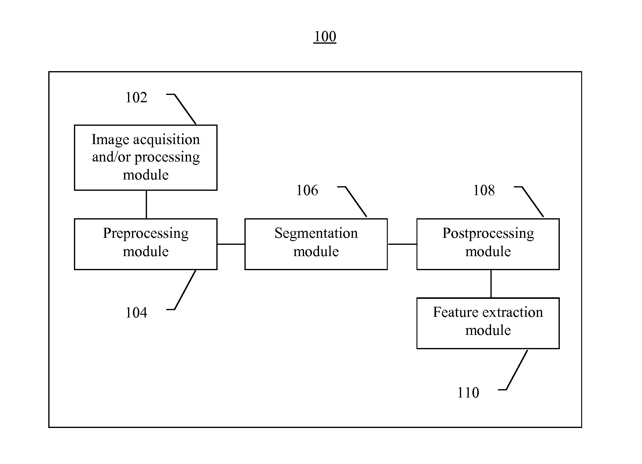
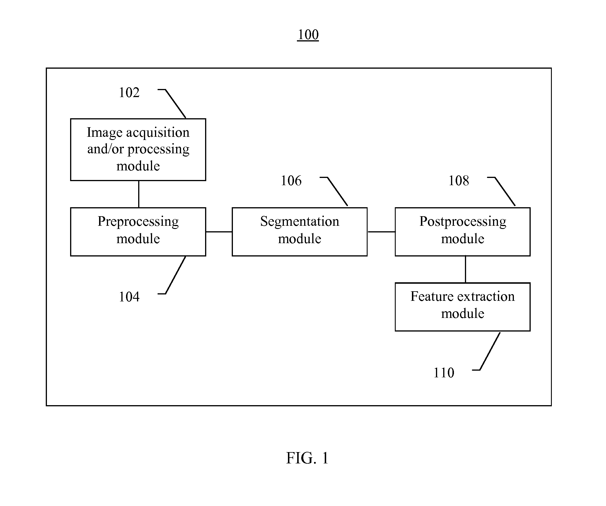
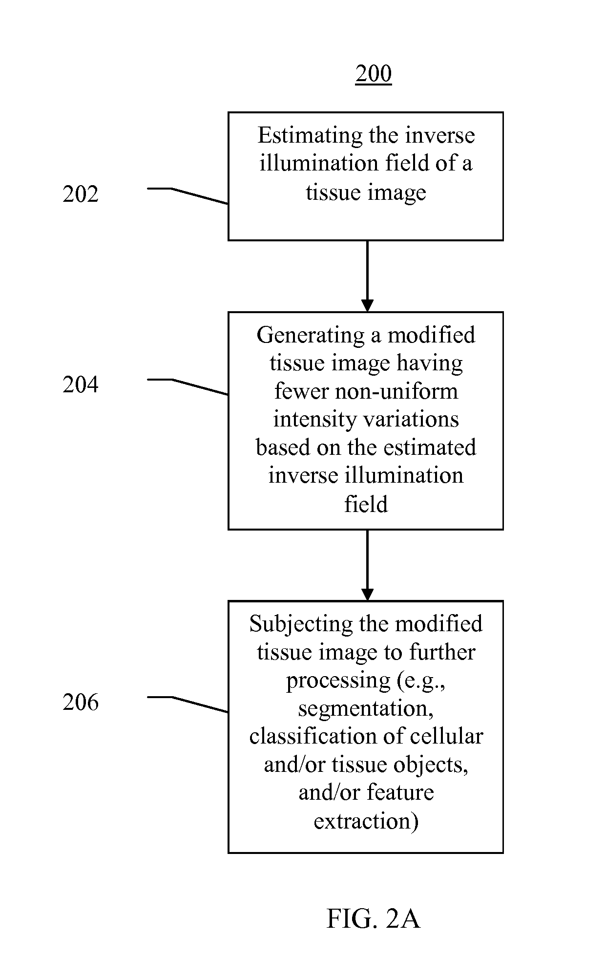
The present invention relates to a method for processing tissue images. The method involves binarizing the tissue image and reducing non-uniform variations in intensity. This is useful because it helps in identifying different cells and structures within the image. The method also involves segmenting the image and extracting marker centers, which are subsequently used to create a new image of intensity valleys and peaks. A transformation is then applied to the new image to generate lines of separation. The resulting image is then labeled and separation boundaries are extracted. This method could be used in conjunction with other methods, such as a boundary initialized process or a watershed transform, to improve the accuracy and efficiency of identifying and extracting epithelial units from tissue images.

Problems solved by technology

Conventional methods for segmentation of tissue images are prone to misclassification of objects in tissue (e.g., epithelial and stromal nuclei) and may produce irregular nuclei, incorrectly identify cytoplasm boundaries, and result in over and under-segmentation of clustered nuclei.
These problems are exacerbated by variations in image acquisition conditions and image artifacts.
However, segmentation of lumens can be difficult as cancer progresses.
However, as cancer progresses, epithelial cells replicate in an uncontrolled way, disrupting regular ring structures.
Existing segmentation systems also have difficulty performing segmentation based on lumens, which have shrunk or disappeared.
The same segmentation difficulty arises for Gleason grade 5, where the tumor loses these structures and becomes undifferentiated sheets of epithelial cells and / or epithelial fragments.

Method used


Image

  • Systems and methods for segmentation and processing of tissue images and feature extraction from same for treating, diagnosing, or predicting medical conditions
  • Systems and methods for segmentation and processing of tissue images and feature extraction from same for treating, diagnosing, or predicting medical conditions
  • Systems and methods for segmentation and processing of tissue images and feature extraction from same for treating, diagnosing, or predicting medical conditions

Examples


example 2

[0226]Localizing per image ARRR70. This involves calculating the weighted average of the ARRR70 per ring feature over the entire image, and the weighted average of the Gleason triangle components per ring over the entire image. The weighting method here uses nuclei area as a weight and normalizes over region types 1 and 2. The weighted average of ARRR70 and each of the three morphology components are multiplied to obtain three values. A least-squares fit of the result to 0 (non-event) or 1 (event) to creates a probability score which is the output feature value.

example 3

[0227]Naïve Bayes per ring ARRR70. This involves multiplying the ring values of ARRR70, by each of the three morphology components (s3, s4f, and s4s). A weighted average of the resultant AR components is then taken over the image, as in Example 1. The three components are separately converted into three probability scores (p3, p4f, p4s) which are then combined assuming the non-event probabilities are independent using the formula:

Probability score=1−(1−p3)(1−p4f)(1−p4s)

example 4

[0228]Naïve Bayes per image ARRR70. This involves calculating the weighted average of the ARRR70 per ring feature over the entire image, and the weighted average of the Gleason triangle components per ring over the entire image as in Example 2. The weighted average of ARRR70 and each of the three Gleason components are multiplied to obtain three values which are separately converted into three probability scores (p3, p4f, p4s). The three probability scores are then combined assuming the non-event probabilities are independent as in Example 3.


PUM


Abstract

Apparatus, methods, and computer-readable media are provided for segmentation, processing (e.g., preprocessing and / or postprocessing), and / or feature extraction from tissue images such as, for example, images of nuclei and / or cytoplasm. Tissue images processed by various embodiments described herein may be generated by Hematoxylin and Eosin (H&E) staining, immunofluorescence (IF) detection, immunohistochemistry (IHC), similar and / or related staining processes, and / or other processes. Predictive features described herein may be provided for use in, for example, one or more predictive models for treating, diagnosing, and / or predicting the occurrence (e.g., recurrence) of one or more medical conditions such as, for example, cancer or other types of disease.

Description

CROSS-REFERENCE TO RELATED APPLICATIONS[0001]This application is a continuation of International Patent Application No. PCT / US2011 / 046149, filed Aug. 1, 2011 which claims priority to U.S. provisional patent application nos. 61 / 400,642, filed Jul. 30, 2010, 61 / 400,657, filed Jul. 30, 2010, 61 / 456,009, filed Oct. 28, 2010, and 61 / 455,988, filed Oct. 28, 2010, all of which are hereby incorporated by reference herein in their entireties.FIELD OF THE INVENTION[0002]Embodiments of the present invention relate to systems and methods for segmentation and processing of images (e.g., tissue images of nuclei and / or cytoplasm) and feature extraction from the same for, for example, treating, diagnosing, and / or predicting the occurrence (e.g., recurrence) of one or more medical conditions (e.g., cancer or other types of disease).BACKGROUND OF THE INVENTION[0003]Conventional methods for segmentation of tissue images are prone to misclassification of objects in tissue (e.g., epithelial and stromal ...

Claims


Application Information

Patent Timeline
IPC IPC(8): G06K9/00
CPCG06T7/0012G06T7/404G06T2207/10056G06T2207/20041G06T7/0091G06T2207/30024G06K9/00147G06T5/008G06T7/0081G06T2207/20212G06T7/11G06T7/155G06T7/45G06T5/94G06V20/698
Inventor AJEMBA, PETERSCOTT, RICHARDRAMACHANDRAN, JANAKIRAMANANZEINEH, JACKDONAVAN, MICHAELFERMANDEZ, GERARDOLIU, QIUHUAKHAN, FAISAL
Owner AUREON LAB INC
Learn More